Ciliary muscle | |
---|---|
Details | |
Pronunciation | UK: /ˈsɪliəri/ , US: /ˈsɪliɛri/ [1] |
Origin | 1) longitudinal fibers → scleral spur; 2) circular fibers → encircle root of iris [2] |
Insertion | 1) longitudinal fibers → ciliary process, 2) circular fibers → encircle root of iris [2] |
Artery | long posterior ciliary arteries |
Vein | Vorticose vein |
Nerve | short ciliary Parasympathetic fibers in the oculomotor nerve (CN-III) synapse in the ciliary ganglion. Parasympathetic postganglionic fibers from the ciliary ganglion travel through short ciliary nerves into the ocular globe. |
Actions | 1) accommodation, 2) regulation of trabecular meshwork pore sizes |
Identifiers | |
Latin | musculus ciliaris |
TA98 | A15.2.03.014 |
TA2 | 6770 |
FMA | 49151 |
Anatomical terms of muscle |
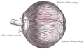
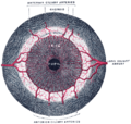
The ciliary muscle is an intrinsic muscle of the eye formed as a ring of smooth muscle [3] [4] in the eye's middle layer, uvea (vascular layer). It controls accommodation for viewing objects at varying distances and regulates the flow of aqueous humor into Schlemm's canal. It also changes the shape of the lens within the eye but not the size of the pupil [5] which is carried out by the sphincter pupillae muscle and dilator pupillae.
The ciliary muscle develops from mesenchyme within the choroid and is considered a cranial neural crest derivative. [6]
The ciliary muscle receives parasympathetic fibers from the short ciliary nerves that arise from the ciliary ganglion. The parasympathetic postganglionic fibers are part of cranial nerve V1 (Nasociliary nerve of the trigeminal), while presynaptic parasympathetic fibers to the ciliary ganglia travel with the oculomotor nerve. [7] The postganglionic parasympathetic innervation arises from the ciliary ganglion. [8]
Presynaptic parasympathetic signals that originate in the Edinger-Westphal nucleus are carried by cranial nerve III (the oculomotor nerve) and travel through the ciliary ganglion via the postganglionic parasympathetic fibers which travel in the short ciliary nerves and supply the ciliary body and iris. Parasympathetic activation of the M3 muscarinic receptors causes ciliary muscle contraction. The effect of contraction is to decrease the diameter of the ring of ciliary muscle causing relaxation of the zonule fibers, the lens becomes more spherical, increasing its power to refract light for near vision.[ citation needed ]
The parasympathetic tone is dominant when a higher degree of accommodation of the lens is required, such as reading a book. [9]
The ciliary fibers have circular (Ivanoff), [10] longitudinal (meridional) and radial orientations. [11]
According to Hermann von Helmholtz's theory, the circular ciliary muscle fibers affect zonular fibers in the eye (fibers that suspend the lens in position during accommodation), enabling changes in lens shape for light focusing. When the ciliary muscle contracts, it pulls itself forward and moves the frontal region toward the axis of the eye. This releases the tension on the lens caused by the zonular fibers (fibers that hold or flatten the lens). This release of tension of the zonular fibers causes the lens to become more spherical, adapting to short range focus. Conversely, relaxation of the ciliary muscle causes the zonular fibers to become taut, flattening the lens, increasing the focal distance, [12] increasing long range focus. Although Helmholtz's theory has been widely accepted since 1855, its mechanism still remains controversial. Alternative theories of accommodation have been proposed by others, including L. Johnson, M. Tscherning, and especially Ronald A. Schachar. [3]
Contraction and relaxation of the longitudinal fibers, which insert into the trabecular meshwork in the anterior chamber of the eye, cause an increase and decrease in the meshwork pore size, respectively, facilitating and impeding aqueous humour flow into the canal of Schlemm. [13]
Open-angle glaucoma (OAG) and closed-angle glaucoma (CAG) may be treated by muscarinic receptor agonists (e.g., pilocarpine), which cause rapid miosis and contraction of the ciliary muscles, opening the trabecular meshwork, facilitating drainage of the aqueous humour into the canal of Schlemm and ultimately decreasing intraocular pressure. [14]
The word ciliary had its origins around 1685–1695. [15] The term cilia originated a few years later in 1705–1715, and is the Neo-Latin plural of cilium meaning eyelash. In Latin, cilia means upper eyelid and is perhaps a back formation from supercilium, meaning eyebrow. The suffix -ary originally occurred in loanwords from Middle English (-arie), Old French (-er, -eer, -ier, -aire, -er), and Latin (-ārius); it can generally mean "pertaining to, connected with", "contributing to", and "for the purpose of". [16] Taken together, cili(a)-ary pertains to various anatomical structures in and around the eye, namely the ciliary body and annular suspension of the lens of the eye. [17]
A ganglion is a group of neuron cell bodies in the peripheral nervous system. In the somatic nervous system this includes dorsal root ganglia and trigeminal ganglia among a few others. In the autonomic nervous system there are both sympathetic and parasympathetic ganglia which contain the cell bodies of postganglionic sympathetic and parasympathetic neurons respectively.
Glaucoma is a group of eye diseases that result in damage to the optic nerve and cause vision loss. The most common type is open-angle glaucoma, in which the drainage angle for fluid within the eye remains open, with less common types including closed-angle glaucoma and normal-tension glaucoma. Open-angle glaucoma develops slowly over time without pain. Peripheral vision may begin to decrease, followed by central vision, resulting in blindness if not treated. Closed-angle glaucoma can present gradually or suddenly. The sudden presentation may involve severe eye pain, blurred vision, mid-dilated pupil, redness of the eye, and nausea. Vision loss from glaucoma, once it has occurred, is permanent. Eyes affected by glaucoma are referred to as being glaucomatous.
In neuroanatomy, the optic nerve, also known as the second cranial nerve, cranial nerve II, or simply CN II, is a paired cranial nerve that transmits visual information from the retina to the brain. In humans, the optic nerve is derived from optic stalks during the seventh week of development and is composed of retinal ganglion cell axons and glial cells; it extends from the optic disc to the optic chiasma and continues as the optic tract to the lateral geniculate nucleus, pretectal nuclei, and superior colliculus.
The autonomic nervous system (ANS), formerly referred to as the vegetative nervous system, is a division of the nervous system that supplies internal organs, smooth muscle and glands. The autonomic nervous system is a control system that acts largely unconsciously and regulates bodily functions, such as the heart rate, its force of contraction, digestion, respiratory rate, pupillary response, urination, and sexual arousal. This system is the primary mechanism in control of the fight-or-flight response.
The parasympathetic nervous system (PSNS) is one of the three divisions of the autonomic nervous system, the others being the sympathetic nervous system and the enteric nervous system. The enteric nervous system is sometimes considered part of the autonomic nervous system, and sometimes considered an independent system.
The oculomotor nerve, also known as the third cranial nerve, cranial nerve III, or simply CN III, is a cranial nerve that enters the orbit through the superior orbital fissure and innervates extraocular muscles that enable most movements of the eye and that raise the eyelid. The nerve also contains fibers that innervate the intrinsic eye muscles that enable pupillary constriction and accommodation. The oculomotor nerve is derived from the basal plate of the embryonic midbrain. Cranial nerves IV and VI also participate in control of eye movement.
The aqueous humour is a transparent water-like fluid similar to blood plasma, but containing low protein concentrations. It is secreted from the ciliary body, a structure supporting the lens of the eyeball. It fills both the anterior and the posterior chambers of the eye, and is not to be confused with the vitreous humour, which is located in the space between the lens and the retina, also known as the posterior cavity or vitreous chamber. Blood cannot normally enter the eyeball.
The ciliary body is a part of the eye that includes the ciliary muscle, which controls the shape of the lens, and the ciliary epithelium, which produces the aqueous humor. The aqueous humor is produced in the non-pigmented portion of the ciliary body. The ciliary body is part of the uvea, the layer of tissue that delivers oxygen and nutrients to the eye tissues. The ciliary body joins the ora serrata of the choroid to the root of the iris.
The accommodation reflex is a reflex action of the eye, in response to focusing on a near object, then looking at a distant object, comprising coordinated changes in vergence, lens shape (accommodation) and pupil size. It is dependent on cranial nerve II, superior centers (interneuron) and cranial nerve III. The change in the shape of the lens is controlled by ciliary muscles inside the eye. Changes in contraction of the ciliary muscles alter the focal distance of the eye, causing nearer or farther images to come into focus on the retina; this process is known as accommodation. The reflex, controlled by the parasympathetic nervous system, involves three responses: pupil constriction, lens accommodation, and convergence.
Accommodation is the process by which the vertebrate eye changes optical power to maintain a clear image or focus on an object as its distance varies. In this, distances vary for individuals from the far point—the maximum distance from the eye for which a clear image of an object can be seen, to the near point—the minimum distance for a clear image. Accommodation usually acts like a reflex, including part of the accommodation-vergence reflex, but it can also be consciously controlled. The main ways animals may change focus are:
The trabecular meshwork is an area of tissue in the eye located around the base of the cornea, near the ciliary body, and is responsible for draining the aqueous humor from the eye via the anterior chamber.
The otic ganglion is a small parasympathetic ganglion located immediately below the foramen ovale in the infratemporal fossa and on the medial surface of the mandibular nerve. It is functionally associated with the glossopharyngeal nerve and innervates the parotid gland for salivation.
The ciliary ganglion is a bundle of nerves, parasympathetic ganglion located just behind the eye in the posterior orbit. It is 1–2 mm in diameter and in humans contains approximately 2,500 neurons. The ganglion contains postganglionic parasympathetic neurons. These neurons supply the pupillary sphincter muscle, which constricts the pupil, and the ciliary muscle which contracts to make the lens more convex. Both of these muscles are involuntary since they are controlled by the parasympathetic division of the autonomic nervous system.
The iris dilator muscle, is a smooth muscle of the eye, running radially in the iris and therefore fit as a dilator. The pupillary dilator consists of a spokelike arrangement of modified contractile cells called myoepithelial cells. These cells are stimulated by the sympathetic nervous system. When stimulated, the cells contract, widening the pupil and allowing more light to enter the eye.
The zonule of Zinn is a ring of fibrous strands forming a zonule that connects the ciliary body with the crystalline lens of the eye. These fibers are sometimes collectively referred to as the suspensory ligaments of the lens, as they act like suspensory ligaments.
The submandibular ganglion is part of the human autonomic nervous system. It is one of four parasympathetic ganglia of the head and neck..
The short ciliary nerves are nerves of the orbit around the eye. They are branches of the ciliary ganglion. They supply parasympathetic and sympathetic nerve fibers to the ciliary muscle, iris, and cornea. Damage to the short ciliary nerve may result in loss of the pupillary light reflex, or mydriasis.
The scleral spur in the visual system is a protrusion of the sclera into the anterior chamber. The spur is an annular structure composed of collagen in the human eye.
Autonomic drugs can either inhibit or enhance the functions of the parasympathetic and sympathetic nervous systems. This type of drug can be used to treat a wide range of diseases, such as glaucoma, asthma, urinary, gastrointestinal and cardiopulmonary disorders.
The ciliary ganglion is a parasympathetic ganglion located just behind the eye in the posterior orbit. Three types of axons enter the ciliary ganglion but only the preganglionic parasympathetic axons synapse there. The entering axons are arranged into three roots of the ciliary ganglion, which join enter the posterior surface of the ganglion.