Accommodation reflex

Last updated
Light from a single point of a distant object and light from a single point of a near object being brought to a focus. Focus in an eye.svg
Light from a single point of a distant object and light from a single point of a near object being brought to a focus.

The accommodation reflex (or accommodation-convergence reflex) is a reflex action of the eye, in response to focusing on a near object, then looking at a distant object (and vice versa), comprising coordinated changes in vergence, lens shape (accommodation) and pupil size. It is dependent on cranial nerve II (afferent limb of reflex), superior centers (interneuron) and cranial nerve III (efferent limb of reflex). The change in the shape of the lens is controlled by ciliary muscles inside the eye. Changes in contraction of the ciliary muscles alter the focal distance of the eye, causing nearer or farther images to come into focus on the retina; this process is known as accommodation. [1] The reflex, controlled by the parasympathetic nervous system, involves three responses: pupil constriction, lens accommodation, and convergence.

Contents

A near object (for example, a computer screen) subtends a large area in the visual field, i.e. the eyes receive light from wide angles. When moving focus from a distant to a near object, the eyes converge. The ciliary muscle constricts making the lens thicker, shortening its focal length. The pupil constricts in order to prevent strongly diverging light rays hitting the periphery of the cornea and the lens from entering the eye and creating a blurred image.

Pathway

Information from the light on each retina is taken to the occipital lobe via the optic nerve and optic radiation (after a synapse in the lateral geniculate body of the posterior thalamus), where it is interpreted as vision. The peristriate area 19 interprets accommodation, and sends signals via the Edinger-Westphal nucleus and the 3rd cranial nerve to the ciliary muscle, the medial rectus muscle and (via parasympathetic fibres) the sphincter pupillae muscle. [2] [3]

Pupil constriction and lens accommodation

The pupil constricts when accommodation is initiated Human eye in bright light.jpg
The pupil constricts when accommodation is initiated

During the accommodation reflex, the pupil constricts to increase the depth of focus of the eye by blocking the light scattered by the periphery of the cornea. The lens then increases its curvature to become more biconvex, thus increasing refractive power. The ciliary muscles are responsible for the lens accommodation response. [4]

Convergence

Convergence is the ability of the eye to simultaneously demonstrate inward rotation of both eyes toward each other. This is helpful in effort to make focus on near objects clearer. Three reactions occur simultaneously; the eyes adduct, the ciliary muscles contract, and the pupils become smaller. [5] This action involves the contraction of the medial rectus muscles of the two eyes and relaxation of the lateral rectus muscles. The medial rectus attaches to the medial aspect of the eye and its contraction adducts the eye. The medial rectus is innervated by motor neurons in the oculomotor nucleus and nerve. [4]

Focus on near objects

The refractive index of the eye's cornea-lens system allows the eye to produce sharply focused images on the retina. The refractive power resides mainly in the cornea, but the finer changes in refractive power of the eye are achieved by the lens changing its shape. [6]

As a distant object is brought closer to the eye, the image moves behind the retina, producing blurring at the retina. This blurring is minimized by squeezing the lens to a more spherical shape, which again moves the image back to the plane of the retina.

In order to fixate on a near object, the ciliary muscle contracts around the lens to decrease its diameter and increase its thickness. The suspensory zonules of Zinn relax and the radial tension around the lens is released. This causes the lens to form a more spherical shape achieving greater refractive power. [6]

Focus on distant objects

When the eye focuses on distant objects, the lens holds itself in a flattened shape due to traction from the suspensory ligaments. Ligaments pull the edges of the elastic lens capsule towards the surrounding ciliary body and by opposing the internal pressure within the elastic lens, keep it relatively flattened. [6]

When viewing a distant object, the ciliary muscle relaxes, the diameter of the lens increases and its thickness decreases. The tension along the suspensory ligaments is increased to flatten the lens and decrease the curvature and achieve a lower refractive power. [6]

Neural circuit

Three regions make up the accommodation neural circuit, the afferent limb, the efferent limb and the ocular motor neurons that are between the afferent and efferent limb.

The afferent limb of the circuit
This limb contains the main structures; the retina that contains the retinal ganglion axons in the optic nerve, chiasm and tract, the lateral geniculate body, and the visual cortex. [4]
The efferent limb of the circuit
This limb includes Edinger-Westphal nucleus and the oculomotor neurons. The main function of the Edinger-Westphal nucleus is to send axons in the oculomotor nerve to control the ciliary ganglion which in turn, sends its axons in the short ciliary nerve to control the iris and the ciliary muscle of the eye. The oculomotor neurons functions to send its axons in the oculomotor nerve, to control the medial rectus, and converge the two eyes. [4]
Ocular motor control neurons
Neurons that are interposed between the afferent and efferent limbs of this circuit and include the visual association cortex, which determines the image is "out-of-focus, and sends corrective signals via the internal capsule and crus cerebri to the supraoculomotor nuclei. It also includes the supraoculomotor nuclei (located immediately superior to the oculomotor nuclei) that generates motor control signals that initiate the accommodation response and sends these control signals bilaterally to the oculomotor complex. [4]

See also

Related Research Articles

<span class="mw-page-title-main">Abducens nerve</span> Cranial nerve VI, for eye movements

The abducens nerve or abducent nerve, also known as the sixth cranial nerve, cranial nerve VI, or simply CN VI, is a cranial nerve in humans and various other animals that controls the movement of the lateral rectus muscle, one of the extraocular muscles responsible for outward gaze. It is a somatic efferent nerve.

Articles related to anatomy include:

<span class="mw-page-title-main">Oculomotor nerve</span> Cranial nerve III, for eye movements

The oculomotor nerve, also known as the third cranial nerve, cranial nerve III, or simply CN III, is a cranial nerve that enters the orbit through the superior orbital fissure and innervates extraocular muscles that enable most movements of the eye and that raise the eyelid. The nerve also contains fibers that innervate the intrinsic eye muscles that enable pupillary constriction and accommodation. The oculomotor nerve is derived from the basal plate of the embryonic midbrain. Cranial nerves IV and VI also participate in control of eye movement.

<span class="mw-page-title-main">Vestibulo–ocular reflex</span> Reflex where rotation of the head causes eye movement to stabilize vision

The vestibulo-ocular reflex (VOR) is a reflex that acts to stabilize gaze during head movement, with eye movement due to activation of the vestibular system. The reflex acts to stabilize images on the retinas of the eye during head movement. Gaze is held steadily on a location by producing eye movements in the direction opposite that of head movement. For example, when the head moves to the right, the eyes move to the left, meaning the image a person sees stays the same even though the head has turned. Since slight head movement is present all the time, VOR is necessary for stabilizing vision: people with an impaired reflex find it difficult to read using print, because the eyes do not stabilise during small head tremors, and also because damage to reflex can cause nystagmus.

<span class="mw-page-title-main">Pupillary light reflex</span> Eye reflex which alters the pupils size in response to light intensity

The pupillary light reflex (PLR) or photopupillary reflex is a reflex that controls the diameter of the pupil, in response to the intensity (luminance) of light that falls on the retinal ganglion cells of the retina in the back of the eye, thereby assisting in adaptation of vision to various levels of lightness/darkness. A greater intensity of light causes the pupil to constrict, whereas a lower intensity of light causes the pupil to dilate. Thus, the pupillary light reflex regulates the intensity of light entering the eye. Light shone into one eye will cause both pupils to constrict.

<span class="mw-page-title-main">Oculomotor nucleus</span>

The fibers of the oculomotor nerve arise from a nucleus in the midbrain, which lies in the gray substance of the floor of the cerebral aqueduct and extends in front of the aqueduct for a short distance into the floor of the third ventricle. From this nucleus the fibers pass forward through the tegmentum, the red nucleus, and the medial part of the substantia nigra, forming a series of curves with a lateral convexity, and emerge from the oculomotor sulcus on the medial side of the cerebral peduncle.

<span class="mw-page-title-main">Ciliary body</span> Part of the eye

The ciliary body is a part of the eye that includes the ciliary muscle, which controls the shape of the lens, and the ciliary epithelium, which produces the aqueous humor. The aqueous humor is produced in the non-pigmented portion of the ciliary body. The ciliary body is part of the uvea, the layer of tissue that delivers oxygen and nutrients to the eye tissues. The ciliary body joins the ora serrata of the choroid to the root of the iris.

<span class="mw-page-title-main">Human eye</span> Sensory organ of vision

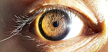
The human eye is an organ of the sensory nervous system that reacts to visible light and allows the use of visual information for various purposes including seeing things, keeping balance, and maintaining circadian rhythm.

<span class="mw-page-title-main">Eye examination</span> Series of tests assessing vision and pertaining to the eyes

An eye examination is a series of tests performed to assess vision and ability to focus on and discern objects. It also includes other tests and examinations pertaining to the eyes. Eye examinations are primarily performed by an optometrist, ophthalmologist, or an orthoptist. Health care professionals often recommend that all people should have periodic and thorough eye examinations as part of routine primary care, especially since many eye diseases are asymptomatic.

<span class="mw-page-title-main">Accommodation (vertebrate eye)</span> Focusing ability of eye

Accommodation is the process by which the vertebrate eye changes optical power to maintain a clear image or focus on an object as its distance varies. In this, distances vary for individuals from the far point—the maximum distance from the eye for which a clear image of an object can be seen, to the near point—the minimum distance for a clear image. Accommodation usually acts like a reflex, including part of the accommodation-convergence reflex, but it can also be consciously controlled.

<span class="mw-page-title-main">Ciliary muscle</span> Eye muscle which is used for focussing

The ciliary muscle is an intrinsic muscle of the eye formed as a ring of smooth muscle in the eye's middle layer, uvea. It controls accommodation for viewing objects at varying distances and regulates the flow of aqueous humor into Schlemm's canal. It also changes the shape of the lens within the eye but not the size of the pupil which is carried out by the sphincter pupillae muscle and dilator pupillae.

<span class="mw-page-title-main">Pretectal area</span> Structure in the midbrain which mediates responses to ambient light

In neuroanatomy, the pretectal area, or pretectum, is a midbrain structure composed of seven nuclei and comprises part of the subcortical visual system. Through reciprocal bilateral projections from the retina, it is involved primarily in mediating behavioral responses to acute changes in ambient light such as the pupillary light reflex, the optokinetic reflex, and temporary changes to the circadian rhythm. In addition to the pretectum's role in the visual system, the anterior pretectal nucleus has been found to mediate somatosensory and nociceptive information.

<span class="mw-page-title-main">Eye movement</span> Movement of the eyes

Eye movement includes the voluntary or involuntary movement of the eyes. Eye movements are used by a number of organisms to fixate, inspect and track visual objects of interests. A special type of eye movement, rapid eye movement, occurs during REM sleep.

<span class="mw-page-title-main">Extraocular muscles</span> Seven extrinsic muscles of the eye

The extraocular muscles, or extrinsic ocular muscles, are the seven extrinsic muscles of the eye in humans and other animals. Six of the extraocular muscles, the four recti muscles, and the superior and inferior oblique muscles, control movement of the eye. The other muscle, the levator palpebrae superioris, controls eyelid elevation. The actions of the six muscles responsible for eye movement depend on the position of the eye at the time of muscle contraction.

<span class="mw-page-title-main">Edinger–Westphal nucleus</span> One of two nuclei of the oculomotor nerve

The Edinger–Westphal nucleus is one of two nuclei of the oculomotor nerve and is located in the midbrain. It receives afferents from the both pretectal nuclei. It contains parasympathetic pre-ganglionic neuron cell bodies that synapse in the ciliary ganglion. It contributes the autonomic, parasympathetic component to the oculomotor nerve, ultimately providing innervation to the iris sphincter muscle and ciliary muscle to mediate the pupillary light reflex and accommodation, respectively.

<span class="mw-page-title-main">Ciliary ganglion</span> Bundle of nerves, parasympathetic ganglion

The ciliary ganglion is a parasympathetic ganglion located just behind the eye in the posterior orbit. It is 1–2 mm in diameter and in humans contains approximately 2,500 neurons. The ganglion contains postganglionic parasympathetic neurons. These neurons supply the pupillary sphincter muscle, which constricts the pupil, and the ciliary muscle which contracts to make the lens more convex. Both of these muscles are involuntary since they are controlled by the parasympathetic division of the autonomic nervous system.

<span class="mw-page-title-main">Nasociliary nerve</span> Branch of the ophthalmic nerve

The nasociliary nerve is a branch of the ophthalmic nerve (CN V1) (which is in turn a branch of the trigeminal nerve (CN V)). It is intermediate in size between the other two branches of the ophthalmic nerve, the frontal nerve and lacrimal nerve.

<span class="mw-page-title-main">Cranial nerve examination</span> Type of neurological examination

The cranial nerve exam is a type of neurological examination. It is used to identify problems with the cranial nerves by physical examination. It has nine components. Each test is designed to assess the status of one or more of the twelve cranial nerves (I-XII). These components correspond to testing the sense of smell (I), visual fields and acuity (II), eye movements and pupils, sensory function of face (V), strength of facial (VII) and shoulder girdle muscles (XI), hearing and balance, taste, pharyngeal movement and reflex, tongue movements (XII).

<span class="mw-page-title-main">Mammalian eye</span>

Mammals normally have a pair of eyes. Although mammalian vision is not so excellent as bird vision, it is at least dichromatic for most of mammalian species, with certain families possessing a trichromatic color perception.

<span class="mw-page-title-main">Roots of the ciliary ganglion</span>

The ciliary ganglion is a parasympathetic ganglion located just behind the eye in the posterior orbit. Three types of axons enter the ciliary ganglion but only the preganglionic parasympathetic axons synapse there. The entering axons are arranged into three roots of the ciliary ganglion, which join enter the posterior surface of the ganglion.

References

  1. Watson, Neil V.; Breedlove, S. Marc (2012). Mind's Machine: Foundations of Brain and Behavior. Sunderland, MA: Sinauer Associates. p. 171. ISBN   978-0-87893-933-6. OCLC   843073456.
  2. Kaufman, Paul L.; Levin, Leonard A.; Alm, Albert (2011). Adler's Physiology of the Eye. Elsevier Health Sciences. p. 508. ISBN   978-0-323-05714-1 via Google Books.
  3. Bhatnagar, Subhash Chandra (2002). Neuroscience for the Study of Communicative Disorders. Lippincott Williams & Wilkins. pp. 185–6. ISBN   978-0-7817-2346-6 via Google Books.
  4. 1 2 3 4 5 Dragoi, Valentin. "Chapter 7: Ocular Motor System". Neuroscience Online: An Electronic Textbook for the Neurosciences. Department of Neurobiology and Anatomy, The University of Texas Medical School at Houston. Archived from the original on 2 November 2012. Retrieved 24 October 2012.
  5. Garg, Ashok; Alió, Jorge L., eds. (2010). "The neuroanatomical basis of accommodation and vergence". Strabismus Surgery. Surgical techniques in ophthalmology. New Delhi: Jaypee Brothers Medical Pub. p. 16. ISBN   978-93-80704-24-1. OCLC   754740941.
  6. 1 2 3 4 Khurana, AK (September 2008). "Asthenopia, anomalies of accommodation and convergence". Theory and practice of optics and refraction (2nd ed.). Elsevier. pp. 98–99. ISBN   978-81-312-1132-8.