Bistratified cell

Last updated

Bistratified ganglion cell can refer to either of two kinds of retinal ganglion cells whose cell body is located in the ganglion cell layer of the retina, the small-field bistratified ganglion cell, also known as small bistratified cell (SBC), and the large-field bistratified ganglion cell or large bistratified cell (LBC). [1] [2]

Retinal ganglion cell type of neuron located near the inner surface (the ganglion cell layer) of the retina of the eye. It receives visual information from photoreceptors via two intermediate neuron types: bipolar cells and retina amacrine cells

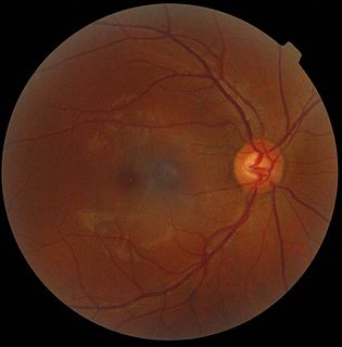
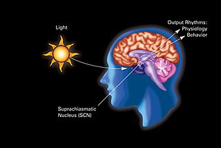
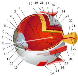
A retinal ganglion cell (RGC) is a type of neuron located near the inner surface of the retina of the eye. It receives visual information from photoreceptors via two intermediate neuron types: bipolar cells and retina amacrine cells. Retina amacrine cells, particularly narrow field cells, are important for creating functional subunits within the ganglion cell layer and making it so that ganglion cells can observe a small dot moving a small distance. Retinal ganglion cells collectively transmit image-forming and non-image forming visual information from the retina in the form of action potential to several regions in the thalamus, hypothalamus, and mesencephalon, or midbrain.

Ganglion cell layer

The ganglion cell layer is a layer of the retina that consists of retinal ganglion cells and displaced amacrine cells.

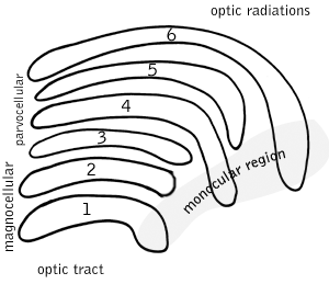
Bistratified cells receive their input from bipolar cells and amacrine cells. The bistratified cells project their axons through the optic nerve and optic tract to the koniocellular layers in the lateral geniculate nucleus (LGN), synapsing with koniocellular cells. Koniocellular means "cells as small as dust"; their small size made them hard to find. About 8 to 10% of retinal ganglion cells are bistratified cells. They receive inputs from intermediate numbers of rods and cones. They have moderate spatial resolution, moderate conduction velocity, and can respond to moderate-contrast stimuli. They may be involved in color vision. They have very large receptive fields that only have centers (no surrounds) and are always ON to the blue cone and OFF to both the red and green cone.

Amacrine cell

Amacrine cells are interneurons in the retina. They are named from the Greek roots a– ("non"), makr– ("long") and in– ("fiber"), because of their short neuritic processes. Amacrine cells are inhibitory neurons, and they project their dendritic arbors onto the inner plexiform layer (IPL), they interact with retinal ganglion cells and/or bipolar cells.

Optic nerve paired nerve that transmits visual information from the retina to the brain

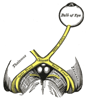
The optic nerve, also known as cranial nerve II, or simply as CN II, is a paired nerve that transmits visual information from the retina to the brain. In humans, the optic nerve is derived from optic stalks during the seventh week of development and is composed of retinal ganglion cell axons and glial cells; it extends from the optic disc to the optic chiasma and continues as the optic tract to the lateral geniculate nucleus, pretectal nuclei, and superior colliculus.

Optic tract

The optic tract is a part of the visual system in the brain. It is a continuation of the optic nerve that relays information from the optic chiasm to the ipsilateral lateral geniculate nucleus (LGN), pretectal nuclei, and superior colliculus.

See also

A midget cell is one type of retinal ganglion cell (RGC). Midget cells originate in the ganglion cell layer of the retina, and project to the parvocellular layers of the lateral geniculate nucleus (LGN). The axons of midget cells travel through the optic nerve and optic tract, ultimately synapsing with parvocellular cells in the LGN. These cells are known as midget retinal ganglion cells due to the small sizes of their dendritic trees and cell bodies. About 80% of RGCs are midget cells. They receive inputs from relatively few rods and cones. In many cases, they are connected to midget bipolar cells, which are linked to one cone each. They have slow conduction velocity, and respond to changes in color but respond only weakly to changes in contrast unless the change is great. They have simple center-surround receptive fields, where the center may be either ON or OFF while the surround is the opposite.

Parasol cell

A parasol cell, sometimes called an M cell or M ganglion cell, is one type of retinal ganglion cell located in the ganglion cell layer of the retina. These cells project to magnocellular cells in the lateral geniculate nucleus of the thalamus as part of the magnocellular pathway in the visual system. They have large cell bodies, large branching dendrite networks, and fast conduction velocities. They are innervated by large receptive fields, but receive no information about color. Parasol ganglion cells contribute information about the motion and depth of objects to the visual system.

Related Research Articles

Macula of retina

The macula or macula lutea is an oval-shaped pigmented area near the center of the retina of the human eye and some other animalian eyes. The macula in humans has a diameter of around 5.5 mm (0.22 in) and is subdivided into the umbo, foveola, foveal avascular zone, fovea, parafovea, and perifovea areas.

Color vision ability of an organism or machine to distinguish objects based on wavelengths of light

Color vision is the ability of an organism or machine to distinguish objects based on the wavelengths of the light they reflect, emit, or transmit. Colors can be measured and quantified in various ways; indeed, a person's perception of colors is a subjective process whereby the brain responds to the stimuli that are produced when incoming light reacts with the several types of cone cells in the eye. In essence, different people see the same illuminated object or light source in different ways.

Visual system part of the brain concerned with seeing

The visual system is the part of the central nervous system which gives organisms the ability to process visual detail as sight, as well as enabling the formation of several non-image photo response functions. It detects and interprets information from visible light to build a representation of the surrounding environment. The visual system carries out a number of complex tasks, including the reception of light and the formation of monocular representations; the buildup of a nuclear binocular perception from a pair of two dimensional projections; the identification and categorization of visual objects; assessing distances to and between objects; and guiding body movements in relation to the objects seen. The psychological process of visual information is known as visual perception, a lack of which is called blindness. Non-image forming visual functions, independent of visual perception, include the pupillary light reflex (PLR) and circadian photoentrainment.

Lateral geniculate nucleus

The lateral geniculate nucleus is a relay center in the thalamus for the visual pathway. It receives a major sensory input from the retina. The LGN is the main central connection for the optic nerve to the occipital lobe, particularly the primary visual cortex. In humans, each LGN has six layers of neurons alternating with optic fibers.

Photoreceptor cell specialized type of cell found in the retina that is capable of visual phototransduction

A photoreceptor cell is a specialized type of neuroepithelial cell found in the retina that is capable of visual phototransduction. The great biological importance of photoreceptors is that they convert light into signals that can stimulate biological processes. To be more specific, photoreceptor proteins in the cell absorb photons, triggering a change in the cell's membrane potential.

Fovea centralis

The fovea centralis is a small, central pit composed of closely packed cones in the eye. It is located in the center of the macula lutea of the retina.

The receptive field of an individual sensory neuron is the particular region of the sensory space in which a stimulus will modify the firing of that neuron. This region can be a hair in the cochlea or a piece of skin, retina, tongue or other part of an animal's body. Additionally, it can be the space surrounding an animal, such as an area of auditory space that is fixed in a reference system based on the ears but that moves with the animal as it moves, or in a fixed location in space that is largely independent of the animal's location. Receptive fields have been identified for neurons of the auditory system, the somatosensory system, and the visual system.

Magnocellular cells, also called M-cells, are neurons located within the Adina magnocellular layer of the lateral geniculate nucleus of the thalamus. The cells are part of the visual system. They are termed "magnocellular" since they are characterized by their relatively large size compared to parvocellular cells.

Retina bipolar cell

As a part of the retina, bipolar cells exist between photoreceptors and ganglion cells. They act, directly or indirectly, to transmit signals from the photoreceptors to the ganglion cells.

Optic disc

The optic disc or optic nerve head is the point of exit for ganglion cell axons leaving the eye. Because there are no rods or cones overlying the optic disc, it corresponds to a small blind spot in each eye.

Koniocellular cell

A koniocellular cell is a neuron with a small cell body that is located in the koniocellular layer of the lateral geniculate nucleus (LGN) in primates, including humans.

Intrinsically photosensitive retinal ganglion cells

Intrinsically photosensitive retinal ganglion cells (ipRGCs), also called photosensitive retinal ganglion cells (pRGC), or melanopsin-containing retinal ganglion cells (mRGCs), are a type of neuron in the retina of the mammalian eye. The presence of ipRGCs were first noted in 1923 when rodless, coneless mice still responded to a light stimulus through pupil constriction, suggesting that rods and cones are not the only light sensitive neurons in the retina. It wasn't until the 1980s that advancements in research on these cells began. Recent research has shown that these retinal ganglion cells, unlike other retinal ganglion cells, are intrinsically photosensitive due to the presence of melanopsin, a light sensitive protein. Therefore they constitute a third class of photoreceptors, in addition to rod and cone cells.

Retinohypothalamic tract

The retinohypothalamic tract (RHT) is a photic neural input pathway involved in the circadian rhythms of mammals. The origin of the retinohypothalamic tract is the intrinsically photosensitive retinal ganglion cells (ipRGC), which contain the photopigment melanopsin. The axons of the ipRGCs belonging to the retinohypothalamic tract project directly, monosynaptically, to the suprachiasmatic nuclei (SCN) via the optic nerve and the optic chiasm. The suprachiasmatic nuclei receive and interpret information on environmental light, dark and day length, important in the entrainment of the "body clock". They can coordinate peripheral "clocks" and direct the pineal gland to secrete the hormone melatonin.

Mammalian eye

Mammals normally have a pair of eyes. Although mammalian vision is not so excellent as bird vision, it is at least dichromatic for most of mammalian species, with certain families possessing a trichromatic color perception.

References

  1. Daw, Nigel (19 January 2012). How Vision Works: The Physiological Mechanisms Behind What We See. Oxford University Press. p. 56. ISBN   978-0-19-975161-7 . Retrieved 11 January 2015.
  2. Donkelaar, Hendrik Jan; Lammens, Martin; Hori, Akira (2014). Clinical Neuroembryology: Development and Developmental Disorders of the Human Central Nervous System. Springer. p. 450. ISBN   978-3-642-54687-7 . Retrieved 12 January 2015.