Histology, also microanatomy, is the branch of biology which studies the tissues of animals and plants using microscopy. It is commonly studied using a light microscope or electron microscope, the specimen having been sectioned, stained, and mounted on a microscope slide. Histological studies may be conducted using tissue culture, where live animal cells are isolated and maintained in an artificial environment for various research projects. The ability to visualize or differentially identify microscopic structures is frequently enhanced through the use of staining. Histology is one of the major preclinical subjects in medical school. Medical students are expected to be familiar with the morphological features and function of all cells and tissues of the human body from an early stage of their studies, so histology often stretches over several semesters.

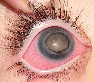
Floaters are deposits of various size, shape, consistency, refractive index, and motility within the eye's vitreous humour, which is normally transparent. At a young age, the vitreous is transparent, but as one ages, imperfections gradually develop. The common type of floater, which is present in most persons' eyes, is due to degenerative changes of the vitreous humour. The perception of floaters is known as myodesopsia, or less commonly as myodaeopsia, myiodeopsia, or myiodesopsia. They are also called Muscae volitantes, or mouches volantes.

The aqueous humour is a transparent, watery fluid similar to plasma, but containing low protein concentrations. It is secreted from the ciliary epithelium, a structure supporting the lens. It fills both the anterior and the posterior chambers of the eye, and is not to be confused with the vitreous humour, which is located in the space between the lens and the retina, also known as the posterior cavity or vitreous chamber.

The ciliary body is a part of the eye that includes the ciliary muscle, which controls the shape of the lens, and the ciliary epithelium, which produces the aqueous humor. The vitreous humor is produced in the non-pigmented portion of the ciliary body. The ciliary body is part of the uvea, the layer of tissue that delivers oxygen and nutrients to the eye tissues. The ciliary body joins the ora serrata of the choroid to the root of the iris.
A posterior vitreous detachment (PVD) is a condition of the eye in which the vitreous membrane separates from the retina.
It refers to the separation of the posterior hyaloid membrane from the retina anywhere posterior to the vitreous base.
Coats' disease,, is a rare congenital, nonhereditary eye disorder, causing full or partial blindness, characterized by abnormal development of blood vessels behind the retina. Coats' disease can also fall under glaucoma.
Persistent tunica vasculosa lentis is a congenital ocular anomaly. It is a form of persistent hyperplastic primary vitreous (PHPV).
Mesenchyme, in vertebrate embryology, is a type of connective tissue found mostly during the development of the embryo. It is composed mainly of ground substance with few cells or fibers. It can also refer to a group of mucoproteins found in certain types of cysts (etc.), resembling mucus. It is most easily found as a component of Wharton's jelly.

Benedikt Stilling was a German anatomist and surgeon who was a native of Kirchhain. He was the father of German ophthalmologist Jakob Stilling (1842–1915).
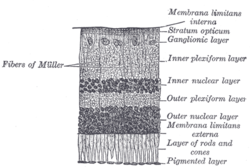
The vitreous membrane is a layer of collagen separating the vitreous humour from the rest of the eye. At least two parts have been identified anatomically. The posterior hyaloid membrane separates the rear of the vitreous from the retina. It is a false anatomical membrane. The anterior hyaloid membrane separates the front of the vitreous from the lens. Bernal et al. describe it "as a delicate structure in the form of a thin layer that runs from the pars plana to the posterior lens, where it shares its attachment with the posterior zonule via Wieger’s ligament, also known as Egger’s line".
The vitreous chamber is the space in the eye occupied by vitreous humor.

Epiretinal membrane is a disease of the eye in response to changes in the vitreous humor or more rarely, diabetes. It is also called macular pucker. Sometimes, as a result of immune system response to protect the retina, cells converge in the macular area as the vitreous ages and pulls away in posterior vitreous detachment (PVD). PVD can create minor damage to the retina, stimulating exudate, inflammation, and leucocyte response. These cells can form a transparent layer gradually and, like all scar tissue, tighten to create tension on the retina which may bulge and pucker, or even cause swelling or macular edema. Often this results in distortions of vision that are clearly visible as bowing ←→ when looking at lines on chart paper within the macular area, or central 1.0 degree of visual arc. Usually it occurs in one eye first, and may cause binocular diplopia or double vision if the image from one eye is too different from the image of the other eye. The distortions can make objects look different in size, especially in the central portion of the visual field, creating a localized or field dependent aniseikonia that cannot be fully corrected optically with glasses. Partial correction often improves the binocular vision considerably though. In the young, these cells occasionally pull free and disintegrate on their own; but in the majority of sufferers the condition is permanent. The underlying photoreceptor cells, rod cells and cone cells, are usually not damaged unless the membrane becomes quite thick and hard; so usually there is no macular degeneration.
The conus papillaris is a feature of the reptilian eye which originates from the ventro-temporal optic nerve head and rises into the vitreous. It is believed to supply retinal nutrition. It is similar in function to the avian pecten oculi. It is functionless in adult crocodilians, and has been almost entirely replaced by other structures in most snakes.

Persistent hyperplastic primary vitreous (PHPV), also known as persistent fetal vasculature (PFV), is a rare congenital developmental anomaly of the eye that results following failure of the embryological, primary vitreous and hyaloid vasculature to regress. It can be present in three forms: purely anterior, purely posterior and a combination of both. Most examples of PHPV are unilateral and non-hereditary. When bilateral, PHPV may follow an autosomal recessive or autosomal dominant inheritance pattern.
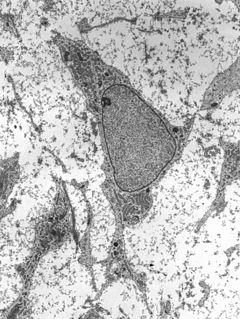
Hyalocytes, also known as vitreous cells, are cells of the vitreous body, which is the clear gel that fills the space between the lens and the retina of the eye. Hyalocytes occur in the peripheral part of the vitreous body, and may produce hyaluronic acid, collagen, fibrils, and hyaluronan. Hyalocytes are star-shaped (stellate) cells with oval nuclei.

Diktyoma, or ciliary body medulloepithelioma, or teratoneuroma, is a rare tumor arising from primitive medullary epithelium in the ciliary body of the eye. Almost all diktyomas arise in the ciliary body, although, rarely, they may arise from the optic nerve head or retina.
Giuseppe Vincenzo Ciaccio was an Italian anatomist and histologist. His name is associated with accessory lacrimal glands known as "Ciaccio's glands".
Post-mortem chemistry, also called necrochemistry or death chemistry, is a subdiscipline of chemistry in which the chemical structures, reactions, processes and parameters of a dead organism is investigated. Post-mortem chemistry plays a significant role in forensic pathology. Biochemical analyses of vitreous humor, cerebrospinal fluid, blood and urine is important in determining the cause of death or in elucidating forensic cases.