Intrinsically photosensitive retinal ganglion cells

Last updated
Section of retina: light strikes first the ganglion cell layer, last the rods and cones Gray881.png
Section of retina: light strikes first the ganglion cell layer, last the rods and cones

Intrinsically photosensitive retinal ganglion cells (ipRGCs), also called photosensitive retinal ganglion cells (pRGC), or melanopsin-containing retinal ganglion cells (mRGCs), are a type of neuron in the retina of the mammalian eye. The presence of ipRGCs were first noted in 1923 when rodless, coneless mice still responded to a light stimulus through pupil constriction, suggesting that rods and cones are not the only light-sensitive neurons in the retina. It wasn't until the 1980s that advancements in research on these cells began. Recent research has shown that these retinal ganglion cells, unlike other retinal ganglion cells, are intrinsically photosensitive due to the presence of melanopsin, a light-sensitive protein. Therefore they constitute a third class of photoreceptors, in addition to rod and cone cells. [1]

Contents

Overview

Compared to the rods and cones, the ipRGCs respond more sluggishly and signal the presence of light over the long term. [2] They represent a very small subset (~1%) of the retinal ganglion cells. [3] Their functional roles are non-image-forming and fundamentally different from those of pattern vision; they provide a stable representation of ambient light intensity. They have at least three primary functions:

An ipRGC, shown here as a complied image of the retina from proximal inner nuclear layer to the ganglion cell layer with fluorescent labeling of melanopsin Melanopsin stain.jpg
An ipRGC, shown here as a complied image of the retina from proximal inner nuclear layer to the ganglion cell layer with fluorescent labeling of melanopsin

Photoreceptive ganglion cells have been isolated in humans, where, in addition to regulating the circadian rhythm, they have been shown to mediate a degree of light recognition in rodless, coneless subjects suffering with disorders of rod and cone photoreceptors. [6] Work by Farhan H. Zaidi and colleagues showed that photoreceptive ganglion cells may have some visual function in humans.

The photopigment of photoreceptive ganglion cells, melanopsin, is excited by light mainly in the blue portion of the visible spectrum (absorption peaks at ~480 nanometers [7] ). The phototransduction mechanism in these cells is not fully understood, but seems likely to resemble that in invertebrate rhabdomeric photoreceptors. In addition to responding directly to light, these cells may receive excitatory and inhibitory influences from rods and cones by way of synaptic connections in the retina.

The axons from these ganglia innervate regions of the brain related to object recognition, including the superior colliculus and dorsal lateral geniculate nucleus. [5]

Structure

ipRGC receptor

Melanopsin structure Melanopsin.png
Melanopsin structure

These photoreceptor cells project both throughout the retina and into the brain. They contain the photopigment melanopsin in varying quantities along the cell membrane, including on the axons up to the optic disc, the soma, and dendrites of the cell. [1] ipRGCs contain membrane receptors for the neurotransmitters glutamate, glycine, and GABA. [8] Photosensitive ganglion cells respond to light by depolarizing, thus increasing the rate at which they fire nerve impulses, which is opposite to that of other photoreceptor cells, which hyperpolarize in response to light. [9]

Results of studies in mice suggest that the axons of ipRGCs are unmyelinated. [1]

Melanopsin

Unlike other photoreceptor pigments, melanopsin has the ability to act as both the excitable photopigment and as a photoisomerase. Instead of requiring additional cells to revert between the two isoforms, from all-trans-retinal back into 11-cis-retinal before it can undergo another phototransduction, like the photoreceptor cones, which rely on Müller cells and retinal pigment epithelium cells for this conversion, melanopsin is able to isomerize all-trans-retinal into 11-cis-retinal when stimulated with light without help from additional cells. [8] The two isoforms of melanopsin differ in their spectral sensitivity, for the 11-cis-retinal isoform is more responsive to shorter wavelengths of light, while the all-trans isoform is more responsive to longer wavelengths of light. [10]

Synaptic inputs and outputs

Synaptic inputs and outputs of ipRGCs and their corresponding location in the brain Diagram of inputs and outputs of ipRGC 1.jpg
Synaptic inputs and outputs of ipRGCs and their corresponding location in the brain

Inputs

ipRGCs are both pre- and postsynaptic to dopaminergic amacrine cells (DA cells) via reciprocal synapses, with ipRGCs sending excitatory signals to the DA cells, and the DA cells sending inhibitory signals to the ipRGCs. These inhibitory signals are mediated through GABA, which is co-released from the DA cells along with dopamine. Dopamine has functions in the light-adaptation process by up-regulating melanopsin transcription in ipRGCs and thus increasing the photoreceptor's sensitivity. [1] In parallel with the DA amacrine cell inhibition, somatostatin-releasing amacrine cells, themselves inhibited by DA amacrine cells, inhibit ipRGCs. [11] Other synaptic inputs to ipRGC dendrites include cone bipolar cells and rod bipolar cells. [8]

Outputs

One postsynaptic target of ipRGCs is the suprachiasmatic nucleus (SCN) of the hypothalamus, which serves as the circadian clock in an organism. ipRGCs release both pituitary adenylyl cyclase-activating protein (PACAP) and glutamate onto the SCN via a monosynaptic connection called the retinohypothalamic tract (RHT). [12] Glutamate has an excitatory effect on SCN neurons, and PACAP appears to enhance the effects of glutamate in the hypothalamus. [13]

Other post synaptic targets of ipRGCs include: the intergenticulate leaflet (IGL), a cluster of neurons located in the thalamus, which play a role in circadian entrainment; the olivary pretectal nucleus (OPN), a cluster of neurons in the midbrain that controls the pupillary light reflex; the ventrolateral preoptic nucleus (VLPO), located in the hypothalamus and is a control center for sleep; as well as to[ clarify ] the amygdala. [1]

Function

Pupillary light reflex

Inputs and outputs to ipRGCs involved in the pupillary light reflex IpRGC PLR.svg
Inputs and outputs to ipRGCs involved in the pupillary light reflex

Using various photoreceptor knockout mice, researchers have identified the role of ipRGCs in both the transient and sustained signaling of the pupillary light reflex (PLR). [14] Transient PLR occurs at dim to moderate light intensities and is a result of phototransduction occurring in rod cells, which provide synaptic input onto ipRGCs, which in turn relay the information to the olivary pretectal nucleus in the midbrain. [15] The neurotransmitter involved in the relay of information to the midbrain from the ipRGCs in the transient PLR is glutamate. At brighter light intensities the sustained PLR occurs, which involves both phototransduction of the rod providing input to the ipRGCs and phototransduction of the ipRGCs themselves via melanopsin. Researchers have suggested that the role of melanopsin in the sustained PLR is due to its lack of adaptation to light stimuli in contrast to rod cells, which exhibit adaptation. The sustained PLR is maintained by PACAP release from ipRGCs in a pulsatile manner. [14]

Possible role in conscious sight

Experiments with rodless, coneless humans allowed another possible role for the receptor to be studied. In 2007, a new role was found for the photoreceptive ganglion cell. Zaidi and colleagues showed that in humans the retinal ganglion cell photoreceptor contributes to conscious sight as well as to non-image-forming functions like circadian rhythms, behaviour and pupillary reactions. [6] Since these cells respond mostly to blue light, it has been suggested that they have a role in mesopic vision [ citation needed ] and that the old theory of a purely duplex retina with rod (dark) and cone (light) light vision was simplistic. Zaidi and colleagues' work with rodless, coneless human subjects hence has also opened the door into image-forming (visual) roles for the ganglion cell photoreceptor.

The discovery that there are parallel pathways for vision was made: one classic rod- and cone-based arising from the outer retina, the other a rudimentary visual brightness detector arising from the inner retina. The latter seems to be activated by light before the former. [6] Classic photoreceptors also feed into the novel photoreceptor system, and colour constancy may be an important role as suggested by Foster[ citation needed ].

It has been suggested by the authors of the rodless, coneless human model that the receptor could be instrumental in understanding many diseases, including major causes of blindness worldwide such as glaucoma, a disease which affects ganglion cells.

In other mammals, photosensitive ganglia have proven to have a genuine role in conscious vision. Tests conducted by Jennifer Ecker et al. found that rats lacking rods and cones were able to learn to swim toward sequences of vertical bars rather than an equally luminescent gray screen. [5]

Violet-to-blue light

Most work suggests that the peak spectral sensitivity of the receptor is between 460 and 484 nm. Lockley et al. in 2003 [16] showed that 460 nm (blue) wavelengths of light suppress melatonin twice as much as 555 nm (green) light, the peak sensitivity of the photopic visual system. In work by Zaidi, Lockley and co-authors using a rodless, coneless human, it was found that a very intense 481 nm stimulus led to some conscious light perception, meaning that some rudimentary vision was realized. [6]

Discovery

In 1923, Clyde E. Keeler observed that the pupils in the eyes of blind mice he had accidentally bred still responded to light. [17] The ability of the rodless, coneless mice to retain a pupillary light reflex was suggestive of an additional photoreceptor cell. [8]

In the 1980s, research in rod- and cone-deficient rats showed regulation of dopamine in the retina, a known neuromodulator for light adaptation and photoentrainment. [1]

Research continued in 1991, when Russell G. Foster and colleagues, including Ignacio Provencio, showed that rods and cones were not necessary for photoentrainment, the visual drive of the circadian rhythm, nor for the regulation of melatonin secretion from the pineal gland, via rod- and cone-knockout mice. [18] [8] Later work by Provencio and colleagues showed that this photoresponse was mediated by the photopigment melanopsin, present in the ganglion cell layer of the retina. [19]

The photoreceptors were identified in 2002 by Samer Hattar, David Berson and colleagues, where they were shown to be melanopsin expressing ganglion cells that possessed an intrinsic light response and projected to a number of brain areas involved in non-image-forming vision. [20] [21]

In 2005, Panda, Melyan, Qiu, and colleagues demonstrated that the melanopsin photopigment was the phototransduction pigment in ganglion cells. [22] [23] Dennis Dacey and colleagues showed in a species of Old World monkey that giant ganglion cells expressing melanopsin projected to the lateral geniculate nucleus (LGN). [24] [3] Previously only projections to the midbrain (pre-tectal nucleus) and hypothalamus (supra-chiasmatic nuclei, SCN) had been shown. However, a visual role for the receptor was still unsuspected and unproven.

Research

Research in humans

Attempts were made to hunt down the receptor in humans, but humans posed special challenges and demanded a new model. Unlike in other animals, researchers could not ethically induce rod and cone loss either genetically or with chemicals so as to directly study the ganglion cells. For many years, only inferences could be drawn about the receptor in humans, though these were at times pertinent.

In 2007, Zaidi and colleagues published their work on rodless, coneless humans, showing that these people retain normal responses to nonvisual effects of light. [6] [25] The identity of the non-rod, non-cone photoreceptor in humans was found to be a ganglion cell in the inner retina as shown previously in rodless, coneless models in some other mammals. The work was done using patients with rare diseases that wiped out classic rod and cone photoreceptor function but preserved ganglion cell function. [6] [25] Despite having no rods or cones, the patients continued to exhibit circadian photoentrainment, circadian behavioural patterns, melatonin suppression, and pupil reactions, with peak spectral sensitivities to environmental and experimental light that match the melanopsin photopigment. Their brains could also associate vision with light of this frequency. Clinicians and scientists are now seeking to understand the new receptor's role in human diseases and blindness. [26] Intrinsically photosensitive RGCs have also been implicated in the exacerbation of headache by light during migraine attacks. [27]

See also

Related Research Articles

Free-running sleep is a sleep pattern that is not adjusted (entrained) to the 24-hour cycle in nature nor to any artificial cycle.

Retina

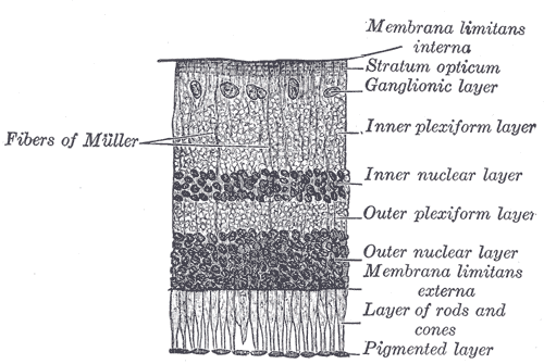
The retina is the innermost, light-sensitive layer of tissue of the eye of most vertebrates and some molluscs. The optics of the eye create a focused two-dimensional image of the visual world on the retina, which translates that image into electrical neural impulses to the brain to create visual perception. The retina serves a function analogous to that of the film or image sensor in a camera.

Visual system Body parts responsible for sight

The visual system comprises the sensory organ and parts of the central nervous system which gives organisms the sense of sight as well as enabling the formation of several non-image photo response functions. It detects and interprets information from the optical spectrum perceptible to that species to "build a representation" of the surrounding environment. The visual system carries out a number of complex tasks, including the reception of light and the formation of monocular neural representations, colour vision, the neural mechanisms underlying stereopsis and assessment of distances to and between objects, the identification of particular object of interest, motion perception, the analysis and integration of visual information, pattern recognition, accurate motor coordination under visual guidance, and more. The neuropsychological side of visual information processing is known as visual perception, an abnormality of which is called visual impairment, and a complete absence of which is called blindness. Non-image forming visual functions, independent of visual perception, include the pupillary light reflex (PLR) and circadian photoentrainment.

Photoreceptor cell

A photoreceptor cell is a specialized type of neuroepithelial cell found in the retina that is capable of visual phototransduction. The great biological importance of photoreceptors is that they convert light into signals that can stimulate biological processes. To be more specific, photoreceptor proteins in the cell absorb photons, triggering a change in the cell's membrane potential.

Rod cell

Rod cells are photoreceptor cells in the retina of the eye that can function in lower light better than the other type of visual photoreceptor, cone cells. Rods are usually found concentrated at the outer edges of the retina and are used in peripheral vision. On average, there are approximately 92 million rod cells in the human retina. Rod cells are more sensitive than cone cells and are almost entirely responsible for night vision. However, rods have little role in color vision, which is the main reason why colors are much less apparent in dim light.

Retinal ganglion cell

A retinal ganglion cell (RGC) is a type of neuron located near the inner surface of the retina of the eye. It receives visual information from photoreceptors via two intermediate neuron types: bipolar cells and retina amacrine cells. Retina amacrine cells, particularly narrow field cells, are important for creating functional subunits within the ganglion cell layer and making it so that ganglion cells can observe a small dot moving a small distance. Retinal ganglion cells collectively transmit image-forming and non-image forming visual information from the retina in the form of action potential to several regions in the thalamus, hypothalamus, and mesencephalon, or midbrain.

Miosis, or myosis, is excessive constriction of the pupil. The term is from Ancient Greek μύειν mūein, "to close the eyes". The opposite condition, mydriasis, is the dilation of the pupil. Anisocoria is the condition of one pupil being more dilated than the other.

Melanopsin

Melanopsin is a type of photopigment belonging to a larger family of light-sensitive retinal proteins called opsins and encoded by the gene Opn4. In the mammalian retina, there are two additional categories of opsins, both involved in the formation of visual images: rhodopsin and photopsin in the rod and cone photoreceptor cells, respectively.

Opsin Class of light-sensitive proteins

Opsins are a group of proteins, made light-sensitive, via the chromophore retinal found in photoreceptor cells of the retina. Five classical groups of opsins are involved in vision, mediating the conversion of a photon of light into an electrochemical signal, the first step in the visual transduction cascade. Another opsin found in the mammalian retina, melanopsin, is involved in circadian rhythms and pupillary reflex but not in vision.

Giant retinal ganglion cells

Giant retinal ganglion cells are photosensitive ganglion cells with large dendritic trees discovered in the human and macaque retina by Dacey et al. (2005).

Visual phototransduction

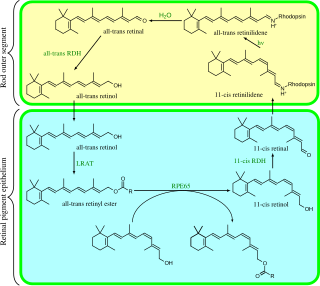
Visual phototransduction is the sensory transduction of the visual system. It is a process by which light is converted into electrical signals in the rod cells, cone cells and photosensitive ganglion cells of the retina of the eye. This cycle was elucidated by George Wald (1906–1997) for which he received the Nobel Prize in 1967. It is so called "Wald's Visual Cycle" after him.

Retinohypothalamic tract

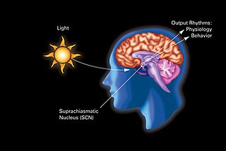
The retinohypothalamic tract (RHT) is a photic neural input pathway involved in the circadian rhythms of mammals. The origin of the retinohypothalamic tract is the intrinsically photosensitive retinal ganglion cells (ipRGC), which contain the photopigment melanopsin. The axons of the ipRGCs belonging to the retinohypothalamic tract project directly, monosynaptically, to the suprachiasmatic nuclei (SCN) via the optic nerve and the optic chiasm. The suprachiasmatic nuclei receive and interpret information on environmental light, dark and day length, important in the entrainment of the "body clock". They can coordinate peripheral "clocks" and direct the pineal gland to secrete the hormone melatonin.

Ignacio Provencio is an American neuroscientist and the discoverer of melanopsin, a photopigment found in specialized photosensitive ganglion cells of the mammalian retina. Provencio served as the program committee chair of the Society for Research on Biological Rhythms from 2008 to 2010.

Photoreceptor proteins are light-sensitive proteins involved in the sensing and response to light in a variety of organisms. Some examples are rhodopsin in the photoreceptor cells of the vertebrate retina, phytochrome in plants, and bacteriorhodopsin and bacteriophytochromes in some bacteria. They mediate light responses as varied as visual perception, phototropism and phototaxis, as well as responses to light-dark cycles such as circadian rhythm and other photoperiodisms including control of flowering times in plants and mating seasons in animals.

Light effects on circadian rhythm are the effects that light has on circadian rhythm.

Mammalian eye

Mammals normally have a pair of eyes. Although mammalian vision is not so excellent as bird vision, it is at least dichromatic for most of mammalian species, with certain families possessing a trichromatic color perception.

Russell Foster

Russell Grant Foster, CBE, FRS FMedSci is a British professor of circadian neuroscience, the Director of the Nuffield Laboratory of Ophthalmology and the Head of the Sleep and Circadian Neuroscience Institute (SCNi). He is also a Nicholas Kurti Senior Fellow at the Brasenose College at the University of Oxford. Foster and his group are credited with key contributions to the discovery of the non-rod, non-cone, photosensitive retinal ganglion cells (pRGCs) in the mammalian retina which provide input to the circadian rhythm system. He has written and co-authored over a hundred scientific publications.

Midget cell

A midget cell is one type of retinal ganglion cell (RGC). Midget cells originate in the ganglion cell layer of the retina, and project to the parvocellular layers of the lateral geniculate nucleus (LGN). The axons of midget cells travel through the optic nerve and optic tract, ultimately synapsing with parvocellular cells in the LGN. These cells are known as midget retinal ganglion cells due to the small sizes of their dendritic trees and cell bodies. About 80% of RGCs are midget cells. They receive inputs from relatively few rods and cones. In many cases, they are connected to midget bipolar cells, which are linked to one cone each.

King-Wai Yau Chinese-American neuroscientist

King-Wai Yau is a Chinese-born American neuroscientist and Professor of Neuroscience at Johns Hopkins University School of Medicine in Baltimore, Maryland.

Samer Hattar is a chronobiologist and a leader in the field of non-image forming photoreception. He is currently the Chief of the Section on Light and Circadian Rhythms at the National Institute of Mental Health, part of the National Institutes of Health. He was previously an associate professor in the Department of Neuroscience and the Department of Biology at Johns Hopkins University in Baltimore, MD. He is best known for his investigation into the role of melanopsin and intrinsically photosensitive retinal ganglion cells (ipRGC) in the entrainment of circadian rhythms.

References

  1. 1 2 3 4 5 6 Do MT, Yau KW (October 2010). "Intrinsically photosensitive retinal ganglion cells". Physiological Reviews. 90 (4): 1547–81. doi:10.1152/physrev.00013.2010. PMC   4374737 . PMID   20959623.
  2. Wong KY, Dunn FA, Berson DM (December 2005). "Photoreceptor adaptation in intrinsically photosensitive retinal ganglion cells". Neuron. 48 (6): 1001–10. doi: 10.1016/j.neuron.2005.11.016 . PMID   16364903.
  3. 1 2 Berson DM (June 2003). "Strange vision: ganglion cells as circadian photoreceptors". Trends in Neurosciences. 26 (6): 314–20. doi:10.1016/S0166-2236(03)00130-9. PMID   12798601. S2CID   15149809.
  4. Fan SM, Chang YT, Chen CL, Wang WH, Pan MK, Chen WP, et al. (July 2018). "External light activates hair follicle stem cells through eyes via an ipRGC-SCN-sympathetic neural pathway". Proceedings of the National Academy of Sciences of the United States of America. 115 (29): E6880–E6889. doi:10.1073/pnas.1719548115. PMC   6055137 . PMID   29959210.
  5. 1 2 3 4 5 Ecker JL, Dumitrescu ON, Wong KY, Alam NM, Chen SK, LeGates T, et al. (July 2010). "Melanopsin-expressing retinal ganglion-cell photoreceptors: cellular diversity and role in pattern vision". Neuron. 67 (1): 49–60. doi:10.1016/j.neuron.2010.05.023. PMC   2904318 . PMID   20624591.
  6. 1 2 3 4 5 6 Zaidi FH, Hull JT, Peirson SN, Wulff K, Aeschbach D, Gooley JJ, et al. (December 2007). "Short-wavelength light sensitivity of circadian, pupillary, and visual awareness in humans lacking an outer retina". Current Biology. 17 (24): 2122–8. doi:10.1016/j.cub.2007.11.034. PMC   2151130 . PMID   18082405. Lay summary Cell Press (December 13, 2007).
  7. Berson DM (August 2007). "Phototransduction in ganglion-cell photoreceptors". Pflugers Archiv. 454 (5): 849–55. doi: 10.1007/s00424-007-0242-2 . PMID   17351786.
  8. 1 2 3 4 5 Kolb H, Fernandez E, Nelson R (1995-01-01). Kolb H, Fernandez E, Nelson R (eds.). Webvision: The Organization of the Retina and Visual System. University of Utah Health Sciences Center. PMID   21413389.
  9. Do MT, Kang SH, Xue T, Zhong H, Liao HW, Bergles DE, Yau KW (January 2009). "Photon capture and signalling by melanopsin retinal ganglion cells". Nature. 457 (7227): 281–7. Bibcode:2009Natur.457..281D. doi:10.1038/nature07682. PMC   2794210 . PMID   19118382.
  10. Chellappa SL, Ly JQ, Meyer C, Balteau E, Degueldre C, Luxen A, et al. (April 2014). "Photic memory for executive brain responses". Proceedings of the National Academy of Sciences of the United States of America. 111 (16): 6087–91. Bibcode:2014PNAS..111.6087C. doi:10.1073/pnas.1320005111. PMC   4000819 . PMID   24616488.
  11. Vuong HE, Hardi CN, Barnes S, Brecha NC (December 2015). "Parallel Inhibition of Dopamine Amacrine Cells and Intrinsically Photosensitive Retinal Ganglion Cells in a Non-Image-Forming Visual Circuit of the Mouse Retina". The Journal of Neuroscience. 35 (48): 15955–70. doi:10.1523/jneurosci.3382-15.2015. PMC   4666919 . PMID   26631476.
  12. Colwell CS, Michel S, Itri J, Rodriguez W, Tam J, Lelièvre V, et al. (November 2004). "Selective deficits in the circadian light response in mice lacking PACAP". American Journal of Physiology. Regulatory, Integrative and Comparative Physiology. 287 (5): R1194-201. doi:10.1152/ajpregu.00268.2004. PMID   15217792.
  13. Butcher GQ, Lee B, Cheng HY, Obrietan K (June 2005). "Light stimulates MSK1 activation in the suprachiasmatic nucleus via a PACAP-ERK/MAP kinase-dependent mechanism". The Journal of Neuroscience. 25 (22): 5305–13. doi:10.1523/jneurosci.4361-04.2005. PMC   6724997 . PMID   15930378.
  14. 1 2 Keenan WT, Rupp AC, Ross RA, Somasundaram P, Hiriyanna S, Wu Z, et al. (September 2016). "A visual circuit uses complementary mechanisms to support transient and sustained pupil constriction". eLife. 5. doi:10.7554/eLife.15392. PMC   5079752 . PMID   27669145.
  15. Gamlin PD, McDougal DH, Pokorny J, Smith VC, Yau KW, Dacey DM (March 2007). "Human and macaque pupil responses driven by melanopsin-containing retinal ganglion cells". Vision Research. 47 (7): 946–54. doi:10.1016/j.visres.2006.12.015. PMC   1945238 . PMID   17320141.
  16. Lockley SW, Brainard GC, Czeisler CA (September 2003). "High sensitivity of the human circadian melatonin rhythm to resetting by short wavelength light". The Journal of Clinical Endocrinology and Metabolism. 88 (9): 4502–5. doi: 10.1210/jc.2003-030570 . PMID   12970330.
  17. Keeler CE (October 1928). "Blind Mice". Journal of Experimental Zoology. 51 (4): 495–508. doi:10.1002/jez.1400510404.
  18. Foster RG, Provencio I, Hudson D, Fiske S, De Grip W, Menaker M (July 1991). "Circadian photoreception in the retinally degenerate mouse (rd/rd)". Journal of Comparative Physiology. A, Sensory, Neural, and Behavioral Physiology. 169 (1): 39–50. doi:10.1007/BF00198171. PMID   1941717. S2CID   1124159.
  19. Provencio I, Rodriguez IR, Jiang G, Hayes WP, Moreira EF, Rollag MD (January 2000). "A novel human opsin in the inner retina". The Journal of Neuroscience. 20 (2): 600–5. doi:10.1523/jneurosci.20-02-00600.2000. PMC   6772411 . PMID   10632589.
  20. Berson DM, Dunn FA, Takao M (February 2002). "Phototransduction by retinal ganglion cells that set the circadian clock". Science. 295 (5557): 1070–3. Bibcode:2002Sci...295.1070B. doi:10.1126/science.1067262. PMID   11834835. S2CID   30745140.
  21. Hattar S, Liao HW, Takao M, Berson DM, Yau KW (February 2002). "Melanopsin-containing retinal ganglion cells: architecture, projections, and intrinsic photosensitivity". Science. 295 (5557): 1065–70. Bibcode:2002Sci...295.1065H. doi:10.1126/science.1069609. PMC   2885915 . PMID   11834834.
  22. Panda S, Nayak SK, Campo B, Walker JR, Hogenesch JB, Jegla T (January 2005). "Illumination of the melanopsin signaling pathway". Science. 307 (5709): 600–4. Bibcode:2005Sci...307..600P. doi:10.1126/science.1105121. PMID   15681390. S2CID   22713904.
  23. Qiu X, Kumbalasiri T, Carlson SM, Wong KY, Krishna V, Provencio I, Berson DM (February 2005). "Induction of photosensitivity by heterologous expression of melanopsin". Nature. 433 (7027): 745–9. Bibcode:2005Natur.433..745Q. doi:10.1038/nature03345. PMID   15674243. S2CID   24999816.
  24. Dacey DM, Liao HW, Peterson BB, Robinson FR, Smith VC, Pokorny J, et al. (February 2005). "Melanopsin-expressing ganglion cells in primate retina signal colour and irradiance and project to the LGN". Nature. 433 (7027): 749–54. Bibcode:2005Natur.433..749D. doi:10.1038/nature03387. PMID   15716953. S2CID   4401722.
  25. 1 2 Coghlan A (2007). "How blind people see sunrise and sunset". New Scientist. 196 (2635–2636): 9. doi:10.1016/S0262-4079(07)63172-8.
  26. Schor J (2008-04-19). "Blue Light and Melatonin" (web page). Morning Light. Retrieved 2008-05-30.[ self-published source? ]
  27. Noseda R (2010). "A neural mechanism for exacerbation of headache by light". Nature Neuroscience. 13: 239–245. doi:10.1038/nn.2475. PMC   2818758 .