Duplex retina

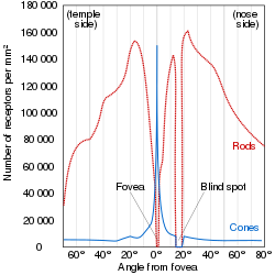
Last updated
The distribution of human photoreceptor cells shows how the photopic and scotopic systems exist in parallel, except in the fovea where the photopic system dominates. Human photoreceptor distribution.svg
The distribution of human photoreceptor cells shows how the photopic and scotopic systems exist in parallel, except in the fovea where the photopic system dominates.

A duplex retina is a retina consisting of both rod cells and cone cells, [1] which are the photoreceptor cells for two parallel but mostly separate visual systems. The rods enable the scotopic visual system, which is active in dim light. The cones enable the photopic visual system, which is active in bright light. While one is active, the other is generally inactive; either the rods are photobleached, or oversaturated, in bright light, or the cones are not sensitive enough to hyperpolarize, or instigate the phototransduction cascade, in dim light. However, at mesopic (twilight) conditions, both visual systems are active. In this region of overlap, both systems are active and combine to contribute to mesopic vision. [2]

Contents

Advantages

Like all sensors, photoreceptors are limited in dynamic range, i.e. the ratio between the lowest and highest signal they can detect. Having two photoreceptors of differing sensitivities can together cover more dynamic range of light. Human rods can detect 7 orders of magnitude between their minimum threshold and saturation and cones can detect 11 orders of magnitude between their minimum threshold and point of damage. However, together, considering their overlap, a human duplex retina can detect 14 orders of magnitude. [3]

For any visual system, there is a tradeoff between sensitivity and spatial/temporal acuity. A duplex retina uses two visual systems, one of which trades acuity for sensitivity (scotopic), and the other which trades sensitivity for high spatial and temporal acuity (photopic), which gives the best of both worlds. [3]

Simplex retina

Most vertebrates exhibit duplex retinas, including all major classes: mammals, birds, reptiles, bony fish, etc. However, some sub-clades will have evolved from the common vertebrate ancestor to lose one of the visual systems and develop a simplex retina, often called a pure-rod or pure-cone retina. [4] Vertebrates that have lost their cone cells and exhibit a pure-rod retina include:

Many vertebrates have lost their rod cells and exhibit a pure-cone retinas, which include:

While most humans possess duplex retinas, some conditions lead to a failure of one of the visual systems. A human lacking cone cells and therefore a photopic system is called an achromat or rod monochromat and experiences day blindness and monochromacy. A human lacking rod cells and therefore a scotopic system has nyctalopia or night blindness.

See also

Related Research Articles

<span class="mw-page-title-main">Retina</span> Part of the eye

The retina is the innermost, light-sensitive layer of tissue of the eye of most vertebrates and some molluscs. The optics of the eye create a focused two-dimensional image of the visual world on the retina, which then processes that image within the retina and sends nerve impulses along the optic nerve to the visual cortex to create visual perception. The retina serves a function which is in many ways analogous to that of the film or image sensor in a camera.

<span class="mw-page-title-main">Eye</span> Organ that detects light and converts it into electro-chemical impulses in neurons

An eye is a sensory organ that allows an organism to perceive visual information. It detects light and converts it into electro-chemical impulses in neurons (neurones). It is part of an organism's visual system.

<span class="mw-page-title-main">Color vision</span> Ability to perceive differences in light frequency

Color vision, a feature of visual perception, is an ability to perceive differences between light composed of different frequencies independently of light intensity.

<span class="mw-page-title-main">Photoreceptor cell</span> Type of neuroepithelial cell

A photoreceptor cell is a specialized type of neuroepithelial cell found in the retina that is capable of visual phototransduction. The great biological importance of photoreceptors is that they convert light into signals that can stimulate biological processes. To be more specific, photoreceptor proteins in the cell absorb photons, triggering a change in the cell's membrane potential.

<span class="mw-page-title-main">Tetrachromacy</span> Type of color vision with four types of cone cells

Tetrachromacy is the condition of possessing four independent channels for conveying color information, or possessing four types of cone cell in the eye. Organisms with tetrachromacy are called tetrachromats.

<span class="mw-page-title-main">Rod cell</span> Photoreceptor cells that can function in lower light better than cone cells

Rod cells are photoreceptor cells in the retina of the eye that can function in lower light better than the other type of visual photoreceptor, cone cells. Rods are usually found concentrated at the outer edges of the retina and are used in peripheral vision. On average, there are approximately 92 million rod cells in the human retina. Rod cells are more sensitive than cone cells and are almost entirely responsible for night vision. However, rods have little role in color vision, which is the main reason why colors are much less apparent in dim light.

<span class="mw-page-title-main">Cone cell</span> Photoreceptor cells responsible for color vision made to function in bright light

Cone cells or cones are photoreceptor cells in the retinas of vertebrates' eyes. They respond differently to light of different wavelengths, and the combination of their responses is responsible for color vision. Cones function best in relatively bright light, called the photopic region, as opposed to rod cells, which work better in dim light, or the scotopic region. Cone cells are densely packed in the fovea centralis, a 0.3 mm diameter rod-free area with very thin, densely packed cones which quickly reduce in number towards the periphery of the retina. Conversely, they are absent from the optic disc, contributing to the blind spot. There are about six to seven million cones in a human eye, with the highest concentration being towards the macula.

In visual physiology, adaptation is the ability of the retina of the eye to adjust to various levels of light. Natural night vision, or scotopic vision, is the ability to see under low-light conditions. In humans, rod cells are exclusively responsible for night vision as cone cells are only able to function at higher illumination levels. Night vision is of lower quality than day vision because it is limited in resolution and colors cannot be discerned; only shades of gray are seen. In order for humans to transition from day to night vision they must undergo a dark adaptation period of up to two hours in which each eye adjusts from a high to a low luminescence "setting", increasing sensitivity hugely, by many orders of magnitude. This adaptation period is different between rod and cone cells and results from the regeneration of photopigments to increase retinal sensitivity. Light adaptation, in contrast, works very quickly, within seconds.

<span class="mw-page-title-main">Monochromacy</span> Type of color vision

Monochromacy is the ability of organisms to perceive only light intensity without respect to spectral composition. Organisms with monochromacy lack color vision and can only see in shades of grey ranging from black to white. Organisms with monochromacy are called monochromats. Many mammals, such as cetaceans, the owl monkey and the Australian sea lion are monochromats. In humans, monochromacy is one among several other symptoms of severe inherited or acquired diseases, including achromatopsia or blue cone monochromacy, together affecting about 1 in 30,000 people.

<span class="mw-page-title-main">Purkinje effect</span> Tendency for sight to shift toward blue colors at low light levels

The Purkinje effect or Purkinje phenomenon is the tendency for the peak luminance sensitivity of the eye to shift toward the blue end of the color spectrum at low illumination levels as part of dark adaptation. In consequence, reds will appear darker relative to other colors as light levels decrease. The effect is named after the Czech anatomist Jan Evangelista Purkyně. While the effect is often described from the perspective of the human eye, it is well established in a number of animals under the same name to describe the general shifting of spectral sensitivity due to pooling of rod and cone output signals as a part of dark/light adaptation.

<span class="mw-page-title-main">Photopic vision</span> Visual perception under well-lit conditions

Photopic vision is the vision of the eye under well-lit conditions (luminance levels from 10 to 108 cd/m2). In humans and many other animals, photopic vision allows color perception, mediated by cone cells, and a significantly higher visual acuity and temporal resolution than available with scotopic vision.

In the study of visual perception, scotopic vision is the vision of the eye under low-light conditions. The term comes from the Greek skotos, meaning 'darkness', and -opia, meaning 'a condition of sight'. In the human eye, cone cells are nonfunctional in low visible light. Scotopic vision is produced exclusively through rod cells, which are most sensitive to wavelengths of around 498 nm and are insensitive to wavelengths longer than about 640 nm. Under scotopic conditions, light incident on the retina is not encoded in terms of the spectral power distribution. Higher visual perception occurs under scotopic vision as it does under photopic vision.

Visual phototransduction is the sensory transduction process of the visual system by which light is detected by photoreceptor cells in the vertebrate retina. A photon is absorbed by a retinal chromophore, which initiates a signal cascade through several intermediate cells, then through the retinal ganglion cells (RGCs) comprising the optic nerve.

Intrinsically photosensitive retinal ganglion cells (ipRGCs), also called photosensitive retinal ganglion cells (pRGC), or melanopsin-containing retinal ganglion cells (mRGCs), are a type of neuron in the retina of the mammalian eye. The presence of an additional photoreceptor was first suspected in 1927 when mice lacking rods and cones still responded to changing light levels through pupil constriction; this suggested that rods and cones are not the only light-sensitive tissue. However, it was unclear whether this light sensitivity arose from an additional retinal photoreceptor or elsewhere in the body. Recent research has shown that these retinal ganglion cells, unlike other retinal ganglion cells, are intrinsically photosensitive due to the presence of melanopsin, a light-sensitive protein. Therefore, they constitute a third class of photoreceptors, in addition to rod and cone cells.

Mesopic vision, sometimes also called twilight vision, is a combination of photopic and scotopic vision under low-light conditions. Mesopic levels range approximately from 0.01 to 3.0 cd/m2 in luminance. Most nighttime outdoor and street lighting conditions are in the mesopic range.

The Stiles–Crawford effect is a property of the human eye that refers to the directional sensitivity of the cone photoreceptors.

<span class="mw-page-title-main">Spectral sensitivity</span> Relative efficiency of detection of a signal as a function of its frequency or wavelength

Spectral sensitivity is the relative efficiency of detection, of light or other signal, as a function of the frequency or wavelength of the signal.

<span class="mw-page-title-main">Vision in fish</span> Sense found in most species of fish

Vision is an important sensory system for most species of fish. Fish eyes are similar to the eyes of terrestrial vertebrates like birds and mammals, but have a more spherical lens. Birds and mammals normally adjust focus by changing the shape of their lens, but fish normally adjust focus by moving the lens closer to or further from the retina. Fish retinas generally have both rod cells and cone cells, and most species have colour vision. Some fish can see ultraviolet and some are sensitive to polarised light.

Blue cone monochromacy (BCM) is an inherited eye disease that causes severe color blindness, poor visual acuity, nystagmus, hemeralopia, and photophobia due to the absence of functional red (L) and green (M) cone photoreceptor cells in the retina. BCM is a recessive X-linked disease and almost exclusively affects XY karyotypes.

<span class="mw-page-title-main">Vertebrate visual opsin</span>

Vertebrate visual opsins are a subclass of ciliary opsins and mediate vision in vertebrates. They include the opsins in human rod and cone cells. They are often abbreviated to opsin, as they were the first opsins discovered and are still the most widely studied opsins.

References

  1. Gruber, Samuel H.; Gulley, Robert L.; Brandon, Janet (1 July 1975). "Duplex Retina in Seven Elasmobranch Species". Bulletin of Marine Science . 25 (3): 353–358. Archived from the original on 18 October 2021. Retrieved 18 October 2021.
  2. "Photopic vs Scotopic Vision". isle.hanover.edu. Archived from the original on 2021-10-18. Retrieved 2021-10-18.
  3. 1 2 Barbur, J.L.; Stockman, A. (2010). "Photopic, Mesopic and Scotopic Vision and Changes in Visual Performance". Encyclopedia of the Eye: 323–331. doi:10.1016/B978-0-12-374203-2.00233-5. ISBN   9780123742032.
  4. Valen, Ragnhild; Eilertsen, Mariann; Edvardsen, Rolf Brudvik; Furmanek, Tomasz; Rønnestad, Ivar; van der Meeren, Terje; Karlsen, Ørjan; Nilsen, Tom Ole; Helvik, Jon Vidar (2016-08-15). "The two-step development of a duplex retina involves distinct events of cone and rod neurogenesis and differentiation". Developmental Biology. 416 (2): 389–401. doi: 10.1016/j.ydbio.2016.06.041 . hdl: 1956/15954 . ISSN   0012-1606. PMID   27374844.
  5. Douglas, R. H.; Partridge, J. C. (January 1997). "On the visual pigments of deep-sea fish". Journal of Fish Biology. 50 (1): 68–85. doi:10.1111/j.1095-8649.1997.tb01340.x.
  6. Meekins, Jessica M.; Moore, Bret A. (2022). "Ophthalmology of Xenarthra: Armadillos, Anteaters, and Sloths". Wild and Exotic Animal Ophthalmology. pp. 39–47. doi:10.1007/978-3-030-81273-7_4. ISBN   978-3-030-81272-0.
  7. 1 2 Davies, Wayne I. L.; Collin, Shaun P.; Hunt, David M. (July 2012). "Molecular ecology and adaptation of visual photopigments in craniates: VISUAL PHOTOPIGMENTS IN CRANIATES". Molecular Ecology. 21 (13): 3121–3158. doi:10.1111/j.1365-294X.2012.05617.x. PMID   22650357. S2CID   9077192.
  8. Jacobs, Gerald H.; Tootell, R. B. H.; Fisher, Steven K.; Anderson, Don H. (1 January 1980). "Rod photoreceptors and scotopic vision in ground squirrels". The Journal of Comparative Neurology. 189 (1): 113–125. doi:10.1002/cne.901890107. PMID   7351444. S2CID   11161113.