Nerve fiber layer

Last updated
Nerve fiber layer
Gray881.png
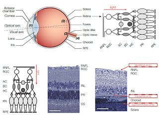
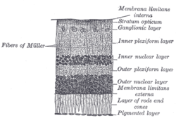
Section of retina. (Stratum opticum labeled at right, second from the top.)
Gray882.png
Plan of retinal neurons. (Stratum opticum labeled at left, second from the top.)
Details
Identifiers
Latin stratum neurofibrarum retinae
TA A15.2.04.017
FMA 58688
Anatomical terminology

The retinal nerve fiber layer (nerve fiber layer, stratum opticum, RNFL) is formed by the expansion of the fibers of the optic nerve; it is thickest near the optic disc, gradually diminishing toward the ora serrata.

Retina light-sensitive organ in the eye

The retina is the innermost, light-sensitive layer of tissue of the eye of most vertebrates and some molluscs. The optics of the eye create a focused two-dimensional image of the visual world on the retina, which translates that image into electrical neural impulses to the brain to create visual perception, the retina serving a function analogous to that of the film or image sensor in a camera.

Optic nerve paired nerve that transmits visual information from the retina to the brain

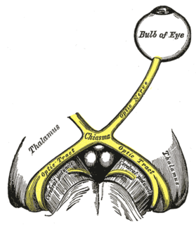
The optic nerve, also known as cranial nerve II, or simply as CN II, is a paired nerve that transmits visual information from the retina to the brain. In humans, the optic nerve is derived from optic stalks during the seventh week of development and is composed of retinal ganglion cell axons and glial cells; it extends from the optic disc to the optic chiasma and continues as the optic tract to the lateral geniculate nucleus, pretectal nuclei, and superior colliculus.

Optic disc

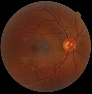
The optic disc or optic nerve head is the point of exit for ganglion cell axons leaving the eye. Because there are no rods or cones overlying the optic disc, it corresponds to a small blind spot in each eye.

As the nerve fibers pass through the lamina cribrosa sclerae they lose their medullary sheaths and are continued onward through the choroid and retina as simple axis-cylinders.

The nerve fibers forming the optic nerve exit the eye posteriorly through a hole in the sclera that is occupied by a mesh-like structure called the lamina cribrosa. It is formed by a multilayered network of collagen fibers that insert into the scleral canal wall. The nerve fibers that comprise the optic nerve run through pores formed by these collagen beams. In humans, a central retinal artery is located slightly off-center in nasal direction.

Choroid vascular layer of the eye, containing connective tissue, and lying between the retina and the sclera

The choroid, also known as the choroidea or choroid coat, is the vascular layer of the eye, containing connective tissues, and lying between the retina and the sclera. The human choroid is thickest at the far extreme rear of the eye, while in the outlying areas it narrows to 0.1 mm. The choroid provides oxygen and nourishment to the outer layers of the retina. Along with the ciliary body and iris, the choroid forms the uveal tract.

When they reach the internal surface of the retina they radiate from their point of entrance over this surface grouped in bundles, and in many places arranged in plexuses.

Most of the fibers are centripetal, and are the direct continuations of the axis-cylinder processes of the cells of the ganglionic layer, but a few of them are centrifugal and ramify in the inner plexiform and inner nuclear layers, where they end in enlarged extremities.

Centrifugal force A force on objects moving within a reference frame that rotates with respect to an inertial frame.

In Newtonian mechanics, the centrifugal force is an inertial force that appears to act on all objects when viewed in a rotating frame of reference. It is directed away from an axis passing through the coordinate system's origin and parallel to the axis of rotation. If the axis of rotation passes through the coordinate system's origin, the centrifugal force is directed radially outwards from that axis. The concept of centrifugal force can be applied in rotating devices, such as centrifuges, centrifugal pumps, centrifugal governors, and centrifugal clutches, and in centrifugal railways, planetary orbits and banked curves, when they are analyzed in a rotating coordinate system. The term has sometimes also been used for the reactive centrifugal force that may be viewed as a reaction to a centripetal force in some circumstances.

Inner nuclear layer

The inner nuclear layer or layer of inner granules, of the retina, is made up of a number of closely packed cells, of which there are three varieties, viz.: bipolar cells, horizontal cells, and amacrine cells.

Patients with retinitis pigmentosa have abnormal thinning of the RNFL which correlates with the severity of the disease. [1] However the thickness of the RNFL also decreases with age and not visual acuity. [2] The sparing of this layer is important in the treatment of the disease as it is the basis for connecting retinal prostheses to the optic nerve, or implanting stem cells that could regenerate the lost photoreceptors.

Retinitis pigmentosa retinal degeneration characterized by the gradual deterioration of the photoreceptors or the retinal pigment epithelium of the retina leading to progressive sight loss

Retinitis pigmentosa (RP) is a genetic disorder of the eyes that causes loss of vision. Symptoms include trouble seeing at night and decreased peripheral vision. Onset of symptoms is generally gradual. As peripheral vision worsens, people may experience "tunnel vision". Complete blindness is uncommon.

RNFL is a sensitive structure. Some process can excites its natural apoptosis. Harmful situation can make some damage on RNFL such as high intraocular pressure, high fluctuation on phase of intraocular pressure, inflammation, vascular disease and any kind of hypoxia. Gede Pardianto (2009) reported 6 cases of RNFL thickness change after the procedures of phacoemulsification. [3] Sudden intraocular fluctuation in any kind of intraocular surgeries maybe harmful to RNFL in accordance with mechanical stress on sudden compression and also ischemic effect of micro emboly as the result of the sudden decompression that may generate micro bubble that can clog micro vessels. [4]

Phacoemulsification

Phacoemulsification is a modern cataract surgery in which the eye's internal lens is emulsified with an ultrasonic handpiece and aspirated from the eye. Aspirated fluids are replaced with irrigation of balanced salt solution to maintain the anterior chamber.

Pattern of retinal nerve fibers Pattern of Retinal Nerve Fibers.jpg
Pattern of retinal nerve fibers

Related Research Articles

Glaucoma eye disease that is characterized by an unstable or a sustained increase in the intraocular pressure which the eye cannot withstand without damage to its structure or impairment of its function

Glaucoma is a group of eye diseases which result in damage to the optic nerve and cause vision loss. The most common type is open-angle glaucoma with less common types including closed-angle glaucoma and normal-tension glaucoma. Open-angle glaucoma develops slowly over time and there is no pain. Peripheral vision may begin to decrease followed by central vision resulting in blindness if not treated. Closed-angle glaucoma can present gradually or suddenly. The sudden presentation may involve severe eye pain, blurred vision, mid-dilated pupil, redness of the eye, and nausea. Vision loss from glaucoma, once it has occurred, is permanent.

Scanning laser ophthalmoscopy

Scanning laser ophthalmoscopy (SLO) is a method of examination of the eye. It uses the technique of confocal laser scanning microscopy for diagnostic imaging of retina or cornea of the human eye.

Intraocular pressure Fluid pressure inside the eye

Intraocular pressure (IOP) is the fluid pressure inside the eye. Tonometry is the method eye care professionals use to determine this. IOP is an important aspect in the evaluation of patients at risk of glaucoma. Most tonometers are calibrated to measure pressure in millimeters of mercury (mmHg).

Electroretinography measurement of electrical responses by cells in the retina

Electroretinography measures the electrical responses of various cell types in the retina, including the photoreceptors, inner retinal cells, and the ganglion cells. Electrodes are usually placed on the surface of the cornea for Full Field/Global/Multifocal ERG's and brass/copper electrodes are placed on the skin near the eye for EOG type testing. During a recording, the patient's eyes are exposed to standardized stimuli and the resulting signal is displayed showing the time course of the signal's amplitude (voltage). Signals are very small, and typically are measured in microvolts or nanovolts. The ERG is composed of electrical potentials contributed by different cell types within the retina, and the stimulus conditions can elicit stronger response from certain components.

Retinal implant

Retinal prostheses for restoration of sight to patients blinded by retinal degeneration are being developed by a number of private companies and research institutions worldwide. The system is meant to partially restore useful vision to people who have lost their photoreceptors due to retinal diseases such as retinitis pigmentosa (RP) or age-related macular degeneration (AMD). Three types of retinal implants are currently in clinical trials: epiretinal, subretinal, and suprachoroidal. Retinal implants introduce visual information into the retina by electrically stimulating the surviving retinal neurons. So far, elicited percepts had rather low resolution, and may be suitable for light perception and recognition of simple objects.

Coats disease Human disease

Coats' disease,, is a rare congenital, nonhereditary eye disorder, causing full or partial blindness, characterized by abnormal development of blood vessels behind the retina. Coats' disease can also fall under glaucoma.

Retinal pigment epithelium

The pigmented layer of retina or retinal pigment epithelium (RPE) is the pigmented cell layer just outside the neurosensory retina that nourishes retinal visual cells, and is firmly attached to the underlying choroid and overlying retinal visual cells.

Optic neuropathy is damage to the optic nerve from any cause. Damage and death of these nerve cells, or neurons, leads to characteristic features of optic neuropathy. The main symptom is loss of vision, with colors appearing subtly washed out in the affected eye. On medical examination, the optic nerve head can be visualised by an ophthalmoscope. A pale disc is characteristic of long-standing optic neuropathy. In many cases, only one eye is affected and patients may not be aware of the loss of color vision until the doctor asks them to cover the healthy eye.

Optic disc drusen Human disease

Optic disc drusen (ODD) or optic nerve head drusen (ONHD) are globules of mucoproteins and mucopolysaccharides that progressively calcify in the optic disc. They are thought to be the remnants of the axonal transport system of degenerated retinal ganglion cells. ODD have also been referred to as congenitally elevated or anomalous discs, pseudopapilledema, pseudoneuritis, buried disc drusen, and disc hyaline bodies.

Scanning laser polarimetry is the use of polarised light to measure the thickness of the retinal nerve fiber layer as part of a glaucoma workup. The GDx-VCC is one example.

Retinal gene therapy holds a promise in treating different forms of non-inherited and inherited blindness.

Flammer syndrome is a recently described clinical entity comprising a complex of clinical features caused mainly by dysregulation of the blood supply which has previously been called vascular dysregulation. It can manifest itself in many symptoms such as cold hands and feet and is often associated with low blood pressure. In certain cases it is associated with or predisposes for the development of diseases such as a normal tension glaucoma. Flammer syndrome is named after the Swiss ophthalmologist Josef Flammer.

Josef Flammer Swiss ophthalmologist

Josef Flammer is a Swiss ophthalmologist and long-time director of the Eye Clinic at Basel University Hospital. Flammer is a glaucoma specialist who developed a new pathogenetic concept of glaucomatous damage according to which unstable blood supply leads to oxidative stress, which in turn plays a major role in apoptosis of cells in optic nerve and retina in glaucoma patients.

Occult macular dystrophy (OMD) is a rare inherited degradation of the retina, characterized by progressive loss of function in the most sensitive part of the central retina (macula), the location of the highest concentration of light-sensitive cells (photoreceptors) but presenting no visible abnormality. "Occult" refers to the degradation in the fundus being difficult to discern. The disorder is called "dystrophy" instead of "degradation" to distinguish its genetic origin from other causes, such as age. OMD was first reported by Y. Miyake et al. in 1989.

References

This article incorporates text in the public domain from page 1015 of the 20th edition of Gray's Anatomy (1918)

  1. Walia S, Fishman GA, Edward DP, Lindeman M. Retinal nerve fiber layer defects in RP patients. Invest Ophthalmol Vis Sci. 2007 Oct;48(10):4748-52. PMID   17898300
  2. Oishi A, Otani A, Sasahara M, Kurimoto M, Nakamura H, Kojima H, Yoshimura N. Retinal nerve fiber layer thickness in patients with retinitis pigmentosa. Eye. 2008 Mar 14, online advance publication. PMID   18344951
  3. Pardianto G, Mastering phacoemulsification in Mimbar Ilmiah Oftalmologi Indonesia.2009;10:26.
  4. Pardianto G, Moeloek N, Reveny J, Wage S, Satari I, Sembiring R et al. Retinal thickness change after phacoemulsification. Clin Ophthalmol. 2013(7):2207-2214 PMC 3821754