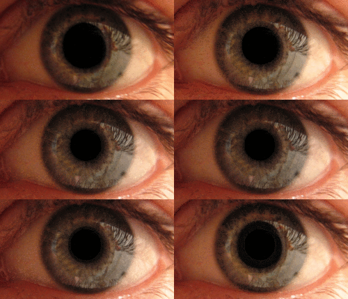
Pupillary response is a physiological response that varies the size of the pupil, via the optic and oculomotor cranial nerve.
A constriction response (miosis), [1] is the narrowing of the pupil, which may be caused by scleral buckles or drugs such as opiates/opioids or anti-hypertension medications. Constriction of the pupil occurs when the circular muscle, controlled by the parasympathetic nervous system (PSNS), contracts, and also to an extent when the radial muscle relaxes.
A dilation response (mydriasis), is the widening of the pupil and may be caused by adrenaline; anticholinergic agents; stimulant drugs such as MDMA, cocaine, and amphetamines; and some hallucinogenics (e.g. LSD). [2] Dilation of the pupil occurs when the smooth cells of the radial muscle, controlled by the sympathetic nervous system (SNS), contract, and also when the cells of the iris sphincter muscle relax.
The responses can have a variety of causes, from an involuntary reflex reaction to exposure or inexposure to light—in low light conditions a dilated pupil lets more light into the eye—or it may indicate interest in the subject of attention or arousal, sexual stimulation, [3] uncertainty, [4] decision conflict, [5] errors, [6] physical activity [7] or increasing cognitive load [8] or demand. The responses correlate strongly with activity in the locus coeruleus neurotransmitter system. [9] [10] [11] The pupils contract immediately before REM sleep begins. [12] A pupillary response can be intentionally conditioned as a Pavlovian response to some stimuli. [13]
Some humans have the ability to exert direct and voluntary control over their iris sphincter muscles and dilator muscles, granting them the ability to dilate and constrict their pupils on command, regardless of lighting condition and/or eye accommodation state. [14] However, this ability is very rare, and its potential use or advantages is are unclear.
The latency of pupillary response (the time in which it takes to occur) increases with age. [15]
In ophthalmology, intensive studies of pupillary response are conducted via videopupillometry. [16]
Anisocoria is the condition of one pupil being more dilated than the other.
Constriction (Parasympathetic) | Dilation (Sympathetic) | |
---|---|---|
Muscular mechanism | Relaxation of iris dilator muscle, activation of iris sphincter muscle | Activation of iris dilator muscle, relaxation of iris sphincter muscle |
Cause in pupillary light reflex | Increased light | Decreased light |
Other physiological causes | Accommodation reflex | Fight-or-flight response, sexual arousal |
Corresponding non-physiological state | Miosis | Mydriasis |
The pupil is a hole located in the center of the iris of the eye that allows light to strike the retina. It appears black because light rays entering the pupil are either absorbed by the tissues inside the eye directly, or absorbed after diffuse reflections within the eye that mostly miss exiting the narrow pupil. The size of the pupil is controlled by the iris, and varies depending on many factors, the most significant being the amount of light in the environment. The term "pupil" was coined by Gerard of Cremona.
The iris is a thin, annular structure in the eye in most mammals and birds, responsible for controlling the diameter and size of the pupil, and thus the amount of light reaching the retina. In optical terms, the pupil is the eye's aperture, while the iris is the diaphragm. Eye color is defined by the iris.
Mydriasis is the dilation of the pupil, usually having a non-physiological cause, or sometimes a physiological pupillary response. Non-physiological causes of mydriasis include disease, trauma, or the use of certain types of drug. It may also be of unknown cause.
The pupillary light reflex (PLR) or photopupillary reflex is a reflex that controls the diameter of the pupil, in response to the intensity (luminance) of light that falls on the retinal ganglion cells of the retina in the back of the eye, thereby assisting in adaptation of vision to various levels of lightness/darkness. A greater intensity of light causes the pupil to constrict, whereas a lower intensity of light causes the pupil to dilate. Thus, the pupillary light reflex regulates the intensity of light entering the eye. Light shone into one eye will cause both pupils to constrict.
Miosis, or myosis, is excessive constriction of the pupil. The opposite condition, mydriasis, is the dilation of the pupil. Anisocoria is the condition of one pupil being more dilated than the other.
The human eye is an organ of the sensory nervous system that reacts to visible light and allows the use of visual information for various purposes including seeing things, keeping balance, and maintaining circadian rhythm.
In neuroanatomy, the pretectal area, or pretectum, is a midbrain structure composed of seven nuclei and comprises part of the subcortical visual system. Through reciprocal bilateral projections from the retina, it is involved primarily in mediating behavioral responses to acute changes in ambient light such as the pupillary light reflex, the optokinetic reflex, and temporary changes to the circadian rhythm. In addition to the pretectum's role in the visual system, the anterior pretectal nucleus has been found to mediate somatosensory and nociceptive information.
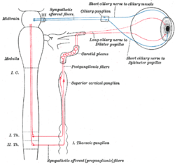
The Edinger–Westphal nucleus is one of two nuclei of the oculomotor nerve and is located in the midbrain. It receives afferents from the both pretectal nuclei. It contains parasympathetic pre-ganglionic neuron cell bodies that synapse in the ciliary ganglion. It contributes the autonomic, parasympathetic component to the oculomotor nerve, ultimately providing innervation to the iris sphincter muscle and ciliary muscle to mediate the pupillary light reflex and accommodation, respectively.
The ciliary ganglion is a parasympathetic ganglion located just behind the eye in the posterior orbit. It is 1–2 mm in diameter and in humans contains approximately 2,500 neurons. The ganglion contains postganglionic parasympathetic neurons. These neurons supply the pupillary sphincter muscle, which constricts the pupil, and the ciliary muscle which contracts to make the lens more convex. Both of these muscles are involuntary since they are controlled by the parasympathetic division of the autonomic nervous system.
The iris dilator muscle, is a smooth muscle of the eye, running radially in the iris and therefore fit as a dilator. The pupillary dilator consists of a spokelike arrangement of modified contractile cells called myoepithelial cells. These cells are stimulated by the sympathetic nervous system. When stimulated, the cells contract, widening the pupil and allowing more light to enter the eye.
The iris sphincter muscle is a muscle in the part of the eye called the iris. It encircles the pupil of the iris, appropriate to its function as a constrictor of the pupil.
Adie syndrome, also known as Holmes–Adie syndrome, is a neurological disorder characterized by a tonically dilated pupil that reacts slowly to light but shows a more definite response to accommodation. It is frequently seen in females with absent knee or ankle jerks and impaired sweating.
Pupillometer, also spelled pupilometer, is a medical device intended to measure by reflected light the size of the pupil of the eye. In addition to measuring pupil size, current automated pupillometers may also be able to characterize pupillary light reflex. Some instruments for measuring pupillary distance (PD) are often, but incorrectly, referred to as pupilometers.
Surprise is a brief mental and physiological state, a startle response experienced by animals and humans as the result of an unexpected event. Surprise can have any valence; that is, it can be neutral/moderate, pleasant, unpleasant, positive, or negative. Surprise can occur in varying levels of intensity ranging from very-surprised, which may induce the fight-or-flight response, or little-surprise that elicits a less intense response to the stimuli.
A relative afferent pupillary defect (RAPD), also known as a Marcus Gunn pupil, is a medical sign observed during the swinging-flashlight test whereupon the patient's pupils dilate when a bright light is swung from the unaffected eye to the affected eye. The affected eye still senses the light and produces pupillary sphincter constriction to some degree, albeit reduced.
Dilated fundus examination (DFE) is a diagnostic procedure that uses mydriatic eye drops to dilate or enlarge the pupil in order to obtain a better view of the fundus of the eye. Once the pupil is dilated, examiners use ophthalmoscopy to view the eye's interior, which makes it easier to assess the retina, optic nerve head, blood vessels, and other important features. DFE has been found to be a more effective method for evaluating eye health when compared to non-dilated examination, and is the best method of evaluating structures behind the iris. It is frequently performed by ophthalmologists and optometrists as part of an eye examination.
Polycoria is a pathological condition of the eye characterized by more than one pupillary opening in the iris. It may be congenital or result from a disease affecting the iris. It results in decreased function of the iris and pupil, affecting the physical eye and visualization.
The oddball paradigm is an experimental design used within psychology research. Presentations of sequences of repetitive stimuli are infrequently interrupted by a deviant stimulus. The reaction of the participant to this "oddball" stimulus is recorded.
Task-invoked pupillary response is a pupillary response caused by a cognitive load imposed on a human and as a result of the decrease in parasympathetic activity in the peripheral nervous system. It is found to result in a linear increase in pupil dilation as the demand a task places on the working memory increases. Beatty evaluated task-invoked pupillary response in different tasks for short-term memory, language processing, reasoning, perception, sustained attention and selective attention and found that it fulfills Kahneman's three criteria for indicating processing load. That is, it can reflect differences in processing load within a task, between different tasks and between individuals. It is used as an indicator of cognitive load levels in psychophysiology research.
Pupillometry, the measurement of pupil size and reactivity, is a key part of the clinical neurological exam for patients with a wide variety of neurological injuries. It is also used in psychology.
{{cite journal}}
: Cite journal requires |journal=
(help)