Short posterior ciliary arteries

Last updated
Short posterior ciliary arteries
Gray873.png
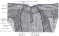
The arteries of the choroid and iris. The greater part of the sclera has been removed.
Gray514.png
Details
Source Ophthalmic artery
Vein Vorticose veins
Supplies Choroid (up to the equator of the eye)
ciliary processes
Identifiers
Latin arteriae ciliares posteriores breves
TA98 A12.2.06.031
TA2 4479
FMA 70777
Anatomical terminology

The short posterior ciliary arteries are a number of branches of the ophthalmic artery. They pass forward with the optic nerve to reach the eyeball, piercing the sclera around the entry of the optic nerve into the eyeball.

Contents

Anatomy

The number of short posterior ciliary arteries varies between individuals; [1] one or more short posterior ciliary arteries initially branch off the ophthalmic artery, [2] subsequently dividing to form up to 20 short posterior ciliary arteries. [2] [3]

Origin

The short posterior ciliary arteries branch off the ophthalmic artery as it crosses the optic nerve medially. [1]

Course and relations

About 7 short posterior ciliary arteries accompany the optic nerve, [3] passing anterior-ward to reach the posterior part of [4] the eyeball, where they divide into 15-20 branches and pierce the sclera [3] around the entrance of the optic nerve. [4] [2]

Distribution

The short posterior ciliary arteries contribute arterial supply to the choroid, ciliary processes, [3] optic disc, the outer retina, and Bruch's membrane. [1]

Some branches of the short posterior ciliary arteries supply the optic disc by means of an anastomotic ring - the circle of Zinn-Haller or circle of Zinn - which is associated with the fibrous extension of the ocular tendons (common tendinous ring (also annulus of Zinn)).[ citation needed ]

Additional images

See also

Related Research Articles

<span class="mw-page-title-main">Oculomotor nerve</span> Cranial nerve III, for eye movements

The oculomotor nerve, also known as the third cranial nerve, cranial nerve III, or simply CN III, is a cranial nerve that enters the orbit through the superior orbital fissure and innervates extraocular muscles that enable most movements of the eye and that raise the eyelid. The nerve also contains fibers that innervate the intrinsic eye muscles that enable pupillary constriction and accommodation. The oculomotor nerve is derived from the basal plate of the embryonic midbrain. Cranial nerves IV and VI also participate in control of eye movement.

<span class="mw-page-title-main">Internal carotid artery</span> Artery of the human brain

The internal carotid artery is an artery in the neck which supplies the anterior circulation of the brain.

<span class="mw-page-title-main">Choroid</span> Vascular layer of the eye, containing connective tissue, and lying between the retina and the sclera

The choroid, also known as the choroidea or choroid coat, is a part of the uvea, the vascular layer of the eye. It contains connective tissues, and lies between the retina and the sclera. The human choroid is thickest at the far extreme rear of the eye, while in the outlying areas it narrows to 0.1 mm. The choroid provides oxygen and nourishment to the outer layers of the retina. Along with the ciliary body and iris, the choroid forms the uveal tract.

<span class="mw-page-title-main">Ophthalmic artery</span> Artery of the head

The ophthalmic artery (OA) is an artery of the head. It is the first branch of the internal carotid artery distal to the cavernous sinus. Branches of the ophthalmic artery supply all the structures in the orbit around the eye, as well as some structures in the nose, face, and meninges. Occlusion of the ophthalmic artery or its branches can produce sight-threatening conditions.

<span class="mw-page-title-main">Central retinal artery</span>

The central retinal artery branches off the ophthalmic artery, running inferior to the optic nerve within its dural sheath to the eyeball.

<span class="mw-page-title-main">Common tendinous ring</span> Ring of fibrous tissue around optic nerve at its entrance to the eye

The common tendinous ring, also known as the annulus of Zinn, or annular tendon, is a ring of fibrous tissue surrounding the optic nerve at its entrance at the apex of the orbit. It is the common origin of the four recti muscles of the group of extraocular muscles.

<span class="mw-page-title-main">Nasociliary nerve</span> Branch of the ophthalmic nerve

The nasociliary nerve is a branch of the ophthalmic nerve (CN V1) (which is in turn a branch of the trigeminal nerve (CN V)). It is intermediate in size between the other two branches of the ophthalmic nerve, the frontal nerve and lacrimal nerve.

<span class="mw-page-title-main">Superior ophthalmic vein</span> Vein of the orbit around the eye

The superior ophthalmic vein is a vein of the orbit that drains venous blood from structures of the upper orbit. It is formed by the union of the angular vein, and supraorbital vein. It passes backwards within the orbit alongside the ophthalmic artery, then exits the orbit through the superior orbital fissure to drain into the cavernous sinus.

<span class="mw-page-title-main">Long ciliary nerves</span>

The long ciliary nerves are 2-3 nerves that arise from the nasociliary nerve (itself a branch of the ophthalmic branch (CN V1) of the trigeminal nerve (CN V)). They enter the eyeball to provide sensory innervation to parts of the eye, and sympathetic visceral motor innervation to the dilator pupillae muscle.

<span class="mw-page-title-main">Lacrimal artery</span> Artery of the orbit

The lacrimal artery is an artery of the orbit. It is a branch of the ophthalmic artery. It accompanies the lacrimal nerve along the upper border of the lateral rectus muscle, travelling forward to reach the lacrimal gland. It supplies the lacrimal gland, two rectus muscles of the eye, the eyelids, and the conjunctiva.

<span class="mw-page-title-main">Posterior ethmoidal artery</span>

The posterior ethmoidal artery is an artery of the head which arises from the ophthalmic artery to supply the posterior ethmoidal air cells, and the meninges. It is smaller than the anterior ethmoidal artery.

<span class="mw-page-title-main">Short ciliary nerves</span> Nerves of the orbit around the eye

The short ciliary nerves are nerves of the orbit around the eye. They are branches of the ciliary ganglion. They supply parasympathetic and sympathetic nerve fibers to the ciliary muscle, iris, and cornea. Damage to the short ciliary nerve may result in loss of the pupillary light reflex, or mydriasis.

<span class="mw-page-title-main">Ciliary arteries</span> Branches of a major artery in the eye

The ciliary arteries are divisible into three groups, the long posterior, short posterior, and the anterior.

<span class="mw-page-title-main">Supraorbital artery</span>

The supraorbital artery is a branch of the ophthalmic artery. It passes anteriorly within the orbit to exit the orbit through the supraorbital foramen or notch alongside the supraorbital nerve, splitting into two terminal branches which go on to form anastomoses with arteries of the head.

<span class="mw-page-title-main">Long posterior ciliary arteries</span>

The long posterior ciliary arteries are arteries of the orbit. There are long posterior ciliary arteries two on each side of the body. They are branches of the ophthalmic artery. They pass forward within the eye to reach the ciliary body where they ramify and anastomose with the anterior ciliary arteries, thus forming the major arterial circle of the iris.The long posterior ciliary arteries contribute arterial supply to the choroid, ciliary body, and iris.

<span class="mw-page-title-main">Anterior ciliary arteries</span> Small arteries in the eye-socket

The anterior ciliary arteries are seven arteries in each eye-socket that arise from muscular branches of the ophthalmic artery and supply the conjunctiva, sclera, rectus muscles, and the ciliary body. The arteries end by anastomosing with branches of the long posterior ciliary arteries to form the circulus arteriosus major.

The Orbital Fascia forms the periosteum of the orbit.

<span class="mw-page-title-main">Central retinal vein</span> Vein in the retina of the eye

The central retinal vein is a vein that drains the retina of the eye. It travels backwards through the centre of the optic nerve accompanied by the central retinal artery before exiting the optic nerve together with the central retinal artery to drain into either the superior ophthalmic vein or the cavernous sinus.

<span class="mw-page-title-main">Outline of human anatomy</span> Overview of and topical guide to human anatomy

The following outline is provided as an overview of and topical guide to human anatomy:

Major arterial circle of the iris" is a circular artery of the eye formed by anastomoses of the anterior ciliary arteries and long posterior ciliary arteries at the ciliary body. It supplies arterial blood to the iris, ciliary processes of the ciliary body, and anterior choroid.

References

  1. 1 2 3 Gupta, Neha; Motlagh, Mahsaw; Singh, Gurdeep (2022), "Anatomy, Head and Neck, Eye Arteries", StatPearls, Treasure Island (FL): StatPearls Publishing, PMID   30725748 , retrieved 2023-01-02
  2. 1 2 3 Remington, Lee Ann (2012). "Orbital Blood Supply". Clinical Anatomy and Physiology of the Visual System. Elsevier. pp. 202–217. doi:10.1016/b978-1-4377-1926-0.10011-6. ISBN   978-1-4377-1926-0.
  3. 1 2 3 4 Standring, Susan (2020). Gray's Anatomy: The Anatomical Basis of Clinical Practice (42nd ed.). [New York]. p. 780. ISBN   978-0-7020-7707-4. OCLC   1201341621.{{cite book}}: CS1 maint: location missing publisher (link)
  4. 1 2 Gray, Henry (1918). Gray's Anatomy (20th ed.). p. 571.