
The external carotid artery is a major artery of the head and neck. It arises from the common carotid artery when it splits into the external and internal carotid artery. It supplies blood to the face and neck.

The middle meningeal artery is typically the third branch of the first part of the maxillary artery, one of the two terminal branches of the external carotid artery. After branching off the maxillary artery in the infratemporal fossa, it runs through the foramen spinosum to supply the dura mater and the calvaria. The middle meningeal artery is the largest of the three (paired) arteries that supply the meninges, the others being the anterior meningeal artery and the posterior meningeal artery.

The procerus muscle is a small pyramidal slip of muscle deep to the superior orbital nerve, artery and vein. Procerus is Latin, meaning tall or extended.
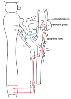
The auriculotemporal nerve is a branch of the mandibular nerve (V3) that runs with the superficial temporal artery and vein, and provides sensory innervation to various regions on the side of the head.

The middle cerebral artery (MCA) is one of the three major paired arteries that supply blood to the cerebrum. The MCA arises from the internal carotid and continues into the lateral sulcus where it then branches and projects to many parts of the lateral cerebral cortex. It also supplies blood to the anterior temporal lobes and the insular cortices.

The cavernous sinus within the human head, is one of the dural venous sinuses creating a cavity called the lateral sellar compartment bordered by the temporal bone of the skull and the sphenoid bone, lateral to the sella turcica.

The greater wing of the sphenoid bone, or alisphenoid, is a bony process of the sphenoid bone; there is one on each side, extending from the side of the body of the sphenoid and curving upward, laterally, and backward.

The pterygoid plexus is a venous plexus of considerable size, and is situated between the temporalis muscle and lateral pterygoid muscle, and partly between the two pterygoid muscles.

The transverse facial artery is an artery that branches from the superficial temporal artery and runs across the face.

The carotid canal is the passageway in the temporal bone through which the internal carotid artery enters the middle cranial fossa from the neck. The canal starts on the inferior surface of the temporal bone at the external opening of the carotid canal. The canal ascends at first superiorly, and then, making a bend, runs anteromedially. The canal's internal opening is near the foramen lacerum, above which the internal carotid artery passes on its way anteriorly to the cavernous sinus.

The middle cranial fossa, deeper than the anterior cranial fossa, is narrow medially and widens laterally to the sides of the skull. It is separated from the posterior fossa by the clivus and the petrous crest.

The lacrimal artery is an artery that arises close to the optic foramen, and is one of the largest branches derived from the ophthalmic artery. Not infrequently it is given off before the artery enters the orbit.

The infraorbital artery is an artery in the head that branches off the maxillary artery, emerging through the infraorbital foramen, just under the orbit of the eye.

The infratemporal fossa is an irregularly shaped cavity, situated below and medial to the zygomatic arch. It is not fully enclosed by bone in all directions, and it contains superficial muscles that are visible during dissection after removing skin and fascia: namely, the lower part of the temporalis muscle, the lateral pterygoid, and the medial pterygoid.
The supraorbital artery is an artery of the head.
The medial palpebral arteries are arteries of the head. They are two in number, superior and inferior, arise from the ophthalmic, opposite the pulley of the Obliquus superior.
The deep temporal arteries, two in number, anterior and posterior, ascend between the temporalis and the pericranium.
The public domain consists of all the creative works to which no exclusive intellectual property rights apply. Those rights may have expired, been forfeited, expressly waived, or may be inapplicable.

Gray's Anatomy is an English language textbook of human anatomy originally written by Henry Gray and illustrated by Henry Vandyke Carter. Earlier editions were called Anatomy: Descriptive and Surgical and Gray's Anatomy: Descriptive and Applied, but the book's name is commonly shortened to, and later editions are titled, Gray's Anatomy. The book is widely regarded as an extremely influential work on the subject, and has continued to be revised and republished from its initial publication in 1858 to the present day. The latest edition of the book, the 41st, was published in September 2015.


