The inner ear is the innermost part of the vertebrate ear. In vertebrates, the inner ear is mainly responsible for sound detection and balance. In mammals, it consists of the bony labyrinth, a hollow cavity in the temporal bone of the skull with a system of passages comprising two main functional parts:

The facial nerve, also known as the seventh cranial nerve, cranial nerve VII, or simply CN VII, is a cranial nerve that emerges from the pons of the brainstem, controls the muscles of facial expression, and functions in the conveyance of taste sensations from the anterior two-thirds of the tongue. The nerve typically travels from the pons through the facial canal in the temporal bone and exits the skull at the stylomastoid foramen. It arises from the brainstem from an area posterior to the cranial nerve VI and anterior to cranial nerve VIII.

The glossopharyngeal nerve, also known as the ninth cranial nerve, cranial nerve IX, or simply CN IX, is a cranial nerve that exits the brainstem from the sides of the upper medulla, just anterior to the vagus nerve. Being a mixed nerve (sensorimotor), it carries afferent sensory and efferent motor information. The motor division of the glossopharyngeal nerve is derived from the basal plate of the embryonic medulla oblongata, whereas the sensory division originates from the cranial neural crest.

The temporal bones are situated at the sides and base of the skull, and lateral to the temporal lobes of the cerebral cortex.

The middle meningeal artery is typically the third branch of the first portion of the maxillary artery. After branching off the maxillary artery in the infratemporal fossa, it runs through the foramen spinosum to supply the dura mater and the calvaria. The middle meningeal artery is the largest of the three (paired) arteries that supply the meninges, the others being the anterior meningeal artery and the posterior meningeal artery.

The tympanic cavity is a small cavity surrounding the bones of the middle ear. Within it sit the ossicles, three small bones that transmit vibrations used in the detection of sound.

The internal auditory meatus is a canal within the petrous part of the temporal bone of the skull between the posterior cranial fossa and the inner ear.

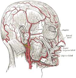
The posterior auricular artery is a small artery that arises from the external carotid artery. It ascends along the side of the head. It supplies several muscles of the neck and several structures of the head.

The ascending pharyngeal artery is an artery of the neck that supplies the pharynx.

The mastoid part of the temporal bone is the posterior (back) part of the temporal bone, one of the bones of the skull. Its rough surface gives attachment to various muscles and it has openings for blood vessels. From its borders, the mastoid part articulates with two other bones.

The petrous part of the temporal bone is pyramid-shaped and is wedged in at the base of the skull between the sphenoid and occipital bones. Directed medially, forward, and a little upward, it presents a base, an apex, three surfaces, and three angles, and houses in its interior, the components of the inner ear. The petrous portion is among the most basal elements of the skull and forms part of the endocranium. Petrous comes from the Latin word petrosus, meaning "stone-like, hard". It is one of the densest bones in the body. In other mammals, it is a separate bone, the petrosal bone.

The facial canal is a Z-shaped canal in the temporal bone of the skull. It extends between the internal acoustic meatus and stylomastoid foramen. It transmits the facial nerve.

The deep auricular artery is a branch of the maxillary artery. The deep auricular artery pierces the external acoustic meatus. It provides arterial supply to the skin of the external acoustic meatus, and contributes arterial supply to the tympanic membrane, and the temporomandibular joint.
The anterior tympanic artery is a branch of the maxillary artery. It passes through the petrotympanic fissure to entre the middle ear where it contributes to the formation of the circular anastomosis around the tympanic membrane. It provides arterial supply to part of the lining of the middle ear. It is accompanied by the chorda tympani nerve.

The infratemporal fossa is an irregularly shaped cavity that is a part of the skull. It is situated below and medial to the zygomatic arch. It is not fully enclosed by bone in all directions. It contains superficial muscles, including the lower part of the temporalis muscle, the lateral pterygoid muscle, and the medial pterygoid muscle. It also contains important blood vessels such as the middle meningeal artery, the pterygoid plexus, and the retromandibular vein, and nerves such as the mandibular nerve (CN V3) and its branches.
The petrotympanic fissure is a fissure in the temporal bone that runs from the temporomandibular joint to the tympanic cavity.

The mastoid cells are air-filled cavities within the mastoid process of the temporal bone of the cranium. The mastoid cells are a form of skeletal pneumaticity. Infection in these cells is called mastoiditis.

The superior tympanic artery is a small artery in the head. It is a branch of the middle meningeal artery. On entering the cranium it runs in the canal for the tensor tympani muscle and supplies this muscle and the lining membrane of the canal.
The caroticotympanic artery is a small, sometimes doubled artery which arises from the internal carotid artery. It leaves the carotid canal through a foramen to reach the tympanic cavity. It contributes arterial supply to the osseous part of the pharyngotympanic tube.
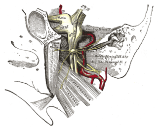
The petrosal branch of middle meningeal artery enters the hiatus for greater petrosal nerve, supplies the facial nerve and anastomoses with the stylomastoid branch of the posterior auricular artery.