Semicircular canals

Last updated
Semicircular canals
Blausen 0329 EarAnatomy InternalEar.png
The internal ear, with "semicircular ducts" at left
Vestibular system's semicircular canal- a cross-section.jpg
Illustration showing a semicircular canal. The osseous structure contains its semicircular duct which is continuous to the membranous ampulla that contains hair cells, a cupula, vestibular nerve endings and endolymph fluid.
Details
Artery Stylomastoid artery, labyrinthine artery
Identifiers
Latin canalis semicircularis
ductus semicircularis
MeSH D012665
TA98 A15.3.03.015
TA2 6954
FMA 60186
Anatomical terminology

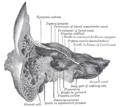
The semicircular canals are three semicircular interconnected tubes located in the innermost part of each ear, the inner ear. The three canals are the lateral, anterior and posterior semicircular canals. They are the part of the bony labyrinth, a periosteum-lined cavity on the petrous part of the temporal bone filled with perilymph.

Contents

Each semicircular canal contains its respective semicircular duct, i.e. the lateral, anterior and posterior semicircular ducts, which provide the sensation of angular acceleration and are part of the membranous labyrinth—therefore filled with endolymph.

Structure

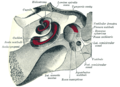
The semicircular canals are a component of the bony labyrinth that are at right angles from each other and contain their respective semicircular duct. At one end of each of the semicircular ducts is a dilated sac called an membranous ampulla, which is more than twice the diameter of the ducts. Each ampulla contains an ampullary crest, the crista ampullaris which consists of a thick gelatinous cap called a cupula and many hair cells. The superior and posterior semicircular ducts are oriented vertically at right angles to each other. The lateral semicircular duct is about a 30-degree angle from the horizontal plane. The orientations of the ducts cause a different duct to be stimulated by movement of the head in different planes, and more than one duct is stimulated at once if the movement is off those planes. The lateral semicircular duct detects angular acceleration of the head when the head is turned and the anterior and posterior semicircular ducts detect vertical head movements when the head is moved up or down. [1] When the head changes position, the endolymph in the ducts lags behind due to inertia and this acts on the cupula which bends the cilia of the hair cells. The stimulation of the hair cells sends the message to the brain that acceleration is taking place. The semicircular canals open into the vestibule by five orifices, one of the apertures being common to two of them.

Among species of mammals, the size of the semicircular canals is correlated with their type of locomotion. Specifically, species that are agile and have fast, jerky locomotion have larger canals relative to their body size than those that move more cautiously. [2]

Lateral semicircular canal

The lateral semicircular canal (also known as horizontal or external semicircular canal) is the shortest of the three canals. Movement of fluid within its duct corresponds to rotation of the head around a vertical axis (i.e. the neck), or in other words, rotation in the transverse plane. This occurs, for example, when one turns the head from side to side (yaw axis).

It measures from 12 to 15 mm (0.47 to 0.59 in), and its arch is directed horizontally backward and laterally; thus each semicircular canal stands at right angles to the other two. Its ampullated end corresponds to the upper and lateral angle of the vestibule, just above the oval window, where it opens close to the ampullated end of the anterior semicircular canal; its opposite end opens at the upper and back part of the vestibule. The lateral canal of one ear is very nearly in the same plane as that of the other.

Anterior semicircular canal

The anterior semicircular canal (also known as superior semicircular canal) contains the part of the vestibular system that detects rotations of the head in around the lateral axis, that is, rotation in the sagittal plane. This occurs, for example, when nodding one's head (pitch axis).

It is 15 to 20 mm (0.59 to 0.79 in) in length, is vertical in direction, and is placed transversely to the long axis of the petrous part of the temporal bone, on the anterior surface of which its arch forms a round projection. It describes about two-thirds of a circle. Its lateral extremity is ampullated, and opens into the upper part of the vestibule; the opposite end joins with the upper part of the posterior semicircular canal to form the crus osseum commune, which opens into the upper and medial part of the vestibule.

Posterior semicircular canal

The posterior semicircular canal contains the part of the vestibular system that detects rotation of the head around the antero-posterior (sagittal) axis, or in other words, rotation in the coronal plane. This occurs, for example, when one moves the head to touch the shoulders, or when doing a cartwheel (roll axis).

It is directed superiorly and posteriorly, as per its nomenclature, nearly parallel to the posterior surface of the petrous part of the temporal bone. The vestibular aqueduct is immediately medial to it. The posterior semicircular canal is part of the bony labyrinth and its duct is used by the vestibular system to detect rotations of the head in the coronal plane. It is the longest of the three semicircular canals, measuring from 18 to 22 mm (0.71 to 0.87 in). Its lower or ampullated end opens into the lower and back part of the vestibule, its upper into the crus osseum commune.

Development

Findings from a 2009 study demonstrated a critical late role for bone morphogenetic protein 2 (BMP-2) in the morphogenesis of semicircular canals in the zebrafish inner ear. It is suspected that the role of BMP-2 in semicircular canal duct outgrowth is likely to be conserved between different vertebrate species. [3]

Additionally, it has been found that the two semicircular canals found in the lamprey inner ear are developmentally similar to the superior and posterior canals found in humans, as the canals of both organisms arise from two depressions in the otic vesicle during early development. These depressions first form in lampreys between the 11 and 42 millimeter larval stages and form in zebrafish 57 hours post-fertilization [4]

Function

Cochlea and vestibular system Cochlea and vestibular system.gif
Cochlea and vestibular system

The semicircular ducts provide sensory input for experiences of rotary movements. They are oriented along the pitch, roll, and yaw axes. The lateral semicircular canal is oriented in the yaw axis, the anterior semicircular canal is oriented in the pitch axis, and the posterior semicircular canal is oriented in the roll axis.

Each duct is filled with a fluid called endolymph and contains motion sensors within the fluids. The base of each duct is enlarged, opening into the utricle, and has a dilated sac at one end called the membranous ampulla. Within the ampulla is a mound of hair cells and supporting cells called crista ampullaris. These hair cells have many cytoplasmic projections on the apical surface called stereocilia which are embedded in a gelatinous structure called the cupula. As the head rotates, the duct moves, but the endolymph lags behind owing to inertia. This deflects the cupula and bends the stereocilia within. The bending of these stereocilia alters an electric signal that is transmitted to the brain. Within approximately 10 seconds of achieving constant motion, the endolymph catches up with the movement of the duct and the cupula is no longer affected, stopping the sensation of acceleration. [1] The specific gravity of the cupula is comparable to that of the surrounding endolymph. Consequently, the cupula is not displaced by gravity, unlike the otolithic membranes of the utricle and saccule. As with macular hair cells, hair cells of the crista ampullaris will depolarise when the stereocilia deflect towards the kinocilium. Deflection in the opposite direction results in hyperpolarisation and inhibition. In the lateral semicircular duct, ampullopetal flow is necessary for hair-cell stimulation, whereas ampullofugal flow is necessary for the anterior and posterior semicircular ducts. [5]

This adjustment period is in part the cause of an illusion known as "the leans" often experienced by pilots. As a pilot enters a turn, hair cells in the semicircular ducts are stimulated, telling the brain that the aircraft, and the pilot, are no longer moving in a straight line but rather making a banked turn. If the pilot were to sustain a constant rate turn, the endolymph would eventually catch up with the ducts and cease to deflect the cupula. The pilot would no longer feel as if the aircraft was in a turn. As the pilot exits the turn, the semicircular ducts are stimulated to make the pilot think that they are now turning in the opposite direction rather than flying straight and level. In response to this, the pilot will often lean in the direction of the original turn in an attempt to compensate for this illusion. A more serious form of this is called a graveyard spiral. Rather than the pilot leaning in the direction of the original turn, they may actually re-enter the turn. As the endolymph stabilizes, the semicircular ducts stop registering the gradual turn and the aircraft slowly loses altitude until impact with the ground. [6]

History

Jean Pierre Flourens, by destroying the horizontal semicircular canal of pigeons, noted that they continue to fly in a circle, showing the purpose of the semicircular canals. [7]

See also

Related Research Articles

<span class="mw-page-title-main">Inner ear</span> Innermost part of the vertebrate ear

The inner ear is the innermost part of the vertebrate ear. In vertebrates, the inner ear is mainly responsible for sound detection and balance. In mammals, it consists of the bony labyrinth, a hollow cavity in the temporal bone of the skull with a system of passages comprising two main functional parts:

<span class="mw-page-title-main">Sense of balance</span> Physiological sense regarding posture

The sense of balance or equilibrioception is the perception of balance and spatial orientation. It helps prevent humans and nonhuman animals from falling over when standing or moving. Equilibrioception is the result of a number of sensory systems working together; the eyes, the inner ears, and the body's sense of where it is in space (proprioception) ideally need to be intact.

<span class="mw-page-title-main">Cochlea</span> Snail-shaped part of inner ear involved in hearing

The cochlea is the part of the inner ear involved in hearing. It is a spiral-shaped cavity in the bony labyrinth, in humans making 2.75 turns around its axis, the modiolus. A core component of the cochlea is the organ of Corti, the sensory organ of hearing, which is distributed along the partition separating the fluid chambers in the coiled tapered tube of the cochlea.

Articles related to anatomy include:

<span class="mw-page-title-main">Utricle (ear)</span> Membranous labyrinth in the vestibule of ear

The utricle and saccule are the two otolith organs in the vertebrate inner ear. They are part of the balancing system in the vestibule of the bony labyrinth. They use small stones and a viscous fluid to stimulate hair cells to detect motion and orientation. The utricle detects linear accelerations and head-tilts in the horizontal plane. The word utricle comes from Latin uter 'leather bag'.

<span class="mw-page-title-main">Saccule</span> Bed of sensory cells in the inner ear

The saccule is a bed of sensory cells in the inner ear. It translates head movements into neural impulses for the brain to interpret. The saccule detects linear accelerations and head tilts in the vertical plane. When the head moves vertically, the sensory cells of the saccule are disturbed and the neurons connected to them begin transmitting impulses to the brain. These impulses travel along the vestibular portion of the eighth cranial nerve to the vestibular nuclei in the brainstem.

<span class="mw-page-title-main">Vestibular system</span> Sensory system that facilitates body balance

The vestibular system, in vertebrates, is a sensory system that creates the sense of balance and spatial orientation for the purpose of coordinating movement with balance. Together with the cochlea, a part of the auditory system, it constitutes the labyrinth of the inner ear in most mammals.

<span class="mw-page-title-main">Ear</span> Organ of hearing and balance

An ear is the organ that enables hearing and body balance using the vestibular system. In mammals, the ear is usually described as having three parts: the outer ear, the middle ear and the inner ear. The outer ear consists of the pinna and the ear canal. Since the outer ear is the only visible portion of the ear in most animals, the word "ear" often refers to the external part alone. The middle ear includes the tympanic cavity and the three ossicles. The inner ear sits in the bony labyrinth, and contains structures which are key to several senses: the semicircular canals, which enable balance and eye tracking when moving; the utricle and saccule, which enable balance when stationary; and the cochlea, which enables hearing. The ear is a self cleaning organ through its relationship with earwax and the ear canals. The ears of vertebrates are placed somewhat symmetrically on either side of the head, an arrangement that aids sound localization.

<span class="mw-page-title-main">Endolymph</span> Inner ear fluid

Endolymph is the fluid contained in the membranous labyrinth of the inner ear. The major cation in endolymph is potassium, with the values of sodium and potassium concentration in the endolymph being 0.91 mM and 154 mM, respectively. It is also called Scarpa's fluid, after Antonio Scarpa.

<span class="mw-page-title-main">Benign paroxysmal positional vertigo</span> Medical condition

Benign paroxysmal positional vertigo (BPPV) is a disorder arising from a problem in the inner ear. Symptoms are repeated, brief periods of vertigo with movement, characterized by a spinning sensation upon changes in the position of the head. This can occur with turning in bed or changing position. Each episode of vertigo typically lasts less than one minute. Nausea is commonly associated. BPPV is one of the most common causes of vertigo.

<span class="mw-page-title-main">Stereocilia (inner ear)</span> Mechanosensing organelles of hair cells

In the inner ear, stereocilia are the mechanosensing organelles of hair cells, which respond to fluid motion in numerous types of animals for various functions, including hearing and balance. They are about 10–50 micrometers in length and share some similar features of microvilli. The hair cells turn the fluid pressure and other mechanical stimuli into electric stimuli via the many microvilli that make up stereocilia rods. Stereocilia exist in the auditory and vestibular systems.

<span class="mw-page-title-main">Vestibular nerve</span> Branch of the vestibulocochlear nerve

The vestibular nerve is one of the two branches of the vestibulocochlear nerve. In humans the vestibular nerve transmits sensory information transmitted by vestibular hair cells located in the two otolith organs and the three semicircular canals via the vestibular ganglion of Scarpa. Information from the otolith organs reflects gravity and linear accelerations of the head. Information from the semicircular canals reflects rotational movement of the head. Both are necessary for the sensation of body position and gaze stability in relation to a moving environment.

<span class="mw-page-title-main">Ampullary cupula</span>

The ampullary cupula, or cupula, is a structure in the vestibular system, providing the sense of spatial orientation.

<span class="mw-page-title-main">Kinocilium</span> Sensory hair in the inner ear

A kinocilium is a special type of cilium on the apex of hair cells located in the sensory epithelium of the vertebrate inner ear. Contrasting with stereocilia, which are numerous, there is only one kinocilium on each hair cell. The kinocilium can be identified by its apical position as well as its enlarged tip.

<span class="mw-page-title-main">Graveyard spiral</span> Spiral dive entered by a pilot due to spatial disorientation

In aviation, a graveyard spiral is a type of dangerous spiral dive entered into accidentally by a pilot who is not trained or not proficient in flying in instrument meteorological conditions (IMC). Other names for this phenomenon include suicide spiral, deadly spiral, death spiral and vicious spiral.

<span class="mw-page-title-main">Crista ampullaris</span> Sensory organ in the inner ear

The crista ampullaris is the sensory organ of rotation. They are found in the ampullae of each of the semicircular canals of the inner ear, meaning that there are three pairs in total. The function of the crista ampullaris is to sense angular acceleration and deceleration.

<span class="mw-page-title-main">Vestibule of the ear</span> Central part of the bony labyrinth

The vestibule is the central part of the bony labyrinth in the inner ear, and is situated medial to the eardrum, behind the cochlea, and in front of the three semicircular canals.

The vestibular evoked myogenic potential is a neurophysiological assessment technique used to determine the function of the otolithic organs of the inner ear. It complements the information provided by caloric testing and other forms of inner ear testing. There are two different types of VEMPs. One is the oVEMP and another is the cVEMP. The oVEMP measures integrity of the utricule and superior vestibular nerve and the cVemp measures the saccule and the inferior vestibular nerve.

The righting reflex, also known as the labyrinthine righting reflex, is a reflex that corrects the orientation of the body when it is taken out of its normal upright position. It is initiated by the vestibular system, which detects that the body is not erect and causes the head to move back into position as the rest of the body follows. The perception of head movement involves the body sensing linear acceleration or the force of gravity through the otoliths, and angular acceleration through the semicircular canals. The reflex uses a combination of visual system inputs, vestibular inputs, and somatosensory inputs to make postural adjustments when the body becomes displaced from its normal vertical position. These inputs are used to create what is called an efference copy. This means that the brain makes comparisons in the cerebellum between expected posture and perceived posture, and corrects for the difference. The reflex takes 6 or 7 weeks to perfect, but can be affected by various types of balance disorders.

The leans is the most common type of spatial disorientation for aviators. Through stabilization of the fluid in the semicircular canals, a pilot may perceive straight and level flight while actually in a banked turn. This is caused by a quick return to level flight after a gradual, prolonged turn that the pilot failed to notice. The phenomenon consists of a false perception of angular displacement about the roll axis and therefore becomes an illusion of bank. This illusion is often associated with a vestibulospinal reflex that results in the pilot actually leaning in the direction of the falsely perceived vertical. Other common explanations of the leans are due to deficiencies of both otolith-organ and semicircular-duct sensory mechanisms.

References

PD-icon.svgThis article incorporates text in the public domain from page 1049 of the 20th edition of Gray's Anatomy (1918)

  1. 1 2 Saladin, Kenneth S. (2012). Anatomy and Physiology: The Unity of Form and Function. New York: McGraw Hill. pp. 607–8. ISBN   978-0-07-337825-1.
  2. Spoor, Fred; Garland, Theodore; Krovitz, Gail; Ryan, Timothy M.; Silcox, Mary T.; Walker, Alan (2007). "The primate semicircular canal system and locomotion". Proceedings of the National Academy of Sciences. 104 (26): 10808–12. Bibcode:2007PNAS..10410808S. doi: 10.1073/pnas.0704250104 . JSTOR   25436020. PMC   1892787 . PMID   17576932.
  3. Hammond, Katherine L.; Loynes, Helen E.; Mowbray, Catriona; Runke, Greg; Hammerschmidt, Matthias; Mullins, Mary C.; Hildreth, Victoria; Chaudhry, Bill; Whitfield, Tanya T. (2009). Hendricks, Michael (ed.). "A Late Role for bmp2b in the Morphogenesis of Semicircular Canal Ducts in the Zebrafish Inner Ear". PLOS ONE. 4 (2): e4368. Bibcode:2009PLoSO...4.4368H. doi: 10.1371/journal.pone.0004368 . PMC   2629815 . PMID   19190757.
  4. Higuchi, S.; Sugahara, F.; Pascual-Anaya, J.; et al. (2019). "Inner ear development in cyclostomes and evolution of the vertebrate semicircular canals". Nature. 565 (7739): 347–350. doi:10.1038/s41586-018-0782-y. PMID   30518864. S2CID   54458839.
  5. Katz, Jack; Chasin, Marshall; English, Kristina; Hood, Linda J.; Tillery, Kim L. (2015). Handbook of Clinical Audiology (7 ed.). Philadelphia, PA: Wolters Kluwer. pp. 383–385. ISBN   978-1-4511-9163-9.
  6. Antunano, Melchor J. "FAA: Medical Facts for Pilots" (PDF). Federal Aviation Administration. Retrieved 8 Dec 2011.
  7. Pearce, J.M.S. (17 March 2009). "Marie-Jean-Pierre Flourens (1794–1867) and Cortical Localization". European Neurology. 61 (5): 311–314. doi: 10.1159/000206858 . PMID   19295220.

Additional images