
The inner ear is the innermost part of the vertebrate ear. In vertebrates, the inner ear is mainly responsible for sound detection and balance. In mammals, it consists of the bony labyrinth, a hollow cavity in the temporal bone of the skull with a system of passages comprising two main functional parts:

The cochlea is the part of the inner ear involved in hearing. It is a spiral-shaped cavity in the bony labyrinth, in humans making 2.75 turns around its axis, the modiolus. A core component of the cochlea is the Organ of Corti, the sensory organ of hearing, which is distributed along the partition separating the fluid chambers in the coiled tapered tube of the cochlea.

The renal medulla is the innermost part of the kidney. The renal medulla is split up into a number of sections, known as the renal pyramids. Blood enters into the kidney via the renal artery, which then splits up to form the segmental arteries which then branch to form interlobar arteries. The interlobar arteries each in turn branch into arcuate arteries, which in turn branch to form interlobular arteries, and these finally reach the glomeruli. At the glomerulus the blood reaches a highly disfavourable pressure gradient and a large exchange surface area, which forces the serum portion of the blood out of the vessel and into the renal tubules. Flow continues through the renal tubules, including the proximal tubule, the Loop of Henle, through the distal tubule and finally leaves the kidney by means of the collecting duct, leading to the renal pelvis, the dilated portion of the ureter.

The paired submandibular glands are major salivary glands located beneath the floor of the mouth. They each weigh about 15 grams and contribute some 60–67% of unstimulated saliva secretion; on stimulation their contribution decreases in proportion as the parotid secretion rises to 50%. The average length of the normal human submandibular salivary gland is approximately 27mm, while the average width is approximately 14.3mm.

The efferent ducts connect the rete testis with the initial section of the epididymis.
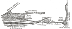
The cochlear nerve is one of two parts of the vestibulocochlear nerve, a cranial nerve present in amniotes, the other part being the vestibular nerve. The cochlear nerve carries auditory sensory information from the cochlea of the inner ear directly to the brain. The other portion of the vestibulocochlear nerve is the vestibular nerve, which carries spatial orientation information to the brain from the semicircular canals, also known as semicircular ducts.
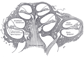
The tympanic duct or scala tympani is one of the perilymph-filled cavities in the inner ear of humans. It is separated from the cochlear duct by the basilar membrane, and it extends from the round window to the helicotrema, where it continues as vestibular duct.
The cochlear duct is an endolymph filled cavity inside the cochlea, located between the tympanic duct and the vestibular duct, separated by the basilar membrane and the vestibular membrane respectively. The cochlear duct houses the organ of Corti.
The vestibular membrane, vestibular wall or Reissner's membrane, is a membrane inside the cochlea of the inner ear. It separates the cochlear duct from the vestibular duct. It helps to transmit vibrations from fluid in the vestibular duct to the cochlear duct. Together with the basilar membrane, it creates a compartment in the cochlea filled with endolymph, which is important for the function of the spiral organ of Corti. It allows nutrients to travel from the perilymph to the endolymph of the membranous labyrinth. It may be damaged in Ménière's disease. It is named after the German anatomist Ernst Reissner.

The submandibular duct or Wharton duct or submaxillary duct, is one of the salivary excretory ducts. It is about 5 cm. long, and its wall is much thinner than that of the parotid duct. It drains saliva from each bilateral submandibular gland and sublingual gland to the sublingual caruncle at the base of the tongue.
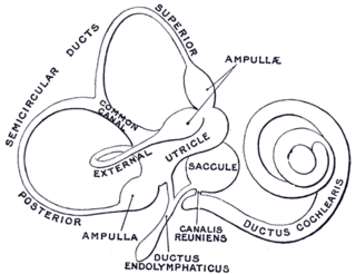
The ductus reuniens also the canalis reuniens of Hensen is part of the human inner ear. It connects the lower part of the saccule to the cochlear duct near its vestibular extremity.
The stria vascularis of the cochlear duct is a capillary loop in the upper portion of the spiral ligament. It produces endolymph for the scala media in the cochlea.

Merocrine is a term used to classify exocrine glands and their secretions in the study of histology. A cell is classified as merocrine if the secretions of that cell are excreted via exocytosis from secretory cells into an epithelial-walled duct or ducts and then onto a bodily surface or into the lumen.

A pseudostratified epithelium is a type of epithelium that, though comprising only a single layer of cells, has its cell nuclei positioned in a manner suggestive of stratified epithelia. As it rarely occurs as squamous or cuboidal epithelia, it is usually considered synonymous with the term pseudostratified columnar epithelium.

In anatomy and physiology, a duct is a circumscribed channel leading from an exocrine gland or organ.

Centroacinar cells are spindle-shaped cells in the exocrine pancreas. They represent an extension of the intercalated duct into each pancreatic acinus. These cells are commonly known as duct cells, and secrete an aqueous bicarbonate solution under stimulation by the hormone secretin. They also secrete mucin.

The crista ampullaris is the sensory organ of rotation. They are found in the ampullae of each of the semicircular canals of the inner ear, meaning that there are 3 pairs in total. The function of the crista ampullaris is to sense angular acceleration and deceleration.

In anatomy, the term nasal meatus can refer to any of the three meatuses (passages) through the skull's nasal cavity: the superior meatus, middle meatus, and inferior meatus.

The reticular membrane is a thin, stiff lamina that extends from the outer hair cells to the Hensen's cells. The RM is composed of "minute-fiddle-shaped cuticular structures" called the phalangeal extensions of the outer hair cells, interspaced with extensions coming from the outer phalangeal cells. The RM separates endolymph in the cochlear duct from underlying corticolymph and perilymph of the scala tympani.
The vestibular duct or scala vestibuli is a perilymph-filled cavity inside the cochlea of the inner ear that conducts sound vibrations to the cochlear duct.