Ampulla of Vater

Last updated

Ampulla of Vater
Biliary system new.svg
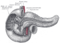
A diagram of the biliary system. Note that the ampulla of Vater is behind the major duodenal papilla.
Ampulla endo.jpg
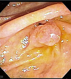
The major duodenal papilla, seen on duodenoscopy at the time of ERCP. This is the protrusion of the ampulla of Vater into the duodenum.
Details
Identifiers
Latin ampulla hepatopancreatica, ampulla Vaterii
MeSH D014670
TA98 A05.8.02.017
TA2 3111
FMA 15076
Anatomical terminology

The ampulla of Vater, hepatopancreatic ampulla or hepatopancreatic duct is the common duct that is usually formed by a union of the common bile duct and the pancreatic duct within the wall of the duodenum. This common duct usually features a dilation ("ampulla"). The common duct then opens medially into the descending part of the duodenum at the major duodenal papilla. The common duct usually measures 2-10mm in length. [1]

Contents

The ampulla of Vater is an important landmark halfway along the second part of the duodenum marking the transition from foregut to midgut.[ citation needed ]

Structure

1. Bile ducts: 2. Intrahepatic bile ducts, 3. Left and right hepatic ducts, 4. Common hepatic duct, 5. Cystic duct, 6. Common bile duct, 7. Ampulla of Vater, 8. Major duodenal papilla
9. Gallbladder.
10-11. Right and left lobes of liver.
12. Spleen.
13. Esophagus.
14. Stomach.
15. Pancreas: 16. Accessory pancreatic duct, 17. Pancreatic duct.
18. Small intestine: 19. Duodenum, 20. Jejunum
21-22. Right and left kidneys.
The front border of the liver has been lifted up (brown arrow). Biliary system multilingual.svg
1. Bile ducts : 2. Intrahepatic bile ducts, 3. Left and right hepatic ducts, 4. Common hepatic duct, 5. Cystic duct, 6. Common bile duct, 7. Ampulla of Vater, 8. Major duodenal papilla
9. Gallbladder.
10–11. Right and left lobes of liver.
12. Spleen.
13. Esophagus.
14. Stomach.
15. Pancreas : 16. Accessory pancreatic duct, 17. Pancreatic duct.
18. Small intestine : 19. Duodenum, 20. Jejunum
21–22. Right and left kidneys.
The front border of the liver has been lifted up (brown arrow).

Sphincters

Various smooth muscle sphincters regulate the flow of bile and pancreatic juice through the ampulla: the sphincter of the pancreatic duct, the sphincter of the bile duct, and the sphincter of Oddi. [3]

Variation

The common bile duct and pancreatic duct may sometimes unite outside the duodenal wall, creating an unusually long common duct. The two ducts may also drain into the duodenum separately, or may fuse yet retain their separate lumens separated by a septum. [1]

Clinical significance

Thomas' sign is the production of silver stools and can be indicative of cancer of the Ampulla of Vater. The silver-colored stool is a combination of the white stool of obstructive jaundice combined with black stool of melena or bleeding. It was first described in the British Medical Journal by Dr. H. Ogilvie in 1955. [4]

Etymology

The eponymic term "ampulla of Vater" is named after Abraham Vater (1684–1751), [5] a German anatomist who first published a description of it in 1723. [6]

Additional images

Related Research Articles

<span class="mw-page-title-main">Pancreas</span> Organ of the digestive system and endocrine system of vertebrates

The pancreas is an organ of the digestive system and endocrine system of vertebrates. In humans, it is located in the abdomen behind the stomach and functions as a gland. The pancreas is a mixed or heterocrine gland, i.e., it has both an endocrine and a digestive exocrine function. 99% of the pancreas is exocrine and 1% is endocrine. As an endocrine gland, it functions mostly to regulate blood sugar levels, secreting the hormones insulin, glucagon, somatostatin and pancreatic polypeptide. As a part of the digestive system, it functions as an exocrine gland secreting pancreatic juice into the duodenum through the pancreatic duct. This juice contains bicarbonate, which neutralizes acid entering the duodenum from the stomach; and digestive enzymes, which break down carbohydrates, proteins and fats in food entering the duodenum from the stomach.

<span class="mw-page-title-main">Duodenum</span> First section of the small intestine

The duodenum is the first section of the small intestine in most higher vertebrates, including mammals, reptiles, and birds. In mammals it may be the principal site for iron absorption. The duodenum precedes the jejunum and ileum and is the shortest part of the small intestine.

<span class="mw-page-title-main">Bile duct</span> Type of organ

A bile duct is any of a number of long tube-like structures that carry bile, and is present in most vertebrates. The bile duct is separated into three main parts the fundus, body, and neck which are superior, middle, and inferior respectively.

<span class="mw-page-title-main">Endoscopic retrograde cholangiopancreatography</span> Use of endoscopy and fluoroscopy to treat and diagnose digestive issues.

Endoscopic retrograde cholangiopancreatography (ERCP) is a technique that combines the use of endoscopy and fluoroscopy to diagnose and treat certain problems of the biliary or pancreatic ductal systems. It is primarily performed by highly skilled and specialty trained gastroenterologists. Through the endoscope, the physician can see the inside of the stomach and duodenum, and inject a contrast medium into the ducts in the biliary tree and pancreas so they can be seen on radiographs.

<span class="mw-page-title-main">Common bile duct</span> Gastrointestinal duct

The bile duct is a part of the biliary tract. It is formed by the union of the common hepatic duct and cystic duct. It ends by uniting with the pancreatic duct to form the hepatopancreatic ampulla. It possesses its own sphincter to enable regulation of bile flow.

<span class="mw-page-title-main">Cystic duct</span> Organ duct

The cystic duct is the duct that (typically) joins the gallbladder and the common hepatic duct; the union of the cystic duct and common hepatic duct forms the bile duct. Its length varies. It is often tortuous.

<span class="mw-page-title-main">Digestive enzyme</span> Class of enzymes

Digestive enzymes are a group of enzymes that break down polymeric macromolecules into their smaller building blocks, in order to facilitate their absorption into the cells of the body. Digestive enzymes are found in the digestive tracts of animals and in the tracts of carnivorous plants, where they aid in the digestion of food, as well as inside cells, especially in their lysosomes, where they function to maintain cellular survival. Digestive enzymes of diverse specificities are found in the saliva secreted by the salivary glands, in the secretions of cells lining the stomach, in the pancreatic juice secreted by pancreatic exocrine cells, and in the secretions of cells lining the small and large intestines.

<span class="mw-page-title-main">Common bile duct stone</span> Medical condition

Common bile duct stone, also known as choledocholithiasis, is the presence of gallstones in the common bile duct (CBD). This condition can cause jaundice and liver cell damage. Treatments include choledocholithotomy and endoscopic retrograde cholangiopancreatography (ERCP).

<span class="mw-page-title-main">Pancreatic duct</span> Duct associated with the human pancreas

The pancreatic duct, or duct of Wirsung, is a duct joining the pancreas to the common bile duct. This supplies it with pancreatic juice from the exocrine pancreas, which aids in digestion.

<span class="mw-page-title-main">Ascending cholangitis</span> Medical condition

Ascending cholangitis, also known as acute cholangitis or simply cholangitis, is inflammation of the bile duct, usually caused by bacteria ascending from its junction with the duodenum. It tends to occur if the bile duct is already partially obstructed by gallstones.

<span class="mw-page-title-main">Hemosuccus pancreaticus</span> Medical condition

Hemosuccus pancreaticus is a rare cause of hemorrhage in the gastrointestinal tract. It is caused by a bleeding source in the pancreas, pancreatic duct, or structures adjacent to the pancreas, such as the splenic artery, that bleed into the pancreatic duct, which is connected with the bowel at the duodenum, the first part of the small intestine. Patients with hemosuccus may develop symptoms of gastrointestinal hemorrhage, such as blood in the stools, maroon stools, or melena, which is a dark, tarry stool caused by digestion of red blood cells. They may also develop abdominal pain. It is associated with pancreatitis, pancreatic cancer and aneurysms of the splenic artery. Hemosuccus may be identified with endoscopy (esophagogastroduodenoscopy), where fresh blood may be seen from the pancreatic duct. Alternatively, angiography may be used to inject the celiac axis to determine the blood vessel that is bleeding. This may also be used to treat hemosuccus, as embolization of the end vessel may terminate the hemorrhage. However, a distal pancreatectomy—surgery to remove of the tail of the pancreas—may be required to stop the hemorrhage.

<span class="mw-page-title-main">Major duodenal papilla</span>

The major duodenal papilla is a rounded projection in the duodenum into which the common bile duct and pancreatic duct drain. The major duodenal papilla is, in most people, the primary mechanism for the secretion of bile and other enzymes that facilitate digestion.

<span class="mw-page-title-main">Biliary tract</span> Organ system

The biliary tract refers to the liver, gallbladder and bile ducts, and how they work together to make, store and secrete bile. Bile consists of water, electrolytes, bile acids, cholesterol, phospholipids and conjugated bilirubin. Some components are synthesized by hepatocytes ; the rest are extracted from the blood by the liver.

The minor duodenal papilla is the opening of the accessory pancreatic duct into the descending second section of the duodenum.

<span class="mw-page-title-main">Periampullary cancer</span> Disease

Periampullary cancer is a cancer that forms near the ampulla of Vater, an enlargement of the ducts from the liver and pancreas where they join and enter the small intestine. It consists of:

  1. ampullary tumour from ampulla of Vater
  2. cancer of lower common bile duct
  3. duodenal cancer adjacent to ampulla
  4. carcinoma head of pancreas
<span class="mw-page-title-main">Sphincter of Oddi dysfunction</span> Medical condition

Sphincter of Oddi dysfunction refers to a group of functional disorders leading to abdominal pain due to dysfunction of the Sphincter of Oddi: functional biliary sphincter of Oddi and functional pancreatic sphincter of Oddi disorder. The sphincter of Oddi is a sphincter muscle, a circular band of muscle at the bottom of the biliary tree which controls the flow of pancreatic juices and bile into the second part of the duodenum. The pathogenesis of this condition is recognized to encompass stenosis or dyskinesia of the sphincter of Oddi ; consequently the terms biliary dyskinesia, papillary stenosis, and postcholecystectomy syndrome have all been used to describe this condition. Both stenosis and dyskinesia can obstruct flow through the sphincter of Oddi and can therefore cause retention of bile in the biliary tree and pancreatic juice in the pancreatic duct.

Sphincter of Oddi Muscular valve that controls the rate of flow of bile and pancreatic juice into the duodenum

The sphincter of Oddi, abbreviated as SO, is a muscular valve that in some animals, including humans, controls the flow of bile and pancreatic juice out of the gallbladder and pancreas respectively through the ampulla of Vater into the second part of the duodenum. It is named after Ruggero Oddi.

<span class="mw-page-title-main">Choledochoduodenostomy</span>

Choledochoduodenostomy (CDD) is a surgical procedure to create an anastomosis, a surgical connection, between the common bile duct (CBD) and an alternative portion of the duodenum. In healthy individuals, the CBD meets the pancreatic duct at the ampulla of Vater, which drains via the major duodenal papilla to the second part of duodenum. In cases of benign conditions such as narrowing of the distal CBD or recurrent CBD stones, performing a CDD provides the diseased patient with CBD drainage and decompression. A side-to-side anastomosis is usually performed.

<span class="mw-page-title-main">Biliary endoscopic sphincterotomy</span> Use of endoscopy and fluoroscopy to treat and diagnose digestive issues.

Biliary endoscopic sphincterotomy is a procedure where the sphincter of Oddi and the segment of the common bile duct where it enters the duodenum are cannulated and then cut with a sphincterotome, a device that includes a wire which cuts with an electric current (electrocautery).

The sphincter of Boyden is a sphincter located in the common bile duct before it joins with the pancreatic duct to form the ampulla of vater. This sphincter controls the flow of bile into the pancreatic duct and it helps in filling up of the gallbladder with bile.

References

  1. 1 2 Standring, Susan (2020). Gray's Anatomy: The Anatomical Basis of Clinical Practice (42nd ed.). New York. p. 1219. ISBN   978-0-7020-7707-4. OCLC   1201341621.{{cite book}}: CS1 maint: location missing publisher (link)
  2. Standring S, Borley NR, eds. (2008). Gray's anatomy : the anatomical basis of clinical practice. Brown JL, Moore LA (40th ed.). London: Churchill Livingstone. pp. 1163, 1177, 1185–6. ISBN   978-0-8089-2371-8.
  3. Allescher, H. D. (1989). "Papilla of Vater: Structure and Function". Endoscopy. 21 (S 1): 324–329. doi:10.1055/s-2007-1012982. PMID   2691236. S2CID   38457896. Archived from the original on June 2, 2018. Retrieved March 27, 2020 via www.thieme-connect.de.
  4. Ogilvie, H (1955). "Thomas's sign, or the silver stool in cancer of the ampulla of Vater". Br Med J. 1 (4907): 208. doi:10.1136/bmj.1.4907.208. PMC   2060824 . PMID   13219383.
  5. Lerch, MM; Domschke, W (2000). "Abraham Vater of the ampulla (papilla) of Vater". Gastroenterology. 118 (2): 379. doi: 10.1016/s0016-5085(00)70243-5 . PMID   10691372.
  6. Vater A. Dissertation in auguralis medica, poes diss. Qua Scirris viscerum dissert, C.S. Ezlerus. Vol 70. 1723.