| Hepatocyte | |
|---|---|
 | |
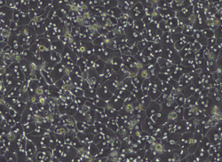
 Human liver stained with hematoxylin and eosin showing hepatocytes organized into plates and lobules | |
| Details | |
| Location | Liver |
| Identifiers | |
| MeSH | D022781 |
| TH | H3.04.05.0.00006 |
| FMA | 14515 |
| Anatomical terms of microanatomy | |
A hepatocyte is a cell of the main parenchymal tissue of the liver. Hepatocytes make up 80% of the liver's mass. These cells are involved in:
Contents
- Structure
- Microanatomy
- Function
- Protein synthesis
- Carbohydrate metabolism
- Lipid metabolism
- Detoxification
- Aging
- Society and culture
- Use in research
- Additional images
- See also
- References
- External links
- Protein synthesis
- Protein storage
- Transformation of carbohydrates
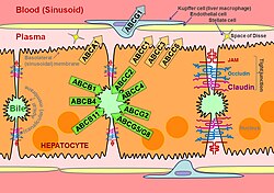
- Synthesis of cholesterol, bile salts and phospholipids
- Detoxification, modification, and excretion of exogenous and endogenous substances
- Initiation of formation and secretion of bile