| Pancreatic duct | |
|---|---|
 The pancreatic duct | |
 | |
| Details | |
| Precursor | Pancreatic bud |
| Identifiers | |
| Latin | ductus pancreaticus |
| MeSH | D010183 |
| TA98 | A05.9.01.015 |
| TA2 | 3129 |
| FMA | 10419 |
| Anatomical terminology | |

2. Intrahepatic bile ducts
3. Left and right hepatic ducts
4. Common hepatic duct
5. Cystic duct
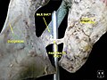
6. Common bile duct
7. Ampulla of Vater
8. Major duodenal papilla
9. Gallbladder
10–11. Right and left lobes of liver
12. Spleen
13. Esophagus
14. Stomach
15. Pancreas :
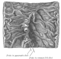
16. Accessory pancreatic duct
17. Pancreatic duct
18. Small intestine :
19. Duodenum
20. Jejunum
21–22. Right and left kidneys
The front border of the liver has been lifted up (brown arrow).
The pancreatic duct or duct of Wirsung (also, the major pancreatic duct due to the existence of an accessory pancreatic duct) is a duct joining the pancreas to the common bile duct. This supplies it with pancreatic juice from the exocrine pancreas, which aids in digestion.