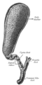
Cystic duct

Last updated

Cystic duct
Illu liver gallbladder.jpg
Details
Artery cystic artery
Identifiers
Latin ductus cysticus
MeSH D003549
TA98 A05.8.02.011
TA2 3101
FMA 14539
Anatomical terminology
1. Bile ducts: 2. Intrahepatic bile ducts, 3. Left and right hepatic ducts, 4. Common hepatic duct, 5. Cystic duct, 6. Common bile duct, 7. Ampulla of Vater, 8. Major duodenal papilla
9. Gallbladder.
10-11. Right and left lobes of liver.
12. Spleen.
13. Esophagus.
14. Stomach.
15. Pancreas: 16. Accessory pancreatic duct, 17. Pancreatic duct.
18. Small intestine: 19. Duodenum, 20. Jejunum
21-22. Right and left kidneys.
The front border of the liver has been lifted up (brown arrow). Biliary system multilingual.svg
1. Bile ducts : 2. Intrahepatic bile ducts, 3. Left and right hepatic ducts, 4. Common hepatic duct, 5. Cystic duct, 6. Common bile duct, 7. Ampulla of Vater, 8. Major duodenal papilla
9. Gallbladder.
10–11. Right and left lobes of liver.
12. Spleen.
13. Esophagus.
14. Stomach.
15. Pancreas : 16. Accessory pancreatic duct, 17. Pancreatic duct.
18. Small intestine : 19. Duodenum, 20. Jejunum
21–22. Right and left kidneys.
The front border of the liver has been lifted up (brown arrow).

The cystic duct is the duct that (typically) joins the gallbladder and the common hepatic duct; the union of the cystic duct and common hepatic duct forms the bile duct (formerly known as the common bile duct). Its length varies. It is often tortuous.

Contents

Anatomy

The cystic duct typically [2] measures (sources differ) 2–4 cm [3] [4] /2–3 cm [2] in length (though its lengh has been known to range from 0.5 cm to 9 cm [3] ), and 2–3 mm in diameter. [2] [4] It is often tortuous. [3] [4]

It is the distal continuation of the neck of the gallbladder, from where it is directed inferoposteriorly and to the left [2] /medially [4] (this occurs in half of individuals [3] ). It typically [2] terminates by uniting with the common hepatic duct to form the bile duct (usually anterior to the right hepatic artery). [2] It usually joins the common bile duct from the right lateral side (forming an oblique angle between the two), [4] and at such a distance that the bile duct is twice as long as the common hepatic duct. [3] [4] It often fuses with the common hepatic duct before actually opening into it after a variable distance [4] [3] (this arrangement may have the purpose of directing bile flow distally instead of back towards the liver [3] ).

Structure

The inner surface of the cystic duct features spiral, crescentic mucosal folds - the spiral folds of cystic duct. [4] [3]

The inner surface of the proximal cystic duct exhibits a network of submicroscopic convoluted folds (rugae), whereas that of the distal cystic duct exhibits submicroscopic parallel longitudinal folds. [3]

Histology

The epithelial lining of the inner surface of the duct is similar to that of the gallbladder and consists mostly of columnar epithelial cells with short microvilli upon their apical surfaces. [3]

The subepithelial layer consists of elastic connective tissue and is highly vascular; vessels that are adjacent to the epithelial basement membrane are fenestrated, possibly to facilitate ion and fluid exchange with the bile as is the case in the gallbladder itself. [3]

The outer fibromuscular layer contains smooth muscle continuous with that of the gallbladder; some of the smooth muscle extends into the spiral valves. [3]

Relations

It usually lies next to the cystic artery.[ citation needed ]

Variation

The cystic duct may rarely be doubled. [2] [3]

An accessory hepatic duct may join the cystic duct. [2] [4]

A pathological diverticulum known as the Hartmann’s pouch may be present at the junction of the neck of bladder and the cystic duct. [2]

Length

The duct may sometimes be extremely short (making cholecystectomy risky) [2] or may rarely be altogether absent (so that the gallbladder is directly attached to the bile duct). [2] [4] [3]

Shape

While most often tortuous, it may occasionally be curved, straight, or S-shaped. [3]

Termination

The cystic duct may unite with the common hepatic duct so that the common hepatic duct is either very short or very long (and the bile duct in turn very long or very short, respectively), [4] or it may instead unite with the a hepatic duct. [3]

Occasionally, the cystic duct may first run alongside the common hepatic duct for some distance [2] [4] within the hepatoduodenal ligament [4] before joining it. [2] [4] It sometimes join the common hepatic duct at its anterior, posterior, or medial side [4] [3] (in the latter case by passing posteriorly around the common bile duct to join it from the other side). [2] It may spiral around the common hepatic duct before joining it. [4]

Very rarely, the cystic duct opens into the duodenum. [3]

Function

Bile can flow in either direction between the gallbladder, and the common bile duct and hepatic duct. [3] In this way, bile is stored in the gallbladder in between meal times. The hormone cholecystokinin, when stimulated by a fatty meal, promotes bile secretion by increased production of hepatic bile, contraction of the gall bladder, and relaxation of the Sphincter of Oddi.

The bile duct was once thought to possess a sphincteric function, however, it is now known that bile flow through the cystic duct proceeds unimpeded and is instead regulated by other mechanisms at other points of the biliary system. [3]

Clinical significance

Gallstones can enter and obstruct the cystic duct, preventing the flow of bile. The increased pressure in the gallbladder leads to swelling and pain. This pain, known as biliary colic, is sometimes referred to as a gallbladder "attack" because of its sudden onset.

During a cholecystectomy, the cystic duct is clipped two or three times and a cut is made between the clips, freeing the gallbladder to be taken out.

See also

Additional images

Related Research Articles

<span class="mw-page-title-main">Gallbladder</span> Organ in humans and other vertebrates

In vertebrates, the gallbladder, also known as the cholecyst, is a small hollow organ where bile is stored and concentrated before it is released into the small intestine. In humans, the pear-shaped gallbladder lies beneath the liver, although the structure and position of the gallbladder can vary significantly among animal species. It receives bile, produced by the liver, via the common hepatic duct, and stores it. The bile is then released via the common bile duct into the duodenum, where the bile helps in the digestion of fats.

<span class="mw-page-title-main">Bile duct</span> Type of organ

A bile duct is any of a number of long tube-like structures that carry bile, and is present in most vertebrates. The bile duct is separated into three main parts: the fundus (superior), the body (middle), and the neck (inferior).

<span class="mw-page-title-main">Cholecystitis</span> Inflammation of the gallbladder

Cholecystitis is inflammation of the gallbladder. Symptoms include right upper abdominal pain, pain in the right shoulder, nausea, vomiting, and occasionally fever. Often gallbladder attacks precede acute cholecystitis. The pain lasts longer in cholecystitis than in a typical gallbladder attack. Without appropriate treatment, recurrent episodes of cholecystitis are common. Complications of acute cholecystitis include gallstone pancreatitis, common bile duct stones, or inflammation of the common bile duct.

<span class="mw-page-title-main">Cholecystectomy</span> Surgical removal of the gallbladder

Cholecystectomy is the surgical removal of the gallbladder. Cholecystectomy is a common treatment of symptomatic gallstones and other gallbladder conditions. In 2011, cholecystectomy was the eighth most common operating room procedure performed in hospitals in the United States. Cholecystectomy can be performed either laparoscopically, or via an open surgical technique.

<span class="mw-page-title-main">Common bile duct</span> Gastrointestinal duct

The bile duct is a part of the biliary tract. It is formed by the union of the common hepatic duct and cystic duct. It ends by uniting with the pancreatic duct to form the hepatopancreatic ampulla. It possesses its own sphincter to enable regulation of bile flow.

Courvoisier's principle states that a painless palpably enlarged gallbladder accompanied with mild jaundice is unlikely to be caused by gallstones. Usually, the term is used to describe the physical examination finding of the right-upper quadrant of the abdomen. This sign implicates possible malignancy of the gallbladder or pancreas and the swelling is unlikely due to gallstones.

<span class="mw-page-title-main">Common hepatic duct</span> Exocrine duct

The common hepatic duct is the first part of the biliary tract. It joins the cystic duct coming from the gallbladder to form the common bile duct.

An accessory bile duct is a conduit that transports bile and is considered to be supernumerary or auxiliary to the biliary tree.

<span class="mw-page-title-main">Cystic artery</span>

The cystic artery is (usually) a branch of the right hepatic artery that provides arterial supply to the gallbladder and contributes arterial supply to the extrahepatic bile ducts.

<span class="mw-page-title-main">Ascending cholangitis</span> Medical condition

Ascending cholangitis, also known as acute cholangitis or simply cholangitis, is inflammation of the bile duct, usually caused by bacteria ascending from its junction with the duodenum. It tends to occur if the bile duct is already partially obstructed by gallstones.

<span class="mw-page-title-main">Cystohepatic triangle</span> Space between the cystic duct, the common hepatic duct, and the liver

The cystohepatic triangle is an anatomic space bordered by the cystic duct inferiorly, the common hepatic duct medially, and the inferior surface of the liver superiorly.

<span class="mw-page-title-main">Biliary tract</span> Organ system

The biliary tract refers to the liver, gallbladder and bile ducts, and how they work together to make, store and secrete bile. Bile consists of water, electrolytes, bile acids, cholesterol, phospholipids and conjugated bilirubin. Some components are synthesized by hepatocytes ; the rest are extracted from the blood by the liver.

<span class="mw-page-title-main">Gallbladder disease</span> Medical condition

Gallbladder diseases are diseases involving the gallbladder and is closely linked to biliary disease, with the most common cause being gallstones (cholelithiasis).

Spiral folds of cystic duct are a series of crescenteric, spirally arranged mucosal folds in the proximal part of the cystic duct.

<span class="mw-page-title-main">Liver</span> Vertebrate organ involved in metabolism

The liver is a major metabolic organ only found in vertebrate animals, which performs many essential biological functions such as detoxification of the organism, and the synthesis of proteins and biochemicals necessary for digestion and growth. In humans, it is located in the right upper quadrant of the abdomen, below the diaphragm and mostly shielded by the lower right rib cage. Its other metabolic roles include carbohydrate metabolism, the production of hormones, conversion and storage of nutrients such as glucose and glycogen, and the decomposition of red blood cells.

Biliary dyskinesia is a disorder of some component of biliary part of the digestive system in which bile cannot physically move in the proper direction through the tubular biliary tract. It most commonly involves abnormal biliary tract peristalsis muscular coordination within the gallbladder in response to dietary stimulation of that organ to squirt the liquid bile through the common bile duct into the duodenum. Ineffective peristaltic contraction of that structure produces postprandial right upper abdominal pain (cholecystodynia) and almost no other problem. When the dyskinesia is localized at the biliary outlet into the duodenum just as increased tonus of that outlet sphincter of Oddi, the backed-up bile can cause pancreatic injury with abdominal pain more toward the upper left side. In general, biliary dyskinesia is the disturbance in the coordination of peristaltic contraction of the biliary ducts, and/or reduction in the speed of emptying of the biliary tree into the duodenum.

The cystic node is the sentinel lymph node of the gall bladder. It is located within the cystohepatic triangle.

<span class="mw-page-title-main">Sphincter of Oddi dysfunction</span> Medical condition

Sphincter of Oddi dysfunction refers to a group of functional disorders leading to abdominal pain due to dysfunction of the Sphincter of Oddi: functional biliary sphincter of Oddi and functional pancreatic sphincter of Oddi disorder. The sphincter of Oddi is a sphincter muscle, a circular band of muscle at the bottom of the biliary tree which controls the flow of pancreatic juices and bile into the second part of the duodenum. The pathogenesis of this condition is recognized to encompass stenosis or dyskinesia of the sphincter of Oddi ; consequently the terms biliary dyskinesia, papillary stenosis, and postcholecystectomy syndrome have all been used to describe this condition. Both stenosis and dyskinesia can obstruct flow through the sphincter of Oddi and can therefore cause retention of bile in the biliary tree and pancreatic juice in the pancreatic duct.

Cholecystostomy or (cholecystotomy) is a medical procedure used to drain the gallbladder through either a percutaneous or endoscopic approach. The procedure involves creating a stoma in the gallbladder, which can facilitate placement of a tube or stent for drainage, first performed by American surgeon, Dr. John Stough Bobbs, in 1867. It is sometimes used in cases of cholecystitis or other gallbladder disease where the person is ill, and there is a need to delay or defer cholecystectomy. The first endoscopic cholecystostomy was performed by Drs. Todd Baron and Mark Topazian in 2007 using ultrasound guidance to puncture the stomach wall and place a plastic biliary catheter for gallbladder drainage.

References

  1. Standring S, Borley NR, eds. (2008). Gray's anatomy : the anatomical basis of clinical practice. Brown JL, Moore LA (40th ed.). London: Churchill Livingstone. pp. 1163, 1177, 1185–6. ISBN   978-0-8089-2371-8.
  2. 1 2 3 4 5 6 7 8 9 10 11 12 13 14 Sinnatamby, Chummy S. (2011). Last's Anatomy (12th ed.). Elsevier Australia. p. 265. ISBN   978-0-7295-3752-0.
  3. 1 2 3 4 5 6 7 8 9 10 11 12 13 14 15 16 17 18 19 20 Dasgupta, D.; Stringer, M. D. (March 2005). "Cystic duct and Heister's "valves"". Clinical Anatomy. 18 (2): 81–87. doi:10.1002/ca.20118. ISSN   0897-3806. PMID   15696536. S2CID   24179512.
  4. 1 2 3 4 5 6 7 8 9 10 11 12 13 14 15 16 Standring, Susan (2020). Gray's Anatomy: The Anatomical Basis of Clinical Practice (42th ed.). New York. pp. 1217–1218. ISBN   978-0-7020-7707-4. OCLC   1201341621.{{cite book}}: CS1 maint: location missing publisher (link)