Common hepatic duct

Last updated

Common hepatic duct
Illu liver gallbladder.jpg
Details
Identifiers
Latin ductus hepaticus communis
MeSH D006500
TA98 A05.8.01.061
TA2 3092
FMA 14668
Anatomical terminology
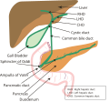
1. Bile ducts: 2. Intrahepatic bile ducts, 3. Left and right hepatic ducts, 4. Common hepatic duct, 5. Cystic duct, 6. Common bile duct, 7. Ampulla of Vater, 8. Major duodenal papilla
9. Gallbladder.
10-11. Right and left lobes of liver.
12. Spleen.
13. Esophagus.
14. Stomach.
15. Pancreas: 16. Accessory pancreatic duct, 17. Pancreatic duct.
18. Small intestine: 19. Duodenum, 20. Jejunum
21-22. Right and left kidneys.
The front border of the liver has been lifted up (brown arrow). Biliary system multilingual.svg
1. Bile ducts : 2. Intrahepatic bile ducts, 3. Left and right hepatic ducts, 4. Common hepatic duct, 5. Cystic duct, 6. Common bile duct, 7. Ampulla of Vater, 8. Major duodenal papilla
9. Gallbladder.
10–11. Right and left lobes of liver.
12. Spleen.
13. Esophagus.
14. Stomach.
15. Pancreas : 16. Accessory pancreatic duct, 17. Pancreatic duct.
18. Small intestine : 19. Duodenum, 20. Jejunum
21–22. Right and left kidneys.
The front border of the liver has been lifted up (brown arrow).

The common hepatic duct is the first part of the biliary tract. It joins the cystic duct coming from the gallbladder to form the common bile duct.
Contents

Structure

The common hepatic duct is the first part of the biliary tract. [2] It is formed by the union of the right hepatic duct (which drains bile from the right functional lobe of the liver) and the left hepatic duct (which drains bile from the left functional lobe of the liver). [3]

The duct is about 3 cm long. [4] The common hepatic duct is about 6 mm in diameter in adults, with some variation. [5]

Termination

The common hepatic duct typically unites with the cystic duct some 1–2 cm superior to the duodenum and anterior to the right hepatic artery, with the cystic duct approaching the common hepatic duct from the right. [4]

Relations

The right branch of the hepatic artery proper usually passes posterior to the duct, but may rarely pass anterior to it instead. [4]

Histology

The inner surface is covered in a simple columnar epithelium. [3]

Variation

Accessory hepatic ducts

Around 1.7% [6] of people have additional accessory hepatic ducts that opens into the common hepatic duct. [6] Accessory hepatic ducts may also instead open into the cystic duct or gallbladder. [4]

Termination

Occasionally, the cystic duct may first run along the right side of the common bile duct for some distance before joining it, or may pass posteriorly around to the common hepatic duct to unite with it from the left side. [4]

Rarely, the common hepatic duct and gallbladder joing directly (with the cystic duct being absent), [6] [4] leading to illness. [6]

Function

The hepatic duct is part of the biliary tract that transports secretions from the liver into the intestines.

Clinical significance

Cholecystectomy

The common hepatic ducts carries a higher volume of bile in people who have had their gallbladder removed.[ citation needed ]

The common hepatic duct is an important anatomic landmark during surgeries such as cholecystectomy. It forms one edge of Calot's triangle, along with the cystic duct and the cystic artery. All constituents of this triangle must be identified to avoid cutting or clipping the wrong structure.

Cholestasis

A diameter of more than 8 mm is regarded as abnormal dilatation, and is a sign of cholestasis. [7]

Mirizzi's syndrome

Mirizzi's syndrome occurs when the common hepatic duct is blocked by gallstones. [8]

Additional images

Related Research Articles

<span class="mw-page-title-main">Duodenum</span> First section of the small intestine

The duodenum is the first section of the small intestine in most higher vertebrates, including mammals, reptiles, and birds. In mammals it may be the principal site for iron absorption. The duodenum precedes the jejunum and ileum and is the shortest part of the small intestine.

<span class="mw-page-title-main">Gallbladder</span> Organ in humans and other vertebrates

In vertebrates, the gallbladder, also known as the cholecyst, is a small hollow organ where bile is stored and concentrated before it is released into the small intestine. In humans, the pear-shaped gallbladder lies beneath the liver, although the structure and position of the gallbladder can vary significantly among animal species. It receives bile, produced by the liver, via the common hepatic duct, and stores it. The bile is then released via the common bile duct into the duodenum, where the bile helps in the digestion of fats.

<span class="mw-page-title-main">Bile duct</span> Type of organ

A bile duct is any of a number of long tube-like structures that carry bile, and is present in most vertebrates. The bile duct is separated into three main parts: the fundus (superior), the body (middle), and the neck (inferior).

<span class="mw-page-title-main">Cholecystectomy</span> Surgical removal of the gallbladder

Cholecystectomy is the surgical removal of the gallbladder. Cholecystectomy is a common treatment of symptomatic gallstones and other gallbladder conditions. In 2011, cholecystectomy was the eighth most common operating room procedure performed in hospitals in the United States. Cholecystectomy can be performed either laparoscopically, or via an open surgical technique.

<span class="mw-page-title-main">Common bile duct</span> Gastrointestinal duct

The bile duct is a part of the biliary tract. It is formed by the union of the common hepatic duct and cystic duct. It ends by uniting with the pancreatic duct to form the hepatopancreatic ampulla. It possesses its own sphincter to enable regulation of bile flow.

<span class="mw-page-title-main">Cystic duct</span> Organ duct

The cystic duct is the duct that (typically) joins the gallbladder and the common hepatic duct; the union of the cystic duct and common hepatic duct forms the bile duct. Its length varies. It is often tortuous.

Courvoisier's principle states that a painless palpably enlarged gallbladder accompanied with mild jaundice is unlikely to be caused by gallstones. Usually, the term is used to describe the physical examination finding of the right-upper quadrant of the abdomen. This sign implicates possible malignancy of the gallbladder or pancreas and the swelling is unlikely due to gallstones.

<span class="mw-page-title-main">Gastrointestinal disease</span> Medical condition

Gastrointestinal diseases refer to diseases involving the gastrointestinal tract, namely the esophagus, stomach, small intestine, large intestine and rectum, and the accessory organs of digestion, the liver, gallbladder, and pancreas.

An accessory bile duct is a conduit that transports bile and is considered to be supernumerary or auxiliary to the biliary tree.

<span class="mw-page-title-main">Cystic artery</span>

The cystic artery is (usually) a branch of the right hepatic artery that provides arterial supply to the gallbladder and contributes arterial supply to the extrahepatic bile ducts.

<span class="mw-page-title-main">Cystohepatic triangle</span> Space between the cystic duct, the common hepatic duct, and the liver

The cystohepatic triangle is an anatomic space bordered by the cystic duct laterally, the common hepatic duct medially, and the inferior surface of the liver superiorly.

<span class="mw-page-title-main">Hepatic plexus</span>

The hepatic plexus is a sympathetic and parasympathetic nerve plexus that provides innervation to the parenchyma of the liver as well as contributing innervation to some other abdominal structures.

<span class="mw-page-title-main">Major duodenal papilla</span> Rounded projection in the duodenum into which the common bile duct and pancreatic duct drain

The major duodenal papilla is a rounded projection in the duodenum into which the common bile duct and pancreatic duct drain. The major duodenal papilla is, in most people, the primary mechanism for the secretion of bile and other enzymes that facilitate digestion.

<span class="mw-page-title-main">Cystic vein</span>

The cystic veins drain venous blood from the gallbladder and the cystic duct. The cystic veins either drain into various branches and tributaries of the hepatic portal vein.

<span class="mw-page-title-main">Biliary tract</span> Organ system

The biliary tract refers to the liver, gallbladder and bile ducts, and how they work together to make, store and secrete bile. Bile consists of water, electrolytes, bile acids, cholesterol, phospholipids and conjugated bilirubin. Some components are synthesized by hepatocytes ; the rest are extracted from the blood by the liver.

<span class="mw-page-title-main">Percutaneous transhepatic cholangiography</span> Medical imaging of the biliary tract

Percutaneous transhepatic cholangiography, percutaneous hepatic cholangiogram (PTHC) is a radiological technique used to visualize the anatomy of the biliary tract. A contrast medium is injected into a bile duct in the liver, after which X-rays are taken. It allows access to the biliary tree in cases where endoscopic retrograde cholangiopancreatography has been unsuccessful. Initially reported in 1937, the procedure became popular in 1952.

<span class="mw-page-title-main">Portal venous system</span> The capillary bed pools into another through veins without first going through the heart

In the circulatory system of vertebrates, a portal venous system occurs when a capillary bed pools into another capillary bed through veins, without first going through the heart. Both capillary beds and the blood vessels that connect them are considered part of the portal venous system.

<span class="mw-page-title-main">Liver</span> Vertebrate organ involved in metabolism

The liver is a major metabolic organ only found in vertebrate animals, which performs many essential biological functions such as detoxification of the organism, and the synthesis of proteins and biochemicals necessary for digestion and growth. In humans, it is located in the right upper quadrant of the abdomen, below the diaphragm and mostly shielded by the lower right rib cage. Its other metabolic roles include carbohydrate metabolism, the production of hormones, conversion and storage of nutrients such as glucose and glycogen, and the decomposition of red blood cells.

The cystic node is the sentinel lymph node of the gall bladder. It is located within the cystohepatic triangle.

<span class="mw-page-title-main">Human digestive system</span> Digestive system in humans

The human digestive system consists of the gastrointestinal tract plus the accessory organs of digestion. Digestion involves the breakdown of food into smaller and smaller components, until they can be absorbed and assimilated into the body. The process of digestion has three stages: the cephalic phase, the gastric phase, and the intestinal phase.

References

  1. Standring S, Borley NR, eds. (2008). Gray's anatomy : the anatomical basis of clinical practice. Brown JL, Moore LA (40th ed.). London: Churchill Livingstone. pp. 1163, 1177, 1185–6. ISBN   978-0-8089-2371-8.
  2. Manohar, Rohan; Lagasse, Eric (2014-01-01), Lanza, Robert; Langer, Robert; Vacanti, Joseph (eds.), "Chapter 45 - Liver Stem Cells", Principles of Tissue Engineering (Fourth Edition), Boston: Academic Press, pp. 935–950, doi:10.1016/b978-0-12-398358-9.00045-8, ISBN   978-0-12-398358-9 , retrieved 2021-01-26
  3. 1 2 Bergman, Simon; Geisinger, Kim R. (2008-01-01), Bibbo, Marluce; Wilbur, David (eds.), "CHAPTER 14 - Alimentary Tract (Esophagus, Stomach, Small Intestine, Colon, Rectum, Anus, Biliary Tract)", Comprehensive Cytopathology (Third Edition), Edinburgh: W.B. Saunders, pp. 373–408, ISBN   978-1-4160-4208-2 , retrieved 2021-01-26
  4. 1 2 3 4 5 6 Sinnatamby, Chummy S. (2011). Last's Anatomy (12th ed.). p. 265. ISBN   978-0-7295-3752-0.
  5. Gray's Anatomy, 39th ed, p. 1228
  6. 1 2 3 4 Portmann, Bernard C.; Roberts, Eve A. (2012-01-01), Burt, Alastair D.; Portmann, Bernard C.; Ferrell, Linda D. (eds.), "3 - Developmental abnormalities and liver disease in childhood", MacSween's Pathology of the Liver (Sixth Edition), Edinburgh: Churchill Livingstone, pp. 101–156, ISBN   978-0-7020-3398-8 , retrieved 2021-01-26
  7. Hoeffel, Christine; Azizi, Louisa; Lewin, Maité; Laurent, Valérie; Aubé, Christophe; Arrivé, Lionel; Tubiana, Jean-Michel (2006). "Normal and Pathologic Features of the Postoperative Biliary Tract at 3D MR Cholangiopancreatography and MR Imaging". RadioGraphics. 26 (6): 1603–1620. doi:10.1148/rg.266055730. ISSN   0271-5333. PMID   17102039.
  8. Katz, Seth S. (2017-01-01), Jarnagin, William R. (ed.), "Chapter 18 - Computed tomography of the liver, biliary tract, and pancreas", Blumgart's Surgery of the Liver, Biliary Tract and Pancreas, 2-Volume Set (Sixth Edition), Philadelphia: Elsevier, pp. 316–357.e6, ISBN   978-0-323-34062-5 , retrieved 2021-01-26