Argon plasma coagulation

Last updated
Argon plasma coagulation
Argon plasma coagulation.jpg
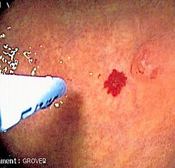
Argon plasma coagulation administered via probe through the colonoscope at an angiodysplasia in the colon. The patient had multiple colonic angiodysplasiae in the setting of aortic stenosis.
Other namesAPC
Specialty Gastroenterology

Argon plasma coagulation (APC) is a medical endoscopic procedure used to control bleeding from certain lesions in the gastrointestinal tract. It is administered during gastrointestinal endoscopy such as esophagogastroduodenoscopy or colonoscopy.

Contents

Medical use

APC involves the use of a jet of ionized argon gas (plasma) directed through a probe passed through the endoscope. The probe is placed at some distance from the bleeding lesion, and argon gas is emitted, then ionized by a high-voltage discharge (approx 6kV). [1] High-frequency electric current is then conducted through the jet of gas, resulting in coagulation of the bleeding lesion. As no physical contact is made with the lesion, the procedure is safe if the bowel has been cleaned of colonic gases, [2] [3] and can be used to treat bleeding in parts of the gastrointestinal tract with thin walls, such as the cecum.

See also

Related Research Articles

<span class="mw-page-title-main">Colonoscopy</span> Examination of the bowel

Colonoscopy or coloscopy is a medical procedure involving the endoscopic examination of the large bowel (colon) and the distal portion of the small bowel. This examination is performed using either a CCD camera or a fiber optic camera, which is mounted on a flexible tube and passed through the anus.

Hematemesis is the vomiting of blood. It can be confused with hemoptysis or epistaxis (nosebleed), which are more common. The source is generally the upper gastrointestinal tract, typically above the suspensory muscle of duodenum. It may be caused by ulcers, tumors of the stomach or esophagus, varices, prolonged and vigorous retching, gastroenteritis, ingested blood, or certain drugs.

<span class="mw-page-title-main">Angiodysplasia</span> Blood vessel malformation in the gut

In medicine (gastroenterology), angiodysplasia is a small vascular malformation of the gut. It is a common cause of otherwise unexplained gastrointestinal bleeding and anemia. Lesions are often multiple, and frequently involve the cecum or ascending colon, although they can occur at other places. Treatment may be with colonoscopic interventions, angiography and embolization, medication, or occasionally surgery.

<span class="mw-page-title-main">Esophagogastroduodenoscopy</span> Diagnostic endoscopic procedure

Esophagogastroduodenoscopy (EGD) or oesophagogastroduodenoscopy (OGD), also called by various other names, is a diagnostic endoscopic procedure that visualizes the upper part of the gastrointestinal tract down to the duodenum. It is considered a minimally invasive procedure since it does not require an incision into one of the major body cavities and does not require any significant recovery after the procedure. However, a sore throat is common.

<span class="mw-page-title-main">Dieulafoy's lesion</span> Medical condition

Dieulafoy's lesion is a medical condition characterized by a large tortuous artery most commonly in the stomach wall (submucosal) that erodes and bleeds. It can present in any part of the gastrointestinal tract. It can cause gastric hemorrhage but is relatively uncommon. It is thought to cause less than 5% of all gastrointestinal bleeds in adults. It was named after French surgeon Paul Georges Dieulafoy, who described this condition in his paper "Exulceratio simplex: Leçons 1-3" in 1898. It is also called "caliber-persistent artery" or "aneurysm" of gastric vessels. However, unlike most other aneurysms, these are thought to be developmental malformations rather than degenerative changes.

<span class="mw-page-title-main">Gastrointestinal bleeding</span> Bleeding in the gastrointestinal tract

Gastrointestinal bleeding, also called gastrointestinal hemorrhage (GIB), is all forms of bleeding in the gastrointestinal tract, from the mouth to the rectum. When there is significant blood loss over a short time, symptoms may include vomiting red blood, vomiting black blood, bloody stool, or black stool. Small amounts of bleeding over a long time may cause iron-deficiency anemia resulting in feeling tired or heart-related chest pain. Other symptoms may include abdominal pain, shortness of breath, pale skin, or passing out. Sometimes in those with small amounts of bleeding no symptoms may be present.

<span class="mw-page-title-main">Mallory–Weiss syndrome</span> Medical condition

Mallory–Weiss syndrome is a condition where high intra-abdominal pressures causes laceration and bleeding of the mucosa called Mallory-Weiss tears. Additionally, Mallory–Weiss syndrome is one of the most common causes of acute upper gastrointestinal bleeding, counting of around 1-15% of all cases in adults and less than 5% in children. It has been found that tears are up to 2 to 4 times more prevalent in men than women. The tears can cause upper gastrointestinal bleeding and predominantly occur where the esophagus meets the stomach. However, the tears can happen anywhere from the middle of the esophagus to the cardia of the stomach. Mallory–Weiss syndrome is often caused by constant vomiting and retching from alcoholism or bulimia. Gastroesophageal reflux disease (GERD) is another risk factor that is often linked with Mallory–Weiss syndrome. However, not every individual with Mallory–Weiss syndrome will have these risk factors. Individuals with Mallory–Weiss syndrome will have hematemesis, however the symptoms can vary.

<span class="mw-page-title-main">Lower gastrointestinal bleeding</span> Bleeding in the small and large intestine

Lower gastrointestinal bleeding (LGIB) is any form of gastrointestinal bleeding in the lower gastrointestinal tract. LGIB is a common reason for seeking medical attention at a hospital's emergency department. LGIB accounts for 30–40% of all gastrointestinal bleeding and is less common than upper gastrointestinal bleeding (UGIB). It is estimated that UGIB accounts for 100–200 per 100,000 cases versus 20–27 per 100,000 cases for LGIB. Approximately 85% of lower gastrointestinal bleeding involves the large intestine, 10% are from bleeds that are actually upper gastrointestinal bleeds, and 3–5% involve the small intestine.

<span class="mw-page-title-main">Radiation proctitis</span> Medical condition

Radiation proctitis or radiation proctopathy is a condition characterized by damage to the rectum after exposure to x-rays or other ionizing radiation as a part of radiation therapy. Radiation proctopathy may occur as acute inflammation called "acute radiation proctitis" or with chronic changes characterized by radiation associated vascular ectasiae (RAVE) and chronic radiation proctopathy. Radiation proctitis most commonly occurs after pelvic radiation treatment for cancers such as cervical cancer, prostate cancer, bladder cancer, and rectal cancer. RAVE and chronic radiation proctopathy involves the lower intestine, primarily the sigmoid colon and the rectum, and was previously called chronic radiation proctitis, pelvic radiation disease and radiation enteropathy.

<span class="mw-page-title-main">Gastric antral vascular ectasia</span> Intestinal bleeding due to widening of blood vessels in the stomach

Gastric antral vascular ectasia (GAVE) is an uncommon cause of chronic gastrointestinal bleeding or iron deficiency anemia. The condition is associated with dilated small blood vessels in the gastric antrum, which is a distal part of the stomach. The dilated vessels result in intestinal bleeding. It is also called watermelon stomach because streaky long red areas that are present in the stomach may resemble the markings on watermelon.

<span class="mw-page-title-main">Colonic polypectomy</span> Removal of colorectal polyps to prevent them turning cancerous

Colonic polypectomy is the removal of colorectal polyps in order to prevent them from turning cancerous.

<span class="mw-page-title-main">Endoclip</span>

An endoclip is a metallic mechanical device used in endoscopy in order to close two mucosal surfaces without the need for surgery and suturing. Its function is similar to a suture in gross surgical applications, as it is used to join together two disjointed surfaces, but, can be applied through the channel of an endoscope under direct visualization. Endoclips have found use in treating gastrointestinal bleeding, in preventing bleeding after therapeutic procedures such as polypectomy, and in closing gastrointestinal perforations. Many forms of endoclips exist of different shapes and sizes, including two and three prong devices, which can be administered using single use and reloadable systems, and may or may not open and close to facilitate placement.

<span class="mw-page-title-main">Capsule endoscopy</span> Medical imaging procedure

Capsule endoscopy is a medical procedure used to record internal images of the gastrointestinal tract for use in disease diagnosis. Newer developments are also able to take biopsies and release medication at specific locations of the entire gastrointestinal tract. Unlike the more widely used endoscope, capsule endoscopy provides the ability to see the middle portion of the small intestine. It can be applied to the detection of various gastrointestinal cancers, digestive diseases, ulcers, unexplained bleedings, and general abdominal pains. After a patient swallows the capsule, it passes along the gastrointestinal tract, taking a number of images per second which are transmitted wirelessly to an array of receivers connected to a portable recording device carried by the patient. General advantages of capsule endoscopy over standard endoscopy include the minimally invasive procedure setup, ability to visualize more of the gastrointestinal tract, and lower cost of the procedure.

<span class="mw-page-title-main">Portal hypertensive gastropathy</span> Changes in the mucosa of the stomach in patients with portal hypertension

Portal hypertensive gastropathy refers to changes in the mucosa of the stomach in patients with portal hypertension; by far the most common cause of this is cirrhosis of the liver. These changes in the mucosa include friability of the mucosa and the presence of ectatic blood vessels at the surface. Patients with portal hypertensive gastropathy may experience bleeding from the stomach, which may uncommonly manifest itself in vomiting blood or melena; however, portal hypertension may cause several other more common sources of upper gastrointestinal bleeding, such as esophageal varices and gastric varices. On endoscopic evaluation of the stomach, this condition shows a characteristic mosaic or "snake-skin" appearance to the mucosa of the stomach.

An intracolonic explosion or colonic gas explosion is an explosion inside the colon of a person due to ignition of explosive gases such as methane. This can happen during colonic exploration, as a result of the electrical nature of a colonoscope.

Therapeutic endoscopy is the medical term for an endoscopic procedure during which treatment is carried out via the endoscope. This contrasts with diagnostic endoscopy, where the aim of the procedure is purely to visualize a part of the gastrointestinal, respiratory or urinary tract in order to aid diagnosis. In practice, a procedure which starts as a diagnostic endoscopy may become a therapeutic endoscopy depending on the findings, such as in cases of upper gastrointestinal bleeding, or the finding of polyps during colonoscopy.

<span class="mw-page-title-main">Blue rubber bleb nevus syndrome</span> Medical condition

Blue rubber bleb nevus syndrome is a rare disorder that consists mainly of abnormal blood vessels affecting the skin or internal organs – usually the gastrointestinal tract. The disease is characterized by the presence of fluid-filled blisters (blebs) as visible, circumscribed, chronic lesions (nevi).

<span class="mw-page-title-main">Endoscopy unit</span>

An endoscopy unit refers to a dedicated area where medical procedures are performed with endoscopes, which are cameras used to visualize structures within the body, such as the digestive tract and genitourinary system. Endoscopy units may be located within a hospital, incorporated within other medical care centres, or may be stand-alone in nature.

Endoscopic mucosal resection is a technique used to remove cancerous or other abnormal lesions found in the digestive tract. It is one method of performing a mucosectomy.

Postpolypectomy coagulation syndrome is a condition that occurs following colonoscopy with electrocautery polypectomy, which results in a burn injury to the wall of the gastrointestinal tract. The condition results in abdominal pain, fever, elevated white blood cell count and elevated serum C-reactive protein.

References

  1. "Post-Radiation Colitis". El Salvador Atlas of Gastrointestinal Video Endoscopy. Archived from the original on 3 July 2014.
  2. Ladas SD, Karamanolis G, Ben-Soussan E (October 2007). "Colonic gas explosion during therapeutic colonoscopy with electrocautery". World Journal of Gastroenterology. 13 (40): 5295–8. doi: 10.3748/wjg.v13.i40.5295 . PMC   4171316 . PMID   17879396.
  3. Ben-Soussan E, Antonietti M, Savoye G, Herve S, Ducrotté P, Lerebours E (November 2004). "Argon plasma coagulation in the treatment of hemorrhagic radiation proctitis is efficient but requires a perfect colonic cleansing to be safe". European Journal of Gastroenterology & Hepatology. 16 (12): 1315–8. doi:10.1097/00042737-200412000-00013. PMID   15618838. S2CID   9655672.

Further reading