Esophagogastroduodenoscopy

Last updated
Esophagogastroduodenoscopy
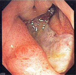
Esophageal varices - post banding.jpg
Endoscopic still of esophageal ulcers seen after banding of esophageal varices, at time of esophagogastroduodenoscopy
Other namesEGD
OGD
Upper endoscopy
ICD-9-CM 45.13
MeSH D016145
OPS-301 code 1-631, 1-632

Esophagogastroduodenoscopy (EGD) or oesophagogastroduodenoscopy (OGD), also called by various other names, is a diagnostic endoscopic procedure that visualizes the upper part of the gastrointestinal tract down to the duodenum. It is considered a minimally invasive procedure since it does not require an incision into one of the major body cavities and does not require any significant recovery after the procedure (unless sedation or anesthesia has been used). However, a sore throat is common. [1] [2] [3]

Contents

Alternative names

The words esophagogastroduodenoscopy (EGD; American English) and oesophagogastroduodenoscopy (OGD; British English; see spelling differences) are both pronounced /ɪˌsɒfəɡˌɡæstrˌddɪˈnɒskpi/ . It is also called panendoscopy (PES) and upper GI endoscopy. It is also often called just upper endoscopy, upper GI, or even just endoscopy; because EGD is the most commonly performed type of endoscopy, the ambiguous term endoscopy is sometimes informally used to refer to EGD by default. The term gastroscopy literally focuses on the stomach alone, but in practice, the usage overlaps.

Medical uses

Gastroscopy or duodenoscopy is performed for a number of indications, one of the most common being in unexplained anemia, where it is used to diagnose gastric or duodenal ulcers, among other things. OSC Microbio 13 04 DuoScope.jpg
Gastroscopy or duodenoscopy is performed for a number of indications, one of the most common being in unexplained anemia, where it is used to diagnose gastric or duodenal ulcers, among other things.

Diagnostic

Surveillance

Confirmation of diagnosis/biopsy

Therapeutic

Newer interventions

Complications

The complication rate is about 1 in 1000. [4] They include:

When used in infants, the esophagogastroduodenoscope may compress the trachealis muscle, which narrows the trachea. [5] This can result in reduced airflow to the lungs. [5] Infants may be intubated to make sure that the trachea is fixed open. [5]

Limitations

Problems of gastrointestinal function are usually not well diagnosed by endoscopy since motion or secretion of the gastrointestinal tract is not easily inspected by EGD. Nonetheless, findings such as excess fluid or poor motion of the gut during endoscopy can be suggestive of disorders of function. Irritable bowel syndrome and functional dyspepsia are not diagnosed with EGD, but EGD may be helpful in excluding other diseases that mimic these common disorders.[ citation needed ]

Procedure

The tip of the endoscope should be lubricated and checked for critical functions including tip angulations, air and water suction, and image quality.

The patient is kept NPO (nil per os) or NBM (nothing by mouth) for at least 4 hours before the procedure. Most patients tolerate the procedure with only topical anesthesia of the oropharynx using lidocaine spray. However, some patients may need sedation and the very anxious/agitated patient may even need a general anesthetic. Informed consent is obtained before the procedure. The main risks are bleeding and perforation. The risk is increased when a biopsy or other intervention is performed.

The patient lies on their left side with the head resting comfortably on a pillow. A mouth-guard is placed between the teeth to prevent the patient from biting on the endoscope. The endoscope is then passed over the tongue and into the oropharynx. This is the most uncomfortable stage for the patient. Quick and gentle manipulation under vision guides the endoscope into the esophagus. The endoscope is gradually advanced down the esophagus making note of any pathology. Excessive insufflation of the stomach is avoided at this stage. The endoscope is quickly passed through the stomach and through the pylorus to examine the first and second parts of the duodenum. Once this has been completed, the endoscope is withdrawn into the stomach and a more thorough examination is performed including a J-maneuver. This involves retroflexing the tip of the scope so it resembles a 'J' shape in order to examine the fundus and gastroesophageal junction. Any additional procedures are performed at this stage. The air in the stomach is aspirated before removing the endoscope. Still photographs can be made during the procedure and later shown to the patient to help explain any findings.

In its most basic use, the endoscope is used to inspect the internal anatomy of the digestive tract. Often inspection alone is sufficient, but biopsy is a valuable adjunct to endoscopy. Small biopsies can be made with a pincer (biopsy forceps) which is passed through the scope and allows sampling of 1 to 3 mm pieces of tissue under direct vision. The intestinal mucosa heals quickly from such biopsies.

Clinical practice varies with respect to routine biopsy for histological analysis of the examined upper gastrointestinal system. A rapid urease test is quick, easy, and cost-effective screening for Helicobacter pylori infection.

Equipment

Indigo carmine staining (stomach) Indigo carmine staining.jpg
Indigo carmine staining (stomach)


See also

Related Research Articles

<span class="mw-page-title-main">Gastroenterology</span> Branch of medicine focused on the digestive system and its disorders

Gastroenterology is the branch of medicine focused on the digestive system and its disorders. The digestive system consists of the gastrointestinal tract, sometimes referred to as the GI tract, which includes the esophagus, stomach, small intestine and large intestine as well as the accessory organs of digestion which include the pancreas, gallbladder, and liver.

<span class="mw-page-title-main">Peptic ulcer disease</span> Ulcer of an area of the gastrointestinal tract

Peptic ulcer disease is a break in the inner lining of the stomach, the first part of the small intestine, or sometimes the lower esophagus. An ulcer in the stomach is called a gastric ulcer, while one in the first part of the intestines is a duodenal ulcer. The most common symptoms of a duodenal ulcer are waking at night with upper abdominal pain, and upper abdominal pain that improves with eating. With a gastric ulcer, the pain may worsen with eating. The pain is often described as a burning or dull ache. Other symptoms include belching, vomiting, weight loss, or poor appetite. About a third of older people with peptic ulcers have no symptoms. Complications may include bleeding, perforation, and blockage of the stomach. Bleeding occurs in as many as 15% of cases.

<span class="mw-page-title-main">Endoscopy</span> Procedure used in medicine to look inside the body

An endoscopy is a procedure used in medicine to look inside the body. The endoscopy procedure uses an endoscope to examine the interior of a hollow organ or cavity of the body. Unlike many other medical imaging techniques, endoscopes are inserted directly into the organ.

<span class="mw-page-title-main">Endoscope</span> Instrument to visually examine the interior of a hollow space

An endoscope is an inspection instrument composed of image sensor, optical lens, light source and mechanical device, which is used to look deep into the body by way of openings such as the mouth or anus. A typical endoscope applies several modern technologies including optics, ergonomics, precision mechanics, electronics, and software engineering. With an endoscope, it is possible to observe lesions that cannot be detected by X-ray, making it useful in medical diagnosis. Endoscopes use tubes which are only a few millimeters thick to transfer illumination in one direction and high-resolution images in real time in the other direction, resulting in minimally invasive surgeries. It is used to examine the internal organs like the throat or esophagus. Specialized instruments are named after their target organ. Examples include the cystoscope (bladder), nephroscope (kidney), bronchoscope (bronchus), arthroscope (joints) and colonoscope (colon), and laparoscope. They can be used to examine visually and diagnose, or assist in surgery such as an arthroscopy.

<span class="mw-page-title-main">Upper gastrointestinal bleeding</span> Medical condition

Upper gastrointestinal bleeding (UGIB) is gastrointestinal bleeding in the upper gastrointestinal tract, commonly defined as bleeding arising from the esophagus, stomach, or duodenum. Blood may be observed in vomit or in altered form as black stool. Depending on the amount of the blood loss, symptoms may include shock.

Hematemesis is the vomiting of blood. It can be confused with hemoptysis or epistaxis (nosebleed), which are more common. The source is generally the upper gastrointestinal tract, typically above the suspensory muscle of duodenum. It may be caused by ulcers, tumors of the stomach or esophagus, varices, prolonged and vigorous retching, gastroenteritis, ingested blood, or certain drugs.

Coffee ground vomitus refers to a particular appearance of vomit. Within organic heme molecules of red blood cells is the element iron, which oxidizes following exposure to gastric acid. This reaction causes the vomitus to look like ground coffee.

<span class="mw-page-title-main">Esophageal varices</span> Dilated veins in the lower oesophagus

Esophageal varices are extremely dilated sub-mucosal veins in the lower third of the esophagus. They are most often a consequence of portal hypertension, commonly due to cirrhosis. People with esophageal varices have a strong tendency to develop severe bleeding which left untreated can be fatal. Esophageal varices are typically diagnosed through an esophagogastroduodenoscopy.

<span class="mw-page-title-main">Gastrointestinal bleeding</span> Medical condition

Gastrointestinal bleeding, also called gastrointestinal hemorrhage (GIB), is all forms of bleeding in the gastrointestinal tract, from the mouth to the rectum. When there is significant blood loss over a short time, symptoms may include vomiting red blood, vomiting black blood, bloody stool, or black stool. Small amounts of bleeding over a long time may cause iron-deficiency anemia resulting in feeling tired or heart-related chest pain. Other symptoms may include abdominal pain, shortness of breath, pale skin, or passing out. Sometimes in those with small amounts of bleeding no symptoms may be present.

<span class="mw-page-title-main">Mallory–Weiss syndrome</span> Medical condition

Mallory–Weiss syndrome is a condition where high intra-abdominal pressures causes laceration and bleeding of the mucosa called Mallory-Weiss tears. Additionally, Mallory–Weiss syndrome is one of the most common causes of acute upper gastrointestinal bleeding, counting of around 1-15% of all cases in adults and less than 5% in children. It has been found that tears are up to 2 to 4 times more prevalent in men than women. The tears can cause upper gastrointestinal bleeding and predominantly occur where the esophagus meets the stomach. However, the tears can happen anywhere from the middle of the esophagus to the cardia of the stomach. Mallory–Weiss syndrome is often caused by constant vomiting and retching from alcoholism or bulimia. Gastroesophageal reflux disease (GERD) is another risk factor that is often linked with Mallory–Weiss syndrome. However, not every individual with Mallory–Weiss syndrome will have these risk factors. Individuals with Mallory–Weiss syndrome will have hematemesis, however the symptoms can vary.

<span class="mw-page-title-main">Gastric varices</span> Dilated submucosal veins in the stomach lining

Gastric varices are dilated submucosal veins in the lining of the stomach, which can be a life-threatening cause of bleeding in the upper gastrointestinal tract. They are most commonly found in patients with portal hypertension, or elevated pressure in the portal vein system, which may be a complication of cirrhosis. Gastric varices may also be found in patients with thrombosis of the splenic vein, into which the short gastric veins that drain the fundus of the stomach flow. The latter may be a complication of acute pancreatitis, pancreatic cancer, or other abdominal tumours, as well as hepatitis C. Gastric varices and associated bleeding are a potential complication of schistosomiasis resulting from portal hypertension.

<span class="mw-page-title-main">Dumping syndrome</span> Medical condition

Dumping syndrome occurs when food, especially sugar, moves too quickly from the stomach to the duodenum—the first part of the small intestine—in the upper gastrointestinal (GI) tract. This condition is also called rapid gastric emptying. It is mostly associated with conditions following gastric or esophageal surgery, though it can also arise secondary to diabetes or to the use of certain medications; it is caused by an absent or insufficiently functioning pyloric sphincter, the valve between the stomach and the duodenum.

<span class="mw-page-title-main">Gastric antral vascular ectasia</span> Intestinal bleeding due to widening of blood vessels in the stomach

Gastric antral vascular ectasia (GAVE) is an uncommon cause of chronic gastrointestinal bleeding or iron deficiency anemia. The condition is associated with dilated small blood vessels in the gastric antrum, which is a distal part of the stomach. The dilated vessels result in intestinal bleeding. It is also called watermelon stomach because streaky long red areas that are present in the stomach may resemble the markings on watermelon.

<span class="mw-page-title-main">Endoscopic ultrasound</span> Medical imaging procedure

Endoscopic ultrasound (EUS) or echo-endoscopy is a medical procedure in which endoscopy is combined with ultrasound to obtain images of the internal organs in the chest, abdomen and colon. It can be used to visualize the walls of these organs, or to look at adjacent structures. Combined with Doppler imaging, nearby blood vessels can also be evaluated.

<span class="mw-page-title-main">Hemosuccus pancreaticus</span> Medical condition

Hemosuccus pancreaticus is a rare cause of hemorrhage in the gastrointestinal tract. It is caused by a bleeding source in the pancreas, pancreatic duct, or structures adjacent to the pancreas, such as the splenic artery, that bleed into the pancreatic duct, which is connected with the bowel at the duodenum, the first part of the small intestine. Patients with hemosuccus may develop symptoms of gastrointestinal hemorrhage, such as blood in the stools, maroon stools, or melena, which is a dark, tarry stool caused by digestion of red blood cells. They may also develop abdominal pain. It is associated with pancreatitis, pancreatic cancer and aneurysms of the splenic artery. Hemosuccus may be identified with endoscopy (esophagogastroduodenoscopy), where fresh blood may be seen from the pancreatic duct. Alternatively, angiography may be used to inject the celiac axis to determine the blood vessel that is bleeding. This may also be used to treat hemosuccus, as embolization of the end vessel may terminate the hemorrhage. However, a distal pancreatectomy—surgery to remove of the tail of the pancreas—may be required to stop the hemorrhage.

<span class="mw-page-title-main">Endoclip</span>

An endoclip is a metallic mechanical device used in endoscopy in order to close two mucosal surfaces without the need for surgery and suturing. Its function is similar to a suture in gross surgical applications, as it is used to join together two disjointed surfaces, but, can be applied through the channel of an endoscope under direct visualization. Endoclips have found use in treating gastrointestinal bleeding, in preventing bleeding after therapeutic procedures such as polypectomy, and in closing gastrointestinal perforations. Many forms of endoclips exist of different shapes and sizes, including two and three prong devices, which can be administered using single use and reloadable systems, and may or may not open and close to facilitate placement.

<span class="mw-page-title-main">Capsule endoscopy</span> Medical imaging procedure

Capsule endoscopy is a medical procedure used to record internal images of the gastrointestinal tract for use in disease diagnosis. Newer developments are also able to take biopsies and release medication at specific locations of the entire gastrointestinal tract. Unlike the more widely used endoscope, capsule endoscopy provides the ability to see the middle portion of the small intestine. It can be applied to the detection of various gastrointestinal cancers, digestive diseases, ulcers, unexplained bleedings, and general abdominal pains. After a patient swallows the capsule, it passes along the gastrointestinal tract, taking a number of images per second which are transmitted wirelessly to an array of receivers connected to a portable recording device carried by the patient. General advantages of capsule endoscopy over standard endoscopy include the minimally invasive procedure setup, ability to visualize more of the gastrointestinal tract, and lower cost of the procedure.

<span class="mw-page-title-main">Portal hypertensive gastropathy</span> Changes in the mucosa of the stomach in patients with portal hypertension

Portal hypertensive gastropathy refers to changes in the mucosa of the stomach in patients with portal hypertension; by far the most common cause of this is cirrhosis of the liver. These changes in the mucosa include friability of the mucosa and the presence of ectatic blood vessels at the surface. Patients with portal hypertensive gastropathy may experience bleeding from the stomach, which may uncommonly manifest itself in vomiting blood or melena; however, portal hypertension may cause several other more common sources of upper gastrointestinal bleeding, such as esophageal varices and gastric varices. On endoscopic evaluation of the stomach, this condition shows a characteristic mosaic or "snake-skin" appearance to the mucosa of the stomach.

<span class="mw-page-title-main">Stomach disease</span> Medical condition

Stomach diseases include gastritis, gastroparesis, Crohn's disease and various cancers.

Therapeutic endoscopy is the medical term for an endoscopic procedure during which treatment is carried out via the endoscope. This contrasts with diagnostic endoscopy, where the aim of the procedure is purely to visualize a part of the gastrointestinal, respiratory or urinary tract in order to aid diagnosis. In practice, a procedure which starts as a diagnostic endoscopy may become a therapeutic endoscopy depending on the findings, such as in cases of upper gastrointestinal bleeding, or the finding of polyps during colonoscopy.

References

  1. "Gastroscopy – examination of oesophagus and stomach by endoscope". BUPA. December 2006. Archived from the original on 2007-10-06. Retrieved 2007-10-07.
  2. National Digestive Diseases Information Clearinghouse (November 2004). "Upper Endoscopy". National Institutes of Health . Retrieved 2007-10-07.
  3. "What is Upper GI Endoscopy?". Patient Center -- Procedures. American Gastroenterological Association. Archived from the original on 2007-09-28. Retrieved 2007-10-07.
  4. "EGD – esophagogastroduodenoscopy".
  5. 1 2 3 Cravero, Joseph P.; Landrigan-Ossar, Mary (2019-01-01), Coté, Charles J.; Lerman, Jerrold; Anderson, Brian J. (eds.), "46 - Anesthesia Outside the Operating Room", A Practice of Anesthesia for Infants and Children (Sixth Edition), Philadelphia: Elsevier, pp. 1077–1094.e4, ISBN   978-0-323-42974-0 , retrieved 2021-01-23