Pyloromyotomy

Last updated
Pyloromyotomy
Specialty gastroenterology

Pyloromyotomy is a surgical procedure in which a portion of the muscle fibers of the pyloric muscle are cut. This is typically done in cases where the contents from the stomach are inappropriately stopped by the pyloric muscle, causing the stomach contents to build up in the stomach and unable to be appropriately digested. The procedure is typically performed in cases of "hypertrophic pyloric stenosis" in young children. [1] In most cases, the procedure can be performed with either an open approach or a laparoscopic approach [2] and the patients typically have good outcomes with minimal complications. [3]

Contents

History and development

The development of the procedure has attributed to Dr. Conrad Ramstedt in 1911, who originally named the procedure Ramstedt's Operation. However, the procedure was truly performed about 17 months earlier by Sir Harold Stiles  in 1910 at the Royal Hospital for sick children. [4] In 1991, the first laparoscopic pyloromyotomy was performed by Dr. Alain and Dr. Grousseau. [4]

Procedure

Drawing showing pyloric stenosis before and after surgery Pyloric Stenosis.png
Drawing showing pyloric stenosis before and after surgery

After pyloric stenosis is identified in a patient, and any electrolyte and fluid imbalances are stabilized, the surgeon will perform the procedure. [4] [1] During which, the surgeon must access the pylorus through the abdominal wall. This can either be done laparoscopically or with an "open" procedure. In either case, the once the pylorus is accessed, the surgeon will visualize the hypertrophied pyloric muscle. Then, the surgeon will carefully cut through the outer layers of tissue and through the pyloric muscle to the mucosa, which is the layer of tissue facing the inside of the gastrointestinal tract. From there, the two portions of the pyloric muscle are tested for mobility and the mucosal layer is inspected for any unintentional damage. Depending on the approach, the pylorus, stomach, and gastrointestinal tract are returned to their appropriate place in the abdominal cavity and the medical equipment is removed. Finally, each of the surgical incisions are stitched closed and the patient is taken back to post-operative area for monitoring. [5] [4]

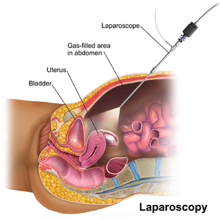
Laparoscopic approach: In the laparoscopic approach, the appropriate area of the gastrointestinal tract is accessed in a minimally invasive manner. [5] This approach may be chosen due to the reduced hospital stay, quicker recovery time, and higher satisfaction with the appearance of the surgical site after the patient has healed when compared to the older open approach. [2] Typically, two to three trocars, a medical device used to penetrate the abdominal wall in laparoscopic medical procedures, are placed in their appropriate positions. This is typically done my making a small cut for each trocar in the abdominal wall before placing the trocar into the cut. The abdomen is then filled with a gas, such as carbon dioxide to increase visibility with the laparoscopic camera and increase working space. [1] Once the laparoscopic instruments and camera are place through the trocars, the hypertrophied pylorus is visualized. Then, the pyloric muscle is cut down to the mucosa and the muscle fibers are spread apart using the laparoscopic instruments. From there, the two pyloric sections are tested independently for appropriate movement. After that, the mucosa is inspected for any unintentional damaged. This is done by inflating the patient's stomach and looking for the formation of bubbles along the mucosa. [4] If a leak is identified it is typically repaired with sutures if determined to be appropriate. Finally, all instruments and trocars are removed before the surgical wound sites are repaired with stitches. [5]

Horizontal surgical wound 10 days after pyloromyotomy in a four-week-old baby PyloricStenosisHorizontal.jpg
Horizontal surgical wound 10 days after pyloromyotomy in a four-week-old baby

Open approach: In the older open pyloromyotomy, the appropriate area of the gastrointestinal tract is accessed by creating a single cut on the abdomen of the patient and the pylorus and stomach are gently pulled through the opening for the procedure. [5] This approach may be chosen due to patient/parent preference or if determined by the surgeon to be more appropriate. [6] Once the initial cut on the abdomen is made, a layer of connective tissue between the abdomen and stomach is cut through. Then, the stomach and pylorus are carefully pulled through the opening created by the initial cut and the hypertrophied pylorus is identified by the surgeon. After that, the pyloric muscle is cut down to the mucosa and the muscle fibers are spread apart using a pyloric spreader. The newly separated pyloric sections are tested for adequate movement and the mucosa is tested for holes or other damage, which are repaired using suture as appropriate. Finally, the stomach and pylorus are carefully placed back into the abdominal cavity and the various tissue layers are repaired with stitches. [5]

Indication

The pyloromyotomy is primarily indicated by the presence of hypertrophic pyloric stenosis. [5] [1] Hypertrophic Pyloric stenosis is a gastrointestinal tract defect, most commonly seen in young children, typically in the first few months of life, caused by enlargement of the tissue in the pyloric muscle. [5] [4] [1] This causes the contents of the stomach to be unable to empty leading to pain after eating, electrolyte abnormalities, and projectile vomiting among other clinical signs and symptoms. [7] [1]

Complications, risks, and outlook

While the procedure has been proven to be highly effective at treating pyloric stenosis, there are still several complications that may occur as a result of the procedure. [1] [3] [7] The rate that any complications may occur as a result of the surgery are estimated to be 4.6–12% of cases. [3] The most common complications include incomplete pyloromyotomy, perforation of the mucosa, and infection of the surgical site. [3] Other complications reported to be related to the procedure, in addition to those stated above, are listed below:

The result of the surgery is typically successful at treating the patient's pyloric stenosis nearly 100% of the time with a quick recovery for most patients. [1] [7] Typically, the patient will have a special liquid diet for a few feedings following the procedure. In most cases the patient can be expected to be able to resume feedings with breast milk within 1 day of the procedure. [1]

See also

Related Research Articles

The term abdominal surgery broadly covers surgical procedures that involve opening the abdomen (laparotomy). Surgery of each abdominal organ is dealt with separately in connection with the description of that organ Diseases affecting the abdominal cavity are dealt with generally under their own names.

<span class="mw-page-title-main">General surgery</span> Medical specialty

General surgery is a surgical specialty that focuses on alimentary canal and abdominal contents including the esophagus, stomach, small intestine, large intestine, liver, pancreas, gallbladder, appendix and bile ducts, and often the thyroid gland. They also deal with diseases involving the skin, breast, soft tissue, trauma, peripheral artery disease and hernias and perform endoscopic as such as gastroscopy, colonoscopy and laparoscopic procedures.

<span class="mw-page-title-main">Laparoscopy</span> Minimally invasive operations within the abdominal or pelvic cavities

Laparoscopy is an operation performed in the abdomen or pelvis using small incisions with the aid of a camera. The laparoscope aids diagnosis or therapeutic interventions with a few small cuts in the abdomen.

Heller myotomy is a surgical procedure in which the muscles of the cardia are cut, allowing food and liquids to pass to the stomach. It is used to treat achalasia, a disorder in which the lower esophageal sphincter fails to relax properly, making it difficult for food and liquids to reach the stomach.

A laparotomy is a surgical procedure involving a surgical incision through the abdominal wall to gain access into the abdominal cavity. It is also known as a celiotomy.

<span class="mw-page-title-main">Pylorus</span> Part of the stomach that connects to the duodenum

The pylorus, or pyloric part, connects the stomach to the duodenum. The pylorus is considered as having two parts, the pyloric antrum and the pyloric canal. The pyloric canal ends as the pyloric orifice, which marks the junction between the stomach and the duodenum. The orifice is surrounded by a sphincter, a band of muscle, called the pyloric sphincter. The word pylorus comes from Greek πυλωρός, via Latin. The word pylorus in Greek means "gatekeeper", related to "gate" and is thus linguistically related to the word "pylon".

<span class="mw-page-title-main">Cholecystectomy</span> Surgical removal of the gallbladder

Cholecystectomy is the surgical removal of the gallbladder. Cholecystectomy is a common treatment of symptomatic gallstones and other gallbladder conditions. In 2011, cholecystectomy was the eighth most common operating room procedure performed in hospitals in the United States. Cholecystectomy can be performed either laparoscopically, or via an open surgical technique.

<span class="mw-page-title-main">Pyloric stenosis</span> Medical condition

Pyloric stenosis is a narrowing of the opening from the stomach to the first part of the small intestine. Symptoms include projectile vomiting without the presence of bile. This most often occurs after the baby is fed. The typical age that symptoms become obvious is two to twelve weeks old.

<span class="mw-page-title-main">Gastric bypass surgery</span> Type of bariatric surgery

Gastric bypass surgery refers to a technique in which the stomach is divided into a small upper pouch and a much larger lower "remnant" pouch and then the small intestine is rearranged to connect to both. Surgeons have developed several different ways to reconnect the intestine, thus leading to several different gastric bypass procedures (GBP). Any GBP leads to a marked reduction in the functional volume of the stomach, accompanied by an altered physiological and physical response to food.

<span class="mw-page-title-main">Pancreaticoduodenectomy</span> Major surgical procedure involving the pancreas, duodenum, and other organs

A pancreaticoduodenectomy, also known as a Whipple procedure, is a major surgical operation most often performed to remove cancerous tumours from the head of the pancreas. It is also used for the treatment of pancreatic or duodenal trauma, or chronic pancreatitis. Due to the shared blood supply of organs in the proximal gastrointestinal system, surgical removal of the head of the pancreas also necessitates removal of the duodenum, proximal jejunum, gallbladder, and, occasionally, part of the stomach.

<span class="mw-page-title-main">Adjustable gastric band</span> Inflatable silicone device

A laparoscopic adjustable gastric band, commonly called a lap-band, A band, or LAGB, is an inflatable silicone device placed around the top portion of the stomach to treat obesity, intended to decrease food consumption.

Digestive system surgery, or gastrointestinal surgery, can be divided into upper GI surgery and lower GI surgery.

<span class="mw-page-title-main">Spigelian hernia</span> Surgical condition

A Spigelian is the type of ventral hernia where aponeurotic fascia pushes through a hole in the junction of the linea semilunaris and the arcuate line creating a bulge. It appears in the abdomen lower quadrant between an area of dense fibrous tissue and abdominal wall muscles causing a.

<span class="mw-page-title-main">Gastric outlet obstruction</span> Medical condition

Gastric outlet obstruction (GOO) is a medical condition where there is an obstruction at the level of the pylorus, which is the outlet of the stomach. Individuals with gastric outlet obstruction will often have recurrent vomiting of food that has accumulated in the stomach, but which cannot pass into the small intestine due to the obstruction. The stomach often dilates to accommodate food intake and secretions. Causes of gastric outlet obstruction include both benign causes, as well as malignant causes, such as gastric cancer.

<span class="mw-page-title-main">Duodenal atresia</span> Medical condition

Duodenal atresia is the congenital absence or complete closure of a portion of the lumen of the duodenum. It causes increased levels of amniotic fluid during pregnancy (polyhydramnios) and intestinal obstruction in newborn babies. Newborns present with bilious or non-bilous vomiting within the first 24 to 48 hours after birth, typically after their first oral feeding. Radiography shows a distended stomach and distended duodenum, which are separated by the pyloric valve, a finding described as the double-bubble sign.

<span class="mw-page-title-main">Obturator hernia</span> Medical condition

An obturator hernia is a rare type of hernia, encompassing 0.07-1% of all hernias, of the pelvic floor in which pelvic or abdominal contents protrudes through the obturator foramen. The obturator foramen is formed by a branch of the ischial as well as the pubic bone. The canal is typically 2-3 centimeters long and 1 centimeters wide, creating a space for pouches of pre-peritoneal fat.

An exploratory laparotomy is a general surgical operation where the abdomen is opened and the abdominal organs are examined for injury or disease. It is the standard of care in various blunt and penetrating trauma situations in which there may be life-threatening internal injuries. It is also used in certain diagnostic situations, in which the operation is undertaken in search of a unifying cause for multiple signs and symptoms of disease, and in the staging of some cancers.

Wilhelm Conrad Ramstedt was a German surgeon remembered for describing Ramstedt's operation.

<span class="mw-page-title-main">Pyloroplasty</span>

Pyloroplasty is a surgery performed to widen the opening at the lower part of the stomach, also known as the pylorus. When the pylorus thickens, it becomes difficult for food to pass through. The surgery is performed to widen the band of muscle known as the pyloric sphincter, a ring of smooth, muscular fibers that surrounds the pylorus and helps to regulate digestion and prevent reflux. The widening of the pyloric sphincter enables the contents of the stomach to pass into the first part of the small intestine known as the duodenum.

The per-oral endoscopic myotomy, or POEM, is a minimally invasive surgical procedure for the treatment of achalasia wherein the inner circular muscle layer of the lower esophageal sphincter is divided through a submucosal tunnel. This enables food and liquids to pass into the stomach, a process that is impaired in achalasia. The tunnel is created, and the myotomy performed, using a flexible endoscope, meaning the entire procedure can be done without external incisions.

References

  1. 1 2 3 4 5 6 7 8 9 "Pyloric Stenosis". www.hopkinsmedicine.org. Retrieved 2021-11-02.
  2. 1 2 Ismail I, Elsherbini R, Elsaied A, Aly K, Sheir H (2020). "Laparoscopic vs. Open Pyloromyotomy in Treatment of Infantile Hypertrophic Pyloric Stenosis". Frontiers in Pediatrics. 8: 426. doi: 10.3389/fped.2020.00426 . PMC   7475708 . PMID   32984197.
  3. 1 2 3 4 5 6 7 8 9 van den Bunder FA, van Heurn E, Derikx JP (January 2020). "Comparison of laparoscopic and open pyloromyotomy: Concerns for omental herniation at port sites after the laparoscopic approach". Scientific Reports. 10 (1): 363. doi:10.1038/s41598-019-57031-4. PMC   6962153 . PMID   31941898.
  4. 1 2 3 4 5 6 MacKinlay GA, Barnhart DC. Laparoscopic Pyloromyotomy. pp. 281–286.{{cite book}}: |work= ignored (help)
  5. 1 2 3 4 5 6 7 Merchant A (2009). "Pyloromyotomy". Surgical Pitfalls. Elsevier. pp. 871–875. doi:10.1016/b978-141602951-9.50106-7. ISBN   9781416029519.
  6. Oomen MW, Hoekstra LT, Bakx R, Ubbink DT, Heij HA (August 2012). "Open versus laparoscopic pyloromyotomy for hypertrophic pyloric stenosis: a systematic review and meta-analysis focusing on major complications". Surgical Endoscopy. 26 (8): 2104–2110. doi:10.1007/s00464-012-2174-y. PMC   3392506 . PMID   22350232.
  7. 1 2 3 4 5 6 7 8 Levi B, George M (2010). "Current Procedures: Surgery. Chapter 46. Operative Management of Pyloric Stenosis: Pyloromyotomy". Access Surgery.