Defecography

Last updated
Defecography
Defproc.JPG
Caulking gun used for defecating proctogram
MeSH D019841

Defecography (also known as proctography, defecating/defecation proctography, evacuating/evacuation proctography or dynamic rectal examination) is a type of medical radiological imaging in which the mechanics of a patient's defecation are visualized in real time using a fluoroscope. [1] The anatomy and function of the anorectum and pelvic floor can be dynamically studied at various stages during defecation. [2]

Contents

History

Defecating proctography was pioneered in 1945, during World War II. The procedure gained popularity at this time in the midst of an outbreak of whipworm, which is known to cause rectal prolapse. [3] It has since become used for diagnosis of various anorectal disorders, including anismus and other causes of obstructed defecation.

It has fallen out of favor due to inadequate training in the technique. It is now only performed at a few institutions. Many radiology residents refer to the procedure as the "Def Proc", "Defogram", or "Stool Finale".[ citation needed ]

Indications

Defecography may be indicated for the following reasons:

Specifically, defecography can differentiate between anterior and posterior rectocele. [4] Also, in external rectal prolapse that was not directly visualized during examination, this radiographic projection will demonstrate its presence.

Technique

In females, pre-procedural preparation involves smearing a small amount of barium contrast agent in the vagina, which will help to identify if anterior rectocele, enterocele or sigmoidocele is present.

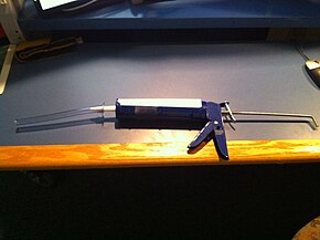
The technique itself involves the insertion of a caulking gun device into the rectum with a subsequent manual infusion of barium paste until there is adequate distension. The patient is then transferred to a portable plastic commode which is situated next to a fluoroscope which records the defecation. Positioning of the X-ray camera is of paramount importance as visualization of the buttocks, rectal vault, and lower pelvis is critical.

Diagnostic yield and interpretation

Anatomical and physiological parameters that can be objectively measured by this investigation include: [2]

Conditions which may be demonstrated include: [2]

The rectum may be seen to prolapse, whether internally or externally. There can be difficulty differentiating between internal intussusception and a normal rectal fold. The thickness of the intussusception is half the width of the intussusception (the intussusception is a doubled over layer of rectal wall). This is most likely to be seen during straining.

Cinedefecography and MRI defecography

Cinedefecography is a technique that is an evolution of defecography. The defecation cycle is recorded as a continuous series rather than individual still radiographs. [2] More recent techniques involve the use of advanced, cross-sectional imaging modalities such as magnetic resonance imaging. [6] This is known as dynamic pelvic MRI, or MRI proctography. [2] The MRI proctography also called MRI defecography is not as efficient as conventional X-ray defecography for some problems.[ citation needed ]

See also

Related Research Articles

<span class="mw-page-title-main">Fecal incontinence</span> Inability to refrain from defecation

Fecal incontinence (FI), or in some forms encopresis, is a lack of control over defecation, leading to involuntary loss of bowel contents, both liquid stool elements and mucus, or solid feces. When this loss includes flatus (gas), it is referred to as anal incontinence. FI is a sign or a symptom, not a diagnosis. Incontinence can result from different causes and might occur with either constipation or diarrhea. Continence is maintained by several interrelated factors, including the anal sampling mechanism, and incontinence usually results from a deficiency of multiple mechanisms. The most common causes are thought to be immediate or delayed damage from childbirth, complications from prior anorectal surgery, altered bowel habits. An estimated 2.2% of community-dwelling adults are affected. However, reported prevalence figures vary. A prevalence of 8.39% among non-institutionalized U.S adults between 2005 and 2010 has been reported, and among institutionalized elders figures come close to 50%.

<span class="mw-page-title-main">Levator ani</span> Broad, thin muscle group, situated on either side of the pelvis

The levator ani is a broad, thin muscle group, situated on either side of the pelvis. It is formed from three muscle components: the pubococcygeus, the iliococcygeus, and the puborectalis.

In medicine, prolapse is a condition in which organs fall down or slip out of place. It is used for organs protruding through the vagina, rectum, or for the misalignment of the valves of the heart. A spinal disc herniation is also sometimes called "disc prolapse". Prolapse means "to fall out of place", from the Latin prolabi meaning "to fall out".

<span class="mw-page-title-main">Rectal prolapse</span> Medical condition

A rectal prolapse occurs when walls of the rectum have prolapsed to such a degree that they protrude out of the anus and are visible outside the body. However, most researchers agree that there are 3 to 5 different types of rectal prolapse, depending on whether the prolapsed section is visible externally, and whether the full or only partial thickness of the rectal wall is involved.

<span class="mw-page-title-main">Pelvic floor</span> Anatomical structure

The pelvic floor or pelvic diaphragm is an anatomical location in the human body, which has an important role in urinary and anal continence, sexual function and support of the pelvic organs. The pelvic floor includes muscles, both skeletal and smooth, ligaments and fascia. and separates between the pelvic cavity from above, and the perineum from below. It is formed by the levator ani muscle and coccygeus muscle, and associated connective tissue.

<span class="mw-page-title-main">Rectocele</span> Bulging of the rectum into the vaginal wall

In gynecology, a rectocele or posterior vaginal wall prolapse results when the rectum bulges (herniates) into the vagina. Two common causes of this defect are childbirth and hysterectomy. Rectocele also tends to occur with other forms of pelvic organ prolapse, such as enterocele, sigmoidocele and cystocele.

<span class="mw-page-title-main">Pelvic floor dysfunction</span> Medical condition

Pelvic floor dysfunction is a term used for a variety of disorders that occur when pelvic floor muscles and ligaments are impaired. The condition affects up to 50 percent of women who have given birth. Although this condition predominantly affects women, up to 16 percent of men are affected as well. Symptoms can include pelvic pain, pressure, pain during sex, urinary incontinence (UI), overactive bladder, bowel incontinence, incomplete emptying of feces, constipation, myofascial pelvic pain and pelvic organ prolapse. When pelvic organ prolapse occurs, there may be visible organ protrusion or a lump felt in the vagina or anus. Research carried out in the UK has shown that symptoms can restrict everyday life for women. However, many people found it difficult to talk about it and to seek care, as they experienced embarassment and stigma.

Rectal tenesmus is a feeling of incomplete defecation. It is the sensation of inability or difficulty to empty the bowel at defecation, even if the bowel contents have already been evacuated. Tenesmus indicates the feeling of a residue, and is not always correlated with the actual presence of residual fecal matter in the rectum. It is frequently painful and may be accompanied by involuntary straining and other gastrointestinal symptoms. Tenesmus has both a nociceptive and a neuropathic component.

<span class="mw-page-title-main">Pelvic organ prolapse</span> Descent of the pelvic organs from their normal positions

Pelvic organ prolapse (POP) is characterized by descent of pelvic organs from their normal positions into the vagina. In women, the condition usually occurs when the pelvic floor collapses after gynecological cancer treatment, childbirth or heavy lifting. Injury incurred to fascia membranes and other connective structures can result in cystocele, rectocele or both. Treatment can involve dietary and lifestyle changes, physical therapy, or surgery.

<span class="mw-page-title-main">Rectum</span> Final portion of the large intestine

The rectum is the final straight portion of the large intestine in humans and some other mammals, and the gut in others. The adult human rectum is about 12 centimetres (4.7 in) long, and begins at the rectosigmoid junction at the level of the third sacral vertebra or the sacral promontory depending upon what definition is used. Its diameter is similar to that of the sigmoid colon at its commencement, but it is dilated near its termination, forming the rectal ampulla. It terminates at the level of the anorectal ring or the dentate line, again depending upon which definition is used. In humans, the rectum is followed by the anal canal which is about 4 centimetres (1.6 in) long, before the gastrointestinal tract terminates at the anal verge. The word rectum comes from the Latin rectumintestinum, meaning straight intestine.

<span class="mw-page-title-main">Human anus</span> External opening of the rectum

In humans, the anus is the external opening of the rectum located inside the intergluteal cleft. Two sphincters control the exit of feces from the body during an act of defecation, which is the primary function of the anus. These are the internal anal sphincter and the external anal sphincter, which are circular muscles that normally maintain constriction of the orifice and which relaxes as required by normal physiological functioning. The inner sphincter is involuntary and the outer is voluntary. Above the anus is the perineum, which is also located beneath the vulva or scrotum.

<span class="mw-page-title-main">Anorectal manometry</span> Medical functional test of the anus and rectum

Anorectal manometry (ARM) is a medical test used to measure pressures in the anus and rectum and to assess their function. The test is performed by inserting a catheter, that contains a probe embedded with pressure sensors, through the anus and into the rectum. Patients may be asked to perform certain maneuvers, such as coughing or attempting to defecate, to assess for pressure changes. Anorectal manometry is a safe and low risk procedure.

Solitary rectal ulcer syndrome or SRUS is a chronic, benign disorder of the rectal mucosa. It commonly occurs with varying degrees of rectal prolapse. The condition is thought to be caused by different factors, such as long term constipation, straining during defecation, and dyssynergic defecation. Treatment is by normalization of bowel habits, biofeedback, and other conservative measures. In more severe cases various surgical procedures may be indicated. The condition is relatively rare, affecting approximately 1 in 100,000 people per year. It affects mainly adults aged 30–50. Females are affected slightly more often than males. The disorder can be confused clinically with rectal cancer or other conditions such as inflammatory bowel disease, even when a biopsy is done.

<span class="mw-page-title-main">Anismus</span> Medical condition

Anismus or dyssynergic defecation is the failure of normal relaxation of pelvic floor muscles during attempted defecation. It can occur in both children and adults, and in both men and women. It can be caused by physical defects or it can occur for other reasons or unknown reasons. Anismus that has a behavioral cause could be viewed as having similarities with parcopresis, or psychogenic fecal retention.

Obstructed defecation syndrome is a major cause of functional constipation, of which it is considered a subtype. It is characterized by difficult and/or incomplete emptying of the rectum with or without an actual reduction in the number of bowel movements per week. Normal definitions of functional constipation include infrequent bowel movements and hard stools. In contrast, ODS may occur with frequent bowel movements and even with soft stools, and the colonic transit time may be normal, but delayed in the rectum and sigmoid colon.

Sigmoidocele refers to a condition where the sigmoid colon descends (prolapses) into the lower pelvic cavity. This can obstruct the rectum and cause symptoms of obstructed defecation.

Descending perineum syndrome refers to a condition where the perineum "balloons" several centimeters below the bony outlet of the pelvis during strain, although this descent may happen without straining. The syndrome was first described in 1966 by Parks et al.

Dyssynergia is any disturbance of muscular coordination, resulting in uncoordinated and abrupt movements. This is also an aspect of ataxia. It is typical for dyssynergic patients to split a movement into several smaller movements. Types of dyssynergia include Ramsay Hunt syndrome type 1, bladder sphincter dyssynergia, and anal sphincter dyssynergia.

In fecal incontinence (FI), surgery may be carried out if conservative measures alone are not sufficient to control symptoms. There are many surgical options described for FI, and they can be considered in 4 general groups.

<span class="mw-page-title-main">Vaginal support structures</span> Structures that maintain the position of the vagina within the pelvic cavity

The vaginal support structures are those muscles, bones, ligaments, tendons, membranes and fascia, of the pelvic floor that maintain the position of the vagina within the pelvic cavity and allow the normal functioning of the vagina and other reproductive structures in the female. Defects or injuries to these support structures in the pelvic floor leads to pelvic organ prolapse. Anatomical and congenital variations of vaginal support structures can predispose a woman to further dysfunction and prolapse later in life. The urethra is part of the anterior wall of the vagina and damage to the support structures there can lead to incontinence and urinary retention.

References

  1. "Defecating proctogram from Bristol Laparoscopic Surgery". Archived from the original on 2016-03-04. Retrieved 2010-07-17.
  2. 1 2 3 4 5 6 7 al.], senior editors, Bruce G. Wolff ... [et (2007). The ASCRS textbook of colon and rectal surgery. New York: Springer. pp. 47–52. ISBN   978-0-387-24846-2.{{cite book}}: |first= has generic name (help)CS1 maint: multiple names: authors list (link)
  3. "ASCRS: Pelvic Floor Dysfunction". Archived from the original on 2015-02-10. Retrieved 2010-07-17.
  4. Wiersma, Tjeerd. "Dynamic rectal examination on the Radiology assistant". Archived from the original on 13 October 2012. Retrieved 19 October 2012.
  5. Voderholzer, WA; Neuhaus, DA; Klauser, AG; Tzavella, K; Müller-Lissner, SA; Schindlbeck, NE (August 1997). "Paradoxical sphincter contraction is rarely indicative of anismus". Gut. 41 (2): 258–62. doi:10.1136/gut.41.2.258. PMC   1891465 . PMID   9301508.
  6. MRI Defecating Proctogram