Colostomy

Last updated
Colostomy
Diagram showing a colostomy with a bag CRUK 061.svg
Diagram showing a colostomy
ICD-9-CM 46.1
MeSH D003125
MedlinePlus 002942

A colostomy is an opening (stoma) in the large intestine (colon), or the surgical procedure that creates one. The opening is formed by drawing the healthy end of the colon through an incision in the anterior abdominal wall and suturing it into place. This opening, often in conjunction with an attached ostomy system, provides an alternative channel for feces to leave the body. Thus if the natural anus is unavailable for that function (for example, in cases where it has been removed as part of treatment for colorectal cancer or ulcerative colitis), an artificial anus takes over. It may be reversible or irreversible, depending on the circumstances.

Contents

Uses

There are many reasons for this procedure. Some common reasons are:

Types

Illustration depicting various types of colostomy Blausen 0247 Colostomy.png
Illustration depicting various types of colostomy

Types of colostomy include: [1] [2]

Colostomy surgery that is planned usually has a higher rate of long-term success than surgery performed in an emergency situation.[ citation needed ]

Duration

A colostomy may be temporary; and reversed at a later date; or permanent.

Alternatives

Colostomy or ileostomy is now rarely performed for rectal cancer, with surgeons usually preferring primary resection and internal anastomosis, [3] e.g. an ileo-anal pouch. In place of an external appliance, an internal ileo-anal pouch is constructed using a portion of the patient's lower intestine, to act as a new rectum to replace the removed original.

Procedure

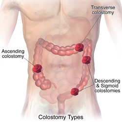
Placement of the stoma on the abdomen can occur at any location along the colon, but the most common placement is on the lower left side near the sigmoid where a majority of colon cancers occur. Other locations include the ascending, transverse, and descending sections of the colon. [4]

Routine care

Pouches and the stick-on appliances to which they attach must be changed regularly. Sometimes an odor neutralizer and lubricant is squirted into a new pouch before it is attached. Two types of pouches are available: one disposable and one drainable. Most pouches are opaque and filter out air through a charcoal filter. The recommended practice is to empty such pouches when one-third full. [5] Appliances, in contrast with pouches, are usually replaced every three to seven days except in cases where their seals have broken contact with the skin, when they should be replaced immediately. [5]

Even as long ago as the 1940s, surgeons conducting a review at the Cleveland Clinic (Jones and Kehm, 1946) [6] could summarize the routine care of the permanent colostomy as usually quite satisfactory, stating that after patients recover from the initial worry prompted by the need for a colostomy, most of them learn to manage their colostomy quite well. [6] "These patients come from all walks of life and carry on their daily work as usual. One patient stated that he could see no advantage of the normal anus over a colostomy. While this may be somewhat overstated, it is true that most people with a permanent colostomy can live a useful, happy life." [6] They found that, just as in anyone else, dietary indiscretion was the usual factor in occasional bowel habit disruption. [6] This historical experience has been borne out, as today the conclusion still stands that most patients can successfully manage a colostomy as part of their activities of daily living.

Jones and Kehm preferred tissue paper as a colostomy cover (held in place with a band or garment) rather than a colostomy bag. [6] They found that irrigation of the colostomy varied with each patient's bowel habit but that most patients developed a routine of every-other-day irrigation, whereas a few needed no irrigation. [6]

People with colostomies must wear an ostomy pouching system to collect intestinal waste. Ordinarily, the pouch must be emptied or changed a couple of times a day depending on the frequency of activity; in general the further from the anus (i.e., the further 'up' the intestinal tract) the ostomy is located the greater the output and more frequent the need to empty or change the pouch. [7]

Colostomy and irrigation

People with colostomies who have ostomies of the sigmoid colon or descending colon may have the option of irrigation, which allows for the person to not wear a pouch, but rather just a gauze cap over the stoma, and to schedule irrigation for times that are convenient. [8] To irrigate, a catheter is placed inside the stoma, and flushed with water, which allows the feces to come out of the body into an irrigation sleeve. [9] Most colostomates irrigate once a day or every other day, though this depends on the person, their food intake, and their health.

Complications

Patient with a colostomy complicated by a large parastomal hernia, which is when tissue protrudes adjacent to the stoma tract. Colostomy and parastomal hernia.JPG
Patient with a colostomy complicated by a large parastomal hernia, which is when tissue protrudes adjacent to the stoma tract.
CT scan of same patient, showing intestines within the hernia. Colostomy and parastomal hernia - CT.png
CT scan of same patient, showing intestines within the hernia.

Parastomal hernia (PH) is the most common late complication of stomata through the abdominal wall, occurring in 10-25% of patients, [10] even up to 50% by some estimates. [11] Prolapse of bowel wall through the stoma occasionally happens and can require reoperation to repair.[ citation needed ]

Clinical pilates-based exercises have been demonstrated to improve patients' core abdominal strength and to reduce the risk of a hernia worsening. [11]

Other common complications of colostomy are high output, skin irritation, prolapse, retraction, and ischemia.

Related Research Articles

<span class="mw-page-title-main">Bowel obstruction</span> Mechanical or functional obstruction of the intestines

Bowel obstruction, also known as intestinal obstruction, is a mechanical or functional obstruction of the intestines which prevents the normal movement of the products of digestion. Either the small bowel or large bowel may be affected. Signs and symptoms include abdominal pain, vomiting, bloating and not passing gas. Mechanical obstruction is the cause of about 5 to 15% of cases of severe abdominal pain of sudden onset requiring admission to hospital.

<span class="mw-page-title-main">Diverticulitis</span> Digestive disease of the large intestine

Diverticulitis, also called colonic diverticulitis, is a gastrointestinal disease characterized by inflammation of abnormal pouches—diverticula—that can develop in the wall of the large intestine. Symptoms typically include lower abdominal pain of sudden onset, but the onset may also occur over a few days. There may also be nausea, diarrhea or constipation. Fever or blood in the stool suggests a complication. People may experience a single attack, repeated attacks, or ongoing "smouldering" diverticulitis.

<span class="mw-page-title-main">Colorectal surgery</span> Field in medicine for disabilities in the rectum

Colorectal surgery is a field in medicine dealing with disorders of the rectum, anus, and colon. The field is also known as proctology, but this term is now used infrequently within medicine and is most often employed to identify practices relating to the anus and rectum in particular. The word proctology is derived from the Greek words πρωκτός proktos, meaning "anus" or "hindparts", and -λογία -logia, meaning "science" or "study".

In anatomy, a stoma is any opening in the body. For example, a mouth, a nose, and an anus are natural stomata. Any hollow organ can be manipulated into an artificial stoma as necessary. This includes the esophagus, stomach, duodenum, ileum, colon, pleural cavity, ureters, urinary bladder, and renal pelvis. Such a stoma may be permanent or temporary.

An abdomino perineal resection, formally known as abdominoperineal resection of the rectum and abdominoperineal excision of the rectum is a surgery for rectal cancer or anal cancer. It is frequently abbreviated as AP resection, APR and APER.

<span class="mw-page-title-main">Ileostomy</span> Surgical procedure

Ileostomy is a stoma constructed by bringing the end or loop of small intestine out onto the surface of the skin, or the surgical procedure which creates this opening. Intestinal waste passes out of the ileostomy and is collected in an external ostomy system which is placed next to the opening. Ileostomies are usually sited above the groin on the right hand side of the abdomen.

In medicine, the ileal pouch–anal anastomosis (IPAA), also known as restorative proctocolectomy (RPC), ileal-anal reservoir (IAR), an ileo-anal pouch, ileal-anal pullthrough, or sometimes referred to as a J-pouch, S-pouch, W-pouch, or a pelvic pouch, is an anastomosis of a reservoir pouch made from ileum to the anus, bypassing the former site of the colon in cases where the colon and rectum have been removed. The pouch retains and restores functionality of the anus, with stools passed under voluntary control of the person, preventing fecal incontinence and serving as an alternative to a total proctocolectomy with ileostomy.

Colic in horses is defined as abdominal pain, but it is a clinical symptom rather than a diagnosis. The term colic can encompass all forms of gastrointestinal conditions which cause pain as well as other causes of abdominal pain not involving the gastrointestinal tract. What makes it tricky is that different causes can manifest with similar signs of distress in the animal. Recognizing and understanding these signs is pivotal, as timely action can spell the difference between a brief moment of discomfort and a life-threatening situation. The most common forms of colic are gastrointestinal in nature and are most often related to colonic disturbance. There are a variety of different causes of colic, some of which can prove fatal without surgical intervention. Colic surgery is usually an expensive procedure as it is major abdominal surgery, often with intensive aftercare. Among domesticated horses, colic is the leading cause of premature death. The incidence of colic in the general horse population has been estimated between 4 and 10 percent over the course of the average lifespan. Clinical signs of colic generally require treatment by a veterinarian. The conditions that cause colic can become life-threatening in a short period of time.

<span class="mw-page-title-main">Intussusception (medical disorder)</span> Folding of part of the intestine into itself

Intussusception is a medical condition in which a part of the intestine folds into the section immediately ahead of it. It typically involves the small intestine and less commonly the large intestine. Symptoms include abdominal pain which may come and go, vomiting, abdominal bloating, and bloody stool. It often results in a small bowel obstruction. Other complications may include peritonitis or bowel perforation.

<span class="mw-page-title-main">Colectomy</span> Surgical removal of any extent of the colon

Colectomy is bowel resection of the large bowel. It consists of the surgical removal of any extent of the colon, usually segmental resection. In extreme cases where the entire large intestine is removed, it is called total colectomy, and proctocolectomy denotes that the rectum is included.

<span class="mw-page-title-main">Imperforate anus</span> Birth defect of malformed rectum

An imperforate anus or anorectal malformations (ARMs) are birth defects in which the rectum is malformed. ARMs are a spectrum of different congenital anomalies which vary from fairly minor lesions to complex anomalies. The cause of ARMs is unknown; the genetic basis of these anomalies is very complex because of their anatomical variability. In 8% of patients, genetic factors are clearly associated with ARMs. Anorectal malformation in Currarino syndrome represents the only association for which the gene HLXB9 has been identified.

An Indiana pouch is a surgically-created urinary diversion used to create a way for the body to store and eliminate urine for patients who have had their urinary bladders removed as a result of bladder cancer, pelvic exenteration, bladder exstrophy or who are not continent due to a congenital, neurogenic bladder. This particular urinary diversion results in a continent reservoir that the patient must catheterize to empty urine. This concept and technique was developed by Drs. Mike Mitchell, Randall Rowland, and Richard Bihrle at Indiana University.

<span class="mw-page-title-main">Bowel resection</span> Surgical procedure in which a part of an intestine is removed

A bowel resection or enterectomy is a surgical procedure in which a part of an intestine (bowel) is removed, from either the small intestine or large intestine. Often the word enterectomy is reserved for the sense of small bowel resection, in distinction from colectomy, which covers the sense of large bowel resection. Bowel resection may be performed to treat gastrointestinal cancer, bowel ischemia, necrosis, or obstruction due to scar tissue, volvulus, and hernias. Some patients require ileostomy or colostomy after this procedure as alternative means of excretion. Complications of the procedure may include anastomotic leak or dehiscence, hernias, or adhesions causing partial or complete bowel obstruction. Depending on which part and how much of the intestines are removed, there may be digestive and metabolic challenges afterward, such as short bowel syndrome.

<span class="mw-page-title-main">Proctocolectomy</span> Surgical removal of the colon and rectum

Proctocolectomy is the surgical removal of the entire colon and rectum from the human body, leaving the patients small intestine disconnected from their anus. It is a major surgery that is performed by colorectal surgeons, however some portions of the surgery, specifically the colectomy may be performed by general surgeons. It was first performed in 1978 and since that time, medical advancements have led to the surgery being less invasive with great improvements in patient outcomes. The procedure is most commonly indicated for severe forms of inflammatory bowel disease such as ulcerative colitis and Crohn's disease. It is also the treatment of choice for patients with familial adenomatous polyposis.

Total mesorectal excision (TME) is a standard surgical technique for treatment of rectal cancer, first described in 1982 by Professor Bill Heald at the UK's Basingstoke District Hospital. It is a precise dissection of the mesorectal envelope comprising rectum containing the tumour together with all the surrounding fatty tissue and the sheet of tissue that contains lymph nodes and blood vessels. Dissection is along the avascular alveolar plane between the presacral and mesorectal fascia, described as holy plane. Dissection along this plane facilitates a straightforward dissection and preserves the sacral vessels and hypogastric nerves and is a sphincter-sparing resection and decreases permanent stoma rates. It is possible to rejoin the two ends of the colon; however, most patients require a temporary ileostomy pouch to bypass the colon, allowing it to heal with less risk of infection, perforation or leakage.

<span class="mw-page-title-main">Ostomy system</span> Pouch system to collect body waste fluids

An ostomy pouching system is a prosthetic medical device that provides a means for the collection of waste from a surgically diverted biological system and the creation of a stoma. Pouching systems are most commonly associated with colostomies, ileostomies, and urostomies.

Rectal discharge is intermittent or continuous expression of liquid from the anus. Normal rectal mucus is needed for proper excretion of waste. Otherwise, this is closely related to types of fecal incontinence but the term rectal discharge does not necessarily imply degrees of incontinence. Types of fecal incontinence that produce a liquid leakage could be thought of as a type of rectal discharge.

In fecal incontinence (FI), surgery may be carried out if conservative measures alone are not sufficient to control symptoms. There are many surgical options described for FI, and they can be considered in 4 general groups.

A rectovestibular fistula, also referred to simply as a vestibular fistula, is an anorectal congenital disorder where an abnormal connection (fistula) exists between the rectum and the vulval vestibule of the female genitalia.

Ralph John Nicholls, FRCS (Eng), EBSQ is a retired British colorectal surgeon, Emeritus Consultant Surgeon at St Mark's Hospital London and Professor of Colorectal Surgery, Imperial College London.

References

  1. Potter et al. Canadian Fundamentals of Nursing 3rd ed.2006, Elsevier Canada. p1393-1394
  2. "Stoma". Archived from the original on 2015-12-10. Retrieved 2015-06-09.
  3. Wolff, Bruce G.; American Society of Colon and Rectal Surgeons, eds. (2007). The ASCRS textbook of colon and rectal surgery. New York: Springer. p. 417. ISBN   978-0-387-24846-2.{{cite book}}: CS1 maint: date and year (link)
  4. Potter et al. Canadian Fundamentals of Nursing 3rd ed.2006, Elsevier Canada.p1393
  5. 1 2 Taylor, C. R., Lillis, C., LeMone, P., Lynn, P. (2011) Fundamentals of nursing: The art and science of nursing care. Philadelphia: Lippincott Williams & Wilkins, page 1327.
  6. 1 2 3 4 5 6 Jones, Thomas E.; Kehm, Ray W. (1946), "Management of the permanent colostomy" (PDF), Cleveland Clinic Quarterly, 13 (4): 198–203, doi:10.3949/ccjm.13.4.198, PMID   20274022, S2CID   76078689.[ permanent dead link ]
  7. "Colostomy irrigation: Colostomy Guide" (PDF). United Ostomy Associations of America. Archived from the original (PDF) on 23 June 2012. Retrieved 4 February 2013.
  8. Rooney, Debra. "Colostomy irrigation: A personal account managing colostomy" (PDF). Ostomy. Retrieved 7 September 2012.
  9. Wax, Arnold. "What is colostomy irrigation?". WebMed. Retrieved 7 September 2012.
  10. Paul H. Sugarbaker (2013). "Paraostomy Hernias: Prosthetic Mesh Repair". Abdominal Surgery. American Society of Abdominal Surgeons. Archived from the original on 2016-01-31. Retrieved 2015-10-28.
  11. 1 2 Taylor, Claire; Munro, Julie; Goodman, William; Russell, Sarah; Oliphant, Raymond; Beeken, Rebecca J; Hubbard, Gill (2023). "Hernia Active Living Trial (HALT): an exercise intervention in people with a parastomal hernia or bulge". British Journal of Nursing. 32 (22): S4–S11. doi:10.12968/bjon.2023.32.22.S4. ISSN   0966-0461. Archived from the original on 11 March 2024.