Prolapse

Last updated

In medicine, prolapse is a condition in which organs fall down or slip out of place. It is used for organs protruding through the vagina, rectum, or for the misalignment of the valves of the heart. A spinal disc herniation is also sometimes called "disc prolapse". Prolapse means "to fall out of place", from the Latin prolabi meaning "to fall out".

Contents

Relating to the uterus, prolapse condition results in an inferior extension of the organ into the vagina, caused by weakened pelvic muscles.

Humans

Heart valve prolapse

The main type of prolapse of heart valves in humans is mitral valve prolapse (MVP), which is a valvular heart disease characterized by the displacement of an abnormally thickened mitral valve leaflet into the left atrium during systole.

Tricuspid valve prolapse can cause tricuspid regurgitation. [1]

Rectal prolapse

Rectal prolapse is a condition in which part of the wall or the entire wall of the rectum falls out of place. Rectal prolapse can be a medical emergency. In some cases, the rectum may protrude.

Symptoms of a rectal prolapse may be:

A surgeon may operate through the abdomen to secure part of the large intestine or rectum to the inside of the abdominal cavity (rectopexy). Sometimes the surgeon removes the affected part of the intestine.

Surgery also can be done through the perineum (the area between the genitals and the anus) to remove the prolapsing tissue.

Surgery is most often successful for people who still have some control over their bowel movements. If the anal sphincter is damaged, surgery may correct the prolapse but not be able to completely correct fecal incontinence (lack of control of bowel movements). Fecal incontinence can both potentially improve or deteriorate after prolapse surgery.

If the lining has fallen out of the anus and is around 7 cm or less, it should eventually retract back inside naturally, though the retraction can take up to four days.

Female genital prolapse

Uterine prolapse (or Pelvic organ prolapse) occurs when the female pelvic organs fall from their normal position, into or through the vagina. Occurring in women of all ages, it is more common as women age, particularly in those who have delivered large babies or had exceedingly long pushing phases of labor. Smoking, obesity, connective tissue disorders, upper respiratory disorders‚ and repetitive strain injuries can all increase prolapse risk. Minor prolapse can be treated with exercises to strengthen the pelvic floor muscles (pelvic physiotherapy); more serious prolapse, e.g., complete procidentia, requires pessary use or reconstructive surgical treatment. Reconstructive pelvic prolapse surgery may be done without resorting to complete hysterectomy by hysteropexy, [2] the resuspension of the prolapsed uterus. Traditional gynecologic practice favors removal of the uterus or ovaries (or both) at the time of prolapse surgery, and one estimate states that of the 600,000 hysterectomies performed in the United States every year, 13 percent are for prolapse. [3] However, there is concern that many of these hysterectomies may be unnecessary and that hysteropexy would suffice as a treatment instead.

Pelvic floor prolapse

The rectum or urinary bladder may prolapse as a result of changes in the integrity of connective tissue in the posterior or anterior vaginal walls, respectively, resulting in pelvic floor prolapse. Symptoms may include a feeling of pressure in the pelvis, or the visible protrusion of organs from the vagina. Prolapse is almost never painful, but the change in position of organs may cause urinary or bowel symptoms.

Pessaries are a treatment option for pelvic organ prolapse. [4]

Umbilical cord prolapse

Umbilical cord prolapse occurs when the umbilical cord comes out of the uterus with or before the presenting part of the fetus. It is a relatively rare condition and occurs in fewer than 1% of pregnancies. Cord prolapse is more common in women who have had rupture of their amniotic sac. Other risk factors include maternal or fetal factors that prevent the fetus from occupying a normal position in the maternal pelvis, such as abnormal fetal lie, too much amniotic fluid, or a premature or small fetus. The concern with cord prolapse is that pressure on the cord from the fetus will cause cord compression that compromises blood flow to the fetus. Whenever there is a sudden decrease in fetal heart rate or abnormal fetal heart tracing, umbilical cord prolapse should be considered. Due to the possibility for fetal death and other complications, umbilical cord prolapse is considered an obstetric emergency during pregnancy or labor. Current management guidelines focus on quick delivery, which usually entails a cesarean section. With appropriate management, the majority of cases have good neonatal outcomes.

Other species

Reptiles

Genital prolapse has been reported in snakes and turtles. [5]

Birds

An egg-bound budgerigar hen with a prolapsed vagina and her mate BudgieProlapse.png
An egg-bound budgerigar hen with a prolapsed vagina and her mate

Oviduct prolapse is an often fatal condition in birds. When an egg is laid, the vagina everts through the cloaca to deliver the egg. Large eggs and avian obesity are contributors to this condition. Immediate veterinary assistance is paramount to the survival of a bird with prolapse. Even with immediate medical intervention the chances for survival are usually uncertain. Untreated birds will begin to tear at the injury site, and other flockmates will begin to cannibalize the prolapse area, a behaviour commonly known as pickout.

Cattle

Prolapsed uterus in cattle, particularly dairy cattle, generally occurs in the first 12 hours post-calving. [6] Frequent causes are hypocalcemia combined with irritation of the birth canal, causing straining. Replacement of the protrusion, which can range from the size of a softball to the hanging of the entire uterus down below the hocks, is performed with the cow in sternal recumbency, an epidural injection, and hindlimbs 'frogged' rearwards to allow the pelvis to tip forward, easing replacement.[ citation needed ] Careful washing and cleaning prior to replacement is important as is ensuring that the horns are completely everted once inside the cow. Often a Buhner suture is placed in the vulva to prevent subsequent reprolapse. Another type of reproductive prolapse in cattle is so-called bovine vaginal prolapse.

Sheep

Same as in cows. [7]

Pigs

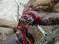
Rectal prolapse is a condition routinely identified in pigs on farms and at slaughterhouses. If not reduced quickly, prolapses in pigs become necrotic and infected, and risk being cannibalized by other pen mates. If the latter happens it normally results in death of the animal by sepsis, shock or faecal peritonitis.

Horses and mules

Rectal prolapse occurring in horse and mule would be better termed anal prolapse, as it only involves mucous membrane moving posteriorly to form a circular protrusion outside the anus  [8] The condition is not painful.

In mares after parturition, it is described as a 10 to 60 mm mucous protrusion. [9]

Anal prolapse in young mules and foals occurs with a frequency similar to that of Gasterophilus haemorrhoidalis . [10] [11] In extensive breeding conditions, the disease is only recognized after some days, leading to intense edema of prolapsed tissues and necrosis of the mucous membrane.

Early cases in should be treated with the application of hygroscopic substances like powdered sugar followed by purse-string suturing. When prolapsed tissues are edematous and necrotic, amputation is performed. The prognosis is fair as the removed tissues do not contain any important organs or large blood vessels.

Related Research Articles

<span class="mw-page-title-main">Uterus</span> Female sex organ in mammals

The uterus or womb is the organ in the reproductive system of most female mammals, including humans, that accommodates the embryonic and fetal development of one or more embryos until birth. The uterus is a hormone-responsive sex organ that contains glands in its lining that secrete uterine milk for embryonic nourishment.

<span class="mw-page-title-main">Hysterectomy</span> Surgical removal of the uterus

Hysterectomy is the partial or total surgical removal of the uterus. It may also involve removal of the cervix, ovaries (oophorectomy), Fallopian tubes (salpingectomy), and other surrounding structures. Partial hysterectomies allow for hormone regulation while total hysterectomies do not.

A pessary is a prosthetic device inserted into the vagina for structural and pharmaceutical purposes. It is most commonly used to treat stress urinary incontinence to stop urinary leakage and to treat pelvic organ prolapse to maintain the location of organs in the pelvic region. It can also be used to administer medications locally in the vagina or as a method of contraception.

<span class="mw-page-title-main">Rectal prolapse</span> Medical condition

A rectal prolapse occurs when walls of the rectum have prolapsed to such a degree that they protrude out of the anus and are visible outside the body. However, most researchers agree that there are 3 to 5 different types of rectal prolapse, depending on whether the prolapsed section is visible externally, and whether the full or only partial thickness of the rectal wall is involved.

<span class="mw-page-title-main">Pelvic floor</span> Anatomical structure

The pelvic floor or pelvic diaphragm is a muscular partition separating the pelvic cavity (above), and perineal region (below). It is formed by the levator ani muscle and coccygeus muscle, and associated connective tissue.

<span class="mw-page-title-main">Rectocele</span> Bulging of the rectum into the vaginal wall

In gynecology, a rectocele or posterior vaginal wall prolapse results when the rectum bulges (herniates) into the vagina. Two common causes of this defect are childbirth and hysterectomy. Rectocele also tends to occur with other forms of pelvic organ prolapse, such as enterocele, sigmoidocele and cystocele.

<span class="mw-page-title-main">Umbilical cord prolapse</span> Complication of pregnancy where the umbilical cord slips out of the uterus prior to birth

Umbilical cord prolapse is when the umbilical cord comes out of the uterus with or before the presenting part of the baby. The concern with cord prolapse is that pressure on the cord from the baby will compromise blood flow to the baby. It usually occurs during labor but can occur anytime after the rupture of membranes.

<span class="mw-page-title-main">Cystocele</span> Medical condition

The cystocele, also known as a prolapsed bladder, is a medical condition in which a woman's bladder bulges into her vagina. Some may have no symptoms. Others may have trouble starting urination, urinary incontinence, or frequent urination. Complications may include recurrent urinary tract infections and urinary retention. Cystocele and a prolapsed urethra often occur together and is called a cystourethrocele. Cystocele can negatively affect quality of life.

<span class="mw-page-title-main">Pelvic organ prolapse</span> Descent of the pelvic organs from their normal positions

Pelvic organ prolapse (POP) is characterized by descent of pelvic organs from their normal positions into the vagina. In women, the condition usually occurs when the pelvic floor collapses after gynecological cancer treatment, childbirth or heavy lifting. Injury incurred to fascia membranes and other connective structures can result in cystocele, rectocele or both. Treatment can involve dietary and lifestyle changes, physical therapy, or surgery.

<span class="mw-page-title-main">Pelvic exenteration</span> Surgical removal of all pelvic organs

Pelvic exenteration is a radical surgical treatment that removes all organs from a person's pelvic cavity. It is used to treat certain advanced or recurrent cancers. The urinary bladder, urethra, rectum, and anus are removed. In women, the vagina, cervix, uterus, Fallopian tubes, ovaries and, in some cases, the vulva are removed. In men, the prostate is removed. The procedure leaves the person with a permanent colostomy and urinary diversion.

<span class="mw-page-title-main">Vaginectomy</span> Surgical removal of the vagina

Vaginectomy is a surgery to remove all or part of the vagina. It is one form of treatment for individuals with vaginal cancer or rectal cancer that is used to remove tissue with cancerous cells. It can also be used in gender affirmation surgery. Some people born with a vagina who identify as trans men or as nonbinary may choose vaginectomy in conjunction with other surgeries to make the clitoris more penis-like (metoidioplasty), construct of a full-size penis (phalloplasty), or create a relatively smooth, featureless genital area.

<span class="mw-page-title-main">Uterine prolapse</span> Medical condition

Uterine prolapse is a form of pelvic organ prolapse in which the uterus and a portion of the upper vagina protrude into the vaginal canal and, in severe cases, through the opening of the vagina. It is most often caused by injury or damage to structures that hold the uterus in place within the pelvic cavity. Symptoms may include vaginal fullness, pain with sexual intercourse, difficulty urinating, and urinary incontinence. Risk factors include older age, pregnancy, vaginal childbirth, obesity, chronic constipation, and chronic cough. Prevalence, based on physical exam alone, is estimated to be approximately 14%.

<span class="mw-page-title-main">Pelvic examination</span> Physical medical examination

A pelvic examination is the physical examination of the external and internal female pelvic organs. It is frequently used in gynecology for the evaluation of symptoms affecting the female reproductive and urinary tract, such as pain, bleeding, discharge, urinary incontinence, or trauma. It can also be used to assess a woman's anatomy in preparation for procedures. The exam can be done awake in the clinic and emergency department, or under anesthesia in the operating room. The most commonly performed components of the exam are 1) the external exam, to evaluate the external genitalia 2) the internal exam with palpation to examine the uterus, ovaries, and fallopian tubes, and 3) the internal exam using the speculum to visualize the vaginal walls and cervix. During the pelvic exam, sample of cells and fluids may be collected to screen for sexually transmitted infections or cancer.

<span class="mw-page-title-main">Rectum</span> Final portion of the large intestine

The rectum is the final straight portion of the large intestine in humans and some other mammals, and the gut in others. The adult human rectum is about 12 centimetres (4.7 in) long, and begins at the rectosigmoid junction at the level of the third sacral vertebra or the sacral promontory depending upon what definition is used. Its diameter is similar to that of the sigmoid colon at its commencement, but it is dilated near its termination, forming the rectal ampulla. It terminates at the level of the anorectal ring or the dentate line, again depending upon which definition is used. In humans, the rectum is followed by the anal canal which is about 4 centimetres (1.6 in) long, before the gastrointestinal tract terminates at the anal verge. The word rectum comes from the Latin rectumintestinum, meaning straight intestine.

An obstetric labor complication is a difficulty or abnormality that arises during the process of labor or delivery.

Obstructed defecation syndrome is a major cause of functional constipation, of which it is considered a subtype. It is characterized by difficult and/or incomplete emptying of the rectum with or without an actual reduction in the number of bowel movements per week. Normal definitions of functional constipation include infrequent bowel movements and hard stools. In contrast, ODS may occur with frequent bowel movements and even with soft stools, and the colonic transit time may be normal, but delayed in the rectum and sigmoid colon.

Sigmoidocele refers to a condition where the sigmoid colon descends (prolapses) into the lower pelvic cavity. This can obstruct the rectum and cause symptoms of obstructed defecation.

A urogenital fistula is an abnormal tract that exists between the urinary tract and bladder, ureters, or urethra. A urogenital fistula can occur between any of the organs and structures of the pelvic region. A fistula allows urine to continually exit through and out the urogenital tract. This can result in significant disability, interference with sexual activity, and other physical health issues, the effects of which may in turn have a negative impact on mental or emotional state, including an increase in social isolation. Urogenital fistulas vary in etiology. Fistulas are usually caused by injury or surgery, but they can also result from malignancy, infection, prolonged and obstructed labor and deliver in childbirth, hysterectomy, radiation therapy or inflammation. Of the fistulas that develop from difficult childbirth, 97 percent occur in developing countries. Congenital urogenital fistulas are rare; only ten cases have been documented. Abnormal passageways can also exist between the vagina and the organs of the gastrointestinal system, and these may also be termed fistulas.

<span class="mw-page-title-main">Vaginal support structures</span> Structures that maintain the position of the vagina within the pelvic cavity

The vaginal support structures are those muscles, bones, ligaments, tendons, membranes and fascia, of the pelvic floor that maintain the position of the vagina within the pelvic cavity and allow the normal functioning of the vagina and other reproductive structures in the female. Defects or injuries to these support structures in the pelvic floor leads to pelvic organ prolapse. Anatomical and congenital variations of vaginal support structures can predispose a woman to further dysfunction and prolapse later in life. The urethra is part of the anterior wall of the vagina and damage to the support structures there can lead to incontinence and urinary retention.

Transvaginal mesh, also known as vaginal mesh implant, is a net-like surgical tool that is used to treat pelvic organ prolapse (POP) and stress urinary incontinence (SUI) among female patients. The surgical mesh is placed transvaginally to reconstruct weakened pelvic muscle walls and to support the urethra or bladder.

References

  1. Page 41 in: Elizabeth D Agabegi; Agabegi, Steven S. (2008). Step-Up to Medicine (Step-Up Series) . Hagerstwon, MD: Lippincott Williams & Wilkins. ISBN   978-0-7817-7153-5.
  2. Price N., Slack A., Jackson S. "Laparoscopic hysteropexy: the initial results of a uterine suspension procedure for uterovaginal prolapse." BJOG 2010;117:62–68. doi : 10.1111/j.1471-0528.2009.02396. www.bjog.org
  3. "Vaginal rejuvenation: sounds great. What is it?". Beautycallbooks.com. Archived from the original on 2010-04-03. Retrieved 2009-12-14.
  4. American Urogynecologic Society (May 5, 2015), "Five Things Physicians and Patients Should Question", Choosing Wisely: an initiative of the ABIM Foundation , American Urogynecologic Society, retrieved June 1, 2015, which cites:
  5. "Male reproductive behaviour of Naja oxiana (Eichwald, 1831) in captivity, with a case of unilateral hemipenile prolapse". Herpetology Notes. 2018 via https://www.biotaxa.org/hn/article/view/34541.{{cite journal}}: External link in |via= (help)
  6. "Calving Time Management for Beef Cows and Heifers - Oklahoma State University". extension.okstate.edu. 1 January 2018. Retrieved 11 May 2022.
  7. "Vaginal and Cervical Prolapse in Cattle and Sheep - Reproductive System". MSD Veterinary Manual. Retrieved 11 May 2022.
  8. The Merck Veterinary Manual, 3rd ed.‚ Merck and co. Inc. Rahway, N.J., USA, 1967
  9. V.L. Tharp, in Cattcott E.J. & Smithcors J.F. Equine Medicine and Surgery, American Veterinary Publications Inc., 2nd ed 1972, French translation Vigot Frères, Paris, France, 1974, ISBN   2-7114-0653-9. OCLC   461509749.
  10. Li, Xin-Yu; Pape, Thomas; Zhang, Dong (2019). "Taxonomic review of Gasterophilus (Oestridae, Gasterophilinae) of the world, with updated nomenclature, keys, biological notes, and distributions". ZooKeys. 891: 119. doi:10.3897/zookeys.891.38560. PMID   31802974.
  11. "sa tử cung" . Retrieved 20 June 2023.