
An echocardiography, echocardiogram, cardiac echo or simply an echo, is an ultrasound of the heart. It is a type of medical imaging of the heart, using standard ultrasound or Doppler ultrasound.

A cardiac stress test is a cardiological test that measures the heart's ability to respond to external stress in a controlled clinical environment. The stress response is induced by exercise or by intravenous pharmacological stimulation.

A transesophageal echocardiogram, or TEE, is an alternative way to perform an echocardiogram. A specialized probe containing an ultrasound transducer at its tip is passed into the patient's esophagus. This allows image and Doppler evaluation which can be recorded. It is commonly used during cardiac surgery and is an excellent modality for assessing the aorta, although there are some limitations.
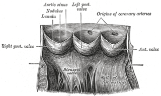
Aneurysm of the aortic sinus, also known as the sinus of Valsalva, is a rare abnormality of the aorta, the largest artery in the body. The aorta normally has three small pouches that sit directly above the aortic valve, and an aneurysm of one of these sinuses is a thin-walled swelling. Aneurysms may affect the right (65–85%), non-coronary (10–30%), or rarely the left coronary sinus. These aneurysms may not cause any symptoms but if large can cause shortness of breath, palpitations or blackouts. Aortic sinus aneurysms can burst or rupture into adjacent cardiac chambers, which can lead to heart failure if untreated.

Radionuclide angiography is an area of nuclear medicine which specialises in imaging to show the functionality of the right and left ventricles of the heart, thus allowing informed diagnostic intervention in heart failure. It involves use of a radiopharmaceutical, injected into a patient, and a gamma camera for acquisition. A MUGA scan involves an acquisition triggered (gated) at different points of the cardiac cycle. MUGA scanning is also called equilibrium radionuclide angiocardiography, radionuclide ventriculography (RNVG), or gated blood pool imaging, as well as SYMA scanning.

The American College of Cardiology (ACC), based in Washington, D.C., is a nonprofit medical association established in 1949. It bestows credentials upon cardiovascular specialists who meet its qualifications. Education is a core component of the college, which is also active in the formulation of health policy and the support of cardiovascular research.
Fate most commonly refers to destiny, a predetermined course of events.
A transthoracic echocardiogram (TTE) is the most common type of echocardiogram, which is a still or moving image of the internal parts of the heart using ultrasound. In this case, the probe is placed on the chest or abdomen of the subject to get various views of the heart. It is used as a non-invasive assessment of the overall health of the heart, including a patient's heart valves and degree of heart muscle contraction. The images are displayed on a monitor for real-time viewing and then recorded.

The valve of the inferior vena cava is a venous valve that lies at the junction of the inferior vena cava and right atrium.

Noncompaction cardiomyopathy (NCC) is a rare congenital disease of heart muscle that affects both children and adults. It results from abnormal prenatal development of heart muscle.
Fetal echocardiography, or Fetal echocardiogram, is the name of the test used to diagnose cardiac conditions in the fetal stage. Cardiac defects are amongst the most common birth defects. Their diagnosis is important in the fetal stage as it might help provide an opportunity to plan and manage the baby as and when the baby is born. Not all pregnancies need to undergo fetal echo.
Cardiothoracic anesthesiology is a subspeciality of the medical practice of anesthesiology, devoted to the preoperative, intraoperative, and postoperative care of adult and pediatric patients undergoing cardiothoracic surgery and related invasive procedures.
Howard Apfel is an American-Israeli Rabbi and Cardiologist practicing medicine at Columbia University Medical Center.

Cardiac imaging refers to non-invasive imaging of the heart using ultrasound, magnetic resonance imaging (MRI), computed tomography (CT), or nuclear medicine (NM) imaging with PET or SPECT. These cardiac techniques are otherwise referred to as echocardiography, Cardiac MRI, Cardiac CT, Cardiac PET and Cardiac SPECT including myocardial perfusion imaging.
Tissue Doppler echocardiography (TDE) is a medical ultrasound technology, specifically a form of echocardiography that measures the velocity of the heart muscle (myocardium) through the phases of one or more heartbeats by the Doppler effect of the reflected ultrasound. The technique is the same as for flow Doppler echocardiography measuring flow velocities. Tissue signals, however, have higher amplitude and lower velocities, and the signals are extracted by using different filter and gain settings. The terms tissue Doppler imaging (TDI) and tissue velocity imaging (TVI) are usually synonymous with TDE because echocardiography is the main use of tissue Doppler.
A quadricuspid aortic valve (QAV) is a rare congenital heart defect characterized by the presence of four cusps, instead of the usual three found normally in the aortic valve. It is a defect that occurs during embryological development of the aortic trunk during gestation. There is an increased risk of developing post-natal aortic regurgitations and other heart-related diseases; therefore patients with the condition should be carefully monitored.
Govindan Vijayaraghavan is a cardiologist from India, credited with establishing the first 2D Echocardiography laboratory in India. He is the vice-chairman & Founder Director of the Kerala Institute of Medical Sciences and the President of the Society for Continuing Medical Education and Research, Trivandrum, Kerala. He was honoured by Government of India in 2009 for his services in the field of medical sciences by awarding him Padmashri.
Numerical manipulation of Doppler parameters obtain during routine Echocardiography has been extensively utilized to non-invasively estimate intra-cardiac pressures, in many cases removing the need for invasive cardiac catheterization.
The Relative Atrial Index (RAI) is a numeric parameter used to assess for cardiac shunt defects. It is calculated from the standard transthoracic Doppler echocardiogram measurements of the right atrial area divided by the left atrial area. RAI = right atrial area / left atrial area. These measurements are made from the apical four chamber view.
Harvey Feigenbaum is an American cardiologist known for his life-long work in the field of echocardiography. He wrote the first textbook on the subject in 1972, which is currently in its 8th edition, and has published over 300 articles. He has trained generations of cardiologists including many of the world's pioneers in the field through his numerous visitors, frequent workshops, annual courses in Indianapolis, Indiana beginning in 1968, the year when he started formal fellowship training He founded the field of cardiac sonography in 1965 and the American Society of Echocardiography in 1975. His seminal article on the diagnosis of pericardial effusions published in 1965 with his technique "brought echocardiography to the attention of thousands of practitioners".


