Left ventricular thrombus

Last updated
Left ventricular thrombus
AC08-07 Ventricular thrombus.JPG
Blood clots in the ventricle found on autopsy

Left ventricular thrombus is a blood clot (thrombus) in the left ventricle of the heart. LVT is a common complication of acute myocardial infarction (AMI). [1] [2] Typically the clot is a mural thrombus, meaning it is on the wall of the ventricle. [3] The primary risk of LVT is the occurrence of cardiac embolism, [1] [4] in which the thrombus detaches from the ventricular wall and travels through the circulation and blocks blood vessels. Blockage can be especially damaging in the heart or brain (stroke). [1] [5]

Contents

Pathophysiology

LVT occurs most often during the first 2 weeks following AMI. [1] AMI patients most at risk display the 3 characteristics of Virchow's triad: [1]

Stagnation of blood

The risk of LVT formation increases as infarction size increases. [5] A larger infarction means a larger area of tissue injury, which may be akinetic or dyskinetic, resulting in stagnation of ventricular blood. [1]

Endothelial injury

Monocytes and macrophages play important roles in healing after myocardial infarction. With the absence of monocytes and macrophages, chances of LVT formation are very high. Failure to clear cellular debris from the infarct compromises the endothelial lining of the left ventricle and exposes the damaged tissue to the blood. [6] The response is to build a thrombus composed of fibrin, red blood cells and platelets. [1]

Hypercoagulable state

For several days after AMI, the levels of tissue factor and D-dimer, which are involved in coagulation, are high, which increases the risk of LVT formation. [7] LVT may be good for the heart when tissues are severely damaged because it acts to thicken the wall, thus protecting it against rupture. [1]

Diagnosis

Echocardiography is the main diagnostic tool for LVT. A distinct mass is visible in the left ventricle. Computed Tomography and Magnetic Resonance Imaging are effective, but less common ways to detect LVT, due to their costs and risks. [1] It is possible to assess whether a thrombus will become an embolus through echocardiography. Mobility and protrusion of the thrombus are two characteristics associated with increased embolic potential. [8]

Prevention

After an AMI, people should be treated to prevent LVT formation. Aspirin plus an oral anticoagulant such as warfarin are suggested for individuals at risk for thromboembolic events. [10] Anticoagulants are also shown to reduce the risk of embolisms [1] [4] when a thrombus is already formed. Heparin, an injectable, fast-acting anticoagulant, is effective in high doses for preventing LVT formation after AMI. [1] [11]

Treatment

Systemic anticoagulation is considered first-line medical therapy for LVT, as it reduces the risk of systemic embolism. [12] [10] There are also surgical procedures for removal of a thrombus (thrombectomy).

Epidemiology

The rate of LVT formation after AMI is thought to be declining [13] [14] due to the use of better therapies and percutaneous coronary intervention used to treat myocardial infarction. [1] [5] [7] [15] In the modern era LVT formation after ST elevation MI treated with percutaneous coronary intervention is low, estimated at only 2.7%. [16] However, incidence of LVT is considered higher in anterior wall AMI, compared with other types. [17]

Related Research Articles

<span class="mw-page-title-main">Embolism</span> Disease of arteries, arterioles and capillaries

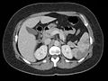
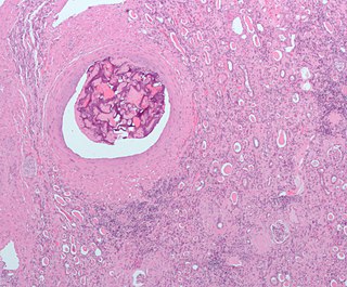
An embolism is the lodging of an embolus, a blockage-causing piece of material, inside a blood vessel. The embolus may be a blood clot (thrombus), a fat globule, a bubble of air or other gas, amniotic fluid, or foreign material. An embolism can cause partial or total blockage of blood flow in the affected vessel. Such a blockage may affect a part of the body distant from the origin of the embolus. An embolism in which the embolus is a piece of thrombus is called a thromboembolism.

<span class="mw-page-title-main">Chest pain</span> Discomfort or pain in the chest as a medical symptom

Chest pain is pain or discomfort in the chest, typically the front of the chest. It may be described as sharp, dull, pressure, heaviness or squeezing. Associated symptoms may include pain in the shoulder, arm, upper abdomen, or jaw, along with nausea, sweating, or shortness of breath. It can be divided into heart-related and non-heart-related pain. Pain due to insufficient blood flow to the heart is also called angina pectoris. Those with diabetes or the elderly may have less clear symptoms.

<span class="mw-page-title-main">Atrial septal defect</span> Human heart defect present at birth

Atrial septal defect (ASD) is a congenital heart defect in which blood flows between the atria of the heart. Some flow is a normal condition both pre-birth and immediately post-birth via the foramen ovale; however, when this does not naturally close after birth it is referred to as a patent (open) foramen ovale (PFO). It is common in patients with a congenital atrial septal aneurysm (ASA).

<span class="mw-page-title-main">Coronary thrombosis</span> Medical condition

Coronary thrombosis is defined as the formation of a blood clot inside a blood vessel of the heart. This blood clot may then restrict blood flow within the heart, leading to heart tissue damage, or a myocardial infarction, also known as a heart attack.

Dressler syndrome is a secondary form of pericarditis that occurs in the setting of injury to the heart or the pericardium. It consists of fever, pleuritic pain, pericarditis and/or pericardial effusion.

<span class="mw-page-title-main">Acute coronary syndrome</span> Medical condition

Acute coronary syndrome (ACS) is a syndrome due to decreased blood flow in the coronary arteries such that part of the heart muscle is unable to function properly or dies. The most common symptom is centrally located pressure-like chest pain, often radiating to the left shoulder or angle of the jaw, and associated with nausea and sweating. Many people with acute coronary syndromes present with symptoms other than chest pain, particularly women, older people, and people with diabetes mellitus.

An embolus, is described as a free-floating mass, located inside blood vessels that can travel from one site in the blood stream to another. An embolus can be made up of solid, liquid, or gas. Once these masses get "stuck" in a different blood vessel, it is then known as an "embolism." An embolism can cause ischemia—damage to an organ from lack of oxygen. A paradoxical embolism is a specific type of embolism in which the embolus travels from the right side of the heart to the left side of the heart and lodges itself in a blood vessel known as an artery. Thus, it is termed "paradoxical" because the embolus lands in an artery, rather than a vein.

The intra-aortic balloon pump (IABP) is a mechanical device that increases myocardial oxygen perfusion and indirectly increases cardiac output through afterload reduction. It consists of a cylindrical polyurethane balloon that sits in the aorta, approximately 2 centimeters (0.79 in) from the left subclavian artery. The balloon inflates and deflates via counter pulsation, meaning it actively deflates in systole and inflates in diastole. Systolic deflation decreases afterload through a vacuum effect and indirectly increases forward flow from the heart. Diastolic inflation increases blood flow to the coronary arteries via retrograde flow. These actions combine to decrease myocardial oxygen demand and increase myocardial oxygen supply.

<span class="mw-page-title-main">Bivalirudin</span>

Bivalirudin (Bivalitroban), sold under the brand names Angiomax and Angiox and manufactured by The Medicines Company, is a direct thrombin inhibitor (DTI).

<span class="mw-page-title-main">Takotsubo cardiomyopathy</span> Sudden temporary weakening of the heart muscle

Takotsubo cardiomyopathy or takotsubo syndrome (TTS), also known as stress cardiomyopathy, is a type of non-ischemic cardiomyopathy in which there is a sudden temporary weakening of the muscular portion of the heart. It usually appears after a significant stressor, either physical or emotional; when caused by the latter, the condition is sometimes called broken heart syndrome. Examples of physical stressors that can cause TTS are sepsis, shock, subarachnoid hemorrhage, and pheochromocytoma. Emotional stressors include bereavement, divorce, or the loss of a job. Reviews suggest that of patients diagnosed with the condition, about 70–80% recently experienced a major stressor, including 41–50% with a physical stressor and 26–30% with an emotional stressor. TTS can also appear in patients who have not experienced major stressors.

<span class="mw-page-title-main">Myocardial rupture</span> Medical condition

Myocardial rupture is a laceration of the ventricles or atria of the heart, of the interatrial or interventricular septum, or of the papillary muscles. It is most commonly seen as a serious sequela of an acute myocardial infarction.

<span class="mw-page-title-main">Myocardial infarction</span> Interruption of blood supply to a part of the heart

A myocardial infarction (MI), commonly known as a heart attack, occurs when blood flow decreases or stops in the coronary artery of the heart, causing damage to the heart muscle. The most common symptom is chest pain or discomfort which may travel into the shoulder, arm, back, neck or jaw. Often it occurs in the center or left side of the chest and lasts for more than a few minutes. The discomfort may occasionally feel like heartburn. Other symptoms may include shortness of breath, nausea, feeling faint, a cold sweat or feeling tired. About 30% of people have atypical symptoms. Women more often present without chest pain and instead have neck pain, arm pain or feel tired. Among those over 75 years old, about 5% have had an MI with little or no history of symptoms. An MI may cause heart failure, an irregular heartbeat, cardiogenic shock or cardiac arrest.

<span class="mw-page-title-main">ST depression</span> Depression of the ST segment on an electrocardiogram

ST depression refers to a finding on an electrocardiogram, wherein the trace in the ST segment is abnormally low below the baseline.

<span class="mw-page-title-main">Primary ventricular fibrillation</span> Heart condition

Primary ventricular fibrillation (PVF) is an unpredictable and potentially fatal arrhythmia occurring during the acute phase of a myocardial infarction leading to immediate collapse and, if left untreated, leads to sudden cardiac death within minutes. In developed countries, PVF is a leading cause of death. Worldwide, the annual number of deaths caused by PVF is comparable to the number of deaths caused by road traffic accidents. A substantial portion of these deaths could be avoided by seeking immediate medical attention when symptoms are noticed.

<span class="mw-page-title-main">Reperfusion therapy</span>

Reperfusion therapy is a medical treatment to restore blood flow, either through or around, blocked arteries, typically after a heart attack. Reperfusion therapy includes drugs and surgery. The drugs are thrombolytics and fibrinolytics used in a process called thrombolysis. Surgeries performed may be minimally-invasive endovascular procedures such as a percutaneous coronary intervention (PCI), which involves coronary angioplasty. The angioplasty uses the insertion of a balloon and/or stents to open up the artery. Other surgeries performed are the more invasive bypass surgeries that graft arteries around blockages.

<span class="mw-page-title-main">Electrocardiography in myocardial infarction</span>

Electrocardiography in suspected myocardial infarction has the main purpose of detecting ischemia or acute coronary injury in emergency department populations coming for symptoms of myocardial infarction (MI). Also, it can distinguish clinically different types of myocardial infarction.

Myocardial infarction complications may occur immediately following a heart attack, or may need time to develop. After an infarction, an obvious complication is a second infarction, which may occur in the domain of another atherosclerotic coronary artery, or in the same zone if there are any live cells left in the infarct.

<span class="mw-page-title-main">Management of acute coronary syndrome</span>

Management of acute coronary syndrome is targeted against the effects of reduced blood flow to the affected area of the heart muscle, usually because of a blood clot in one of the coronary arteries, the vessels that supply oxygenated blood to the myocardium. This is achieved with urgent hospitalization and medical therapy, including drugs that relieve chest pain and reduce the size of the infarct, and drugs that inhibit clot formation; for a subset of patients invasive measures are also employed. Basic principles of management are the same for all types of acute coronary syndrome. However, some important aspects of treatment depend on the presence or absence of elevation of the ST segment on the electrocardiogram, which classifies cases upon presentation to either ST segment elevation myocardial infarction (STEMI) or non-ST elevation acute coronary syndrome (NST-ACS); the latter includes unstable angina and non-ST elevation myocardial infarction (NSTEMI). Treatment is generally more aggressive for STEMI patients, and reperfusion therapy is more often reserved for them. Long-term therapy is necessary for prevention of recurrent events and complications.

Impella is a family of medical devices used for temporary ventricular support in patients with depressed heart function. Some versions of the device can provide left heart support during other forms of mechanical circulatory support including ECMO and Centrimag.

The volume of the heart's left atrium is an important biomarker for cardiovascular physiology and clinical cardiology. It is usually calculated as left atrial volume index in terms of body surface area.

References

  1. 1 2 3 4 5 6 7 8 9 10 11 12 Delewi R.; Zijlstra F.; Piek J.J (2012). "Left Ventricular Thrombus Formation after Acute Myocardial Infarction". Heart. 98 (23): 1743–1749. doi:10.1136/heartjnl-2012-301962. PMC   3505867 . PMID   23151669.
  2. Visser C. A.; Kan G.; David G. K.; Lie K. I.; Durrer D. (1983). "Two dimensional echocardiography in the diagnosis of left ventricular thrombus. A prospective study of 67 patients with anatomic validation". Chest. 83 (2): 228–232. doi:10.1378/chest.83.2.228. PMID   6822107.
  3. Reeder G. S.; Tajik A. J.; Seward J. B. (1981). "Left ventricular mural thrombus: Two-dimensional echocardiographic diagnosis". Mayo Clinic Proceedings. 56 (2): 82–86. PMID   7464235.
  4. 1 2 Vaitkus PT, Barnathan ES (1994). "Embolic potential, prevention and management of mural thrombus complicating anterior myocardial infarction: a meta-analysis". J Am Coll Cardiol. 22 (4): 1004–9. doi: 10.1016/0735-1097(93)90409-t . PMID   8409034.
  5. 1 2 3 Solheim S.; Seljeflot I.; Lunde K.; Bjørnerheim R.; Aakhus S.; Forfang K.; Arnesen H. (2010). "Frequency of left ventricular thrombus in patients with anterior wall acute myocardial infarction treated with percutaneous coronary intervention and dual antiplatelet therapy". The American Journal of Cardiology. 106 (9): 1197–1200. doi:10.1016/j.amjcard.2010.06.043. PMID   21029812.
  6. Frantz, S., Hofmann, U., Fraccarollo, D., Schäfer, A., Kranepuhl, S., Hagedorn, I., Nieswandt, B., Nahrendorf, M., Wagner, H., Bayer, B., Pachel, C., Schon, M., Kneitz, S., Bobinger, T., Weidemann, F., Ertl, G., Bauersachs, J. (2012). Monocytes/ macrophages prevent healing defects and left ventricular thrombus formation after myocardial infarction. The FASEB Journal.
  7. 1 2 Solheim S.; Seljeflot I.; Lunde K.; Bratseth V.; Aakhus S.; Forfang K.; Arnesen H. (2013). "Prothrombotic markers in patients with acute myocardial infarction and left ventricular thrombus formation treated with pci and dual antiplatelet therapy". Thrombosis Journal. 11 (1): 1–6. doi:10.1186/1477-9560-11-1. PMC   3554510 . PMID   23311309.
  8. Haugland JM, Asinger RW, Mikell FL, Elsperger J, Hodges M (1984). "Embolic potential of left ventricular thrombi detected by two-dimensional echocardiography". Circulation. 70 (4): 588–598. doi: 10.1161/01.cir.70.4.588 . PMID   6478564.
  9. 1 2 3 4 "UOTW #24 - Ultrasound of the Week". Ultrasound of the Week. 6 November 2014. Retrieved 27 May 2017.
  10. 1 2 Van de Werf F., Staff E. S. C. (2008). "ESC Guidelines on the Management of Acute Myocardial Infarction in Patients Presenting with STEMI". European Heart Journal. 29 (23): 2909–2945. doi: 10.1093/eurheartj/ehn416 . PMID   19004841.
  11. Turpie A. G.; Robinson J. G.; Doyle D. J.; Mulji A. S.; Mishkel G. J.; Sealey B. J.; Cairns J.A.; Skingley L.; Hirsh J.; Gent M. (1989). "Comparison of high-dose with low-dose subcutaneous heparin to prevent left ventricular mural thrombosis in patients with acute transmural anterior myocardial infarction". The New England Journal of Medicine. 320 (6): 352–357. doi:10.1056/nejm198902093200604. PMID   2643772.
  12. Vaitkus, P. T.; Barnathan, E. S. (October 1993). "Embolic potential, prevention and management of mural thrombus complicating anterior myocardial infarction: a meta-analysis". Journal of the American College of Cardiology. 22 (4): 1004–1009. doi: 10.1016/0735-1097(93)90409-t . ISSN   0735-1097. PMID   8409034.
  13. Asinger R. W.; Mikell F. L.; Elsperger J.; Hodges M. (1981). "Incidence of left- ventricular thrombosis after acute transmural myocardial infarction. Serial evaluation by two-dimensional echocardiography". The New England Journal of Medicine. 305 (6): 297–302. doi:10.1056/nejm198108063050601. PMID   7242633.
  14. Stokman P. J.; Nandra C. S.; Asinger R. W. (2001). "Left ventricular thrombus". Current Treatment Options in Cardiovascular Medicine. 3 (6): 515–521. doi:10.1007/s11936-001-0025-6. PMID   11696271. S2CID   3734965.
  15. Osherov A. B.; Borovik-Raz M.; Aronson D.; Agmon Y.; Kapeliovich M.; Kerner A.; Grenadier E.; Hammerman H.; Nikolsky E.; Roguin A. (2009). "Incidence of early left ventricular thrombus after acute anterior wall myocardial infarction in the primary coronary intervention era". American Heart Journal. 157 (6): 1074–1080. doi:10.1016/j.ahj.2009.03.020. PMID   19464419.
  16. Robinson, Austin A.; Jain, Amit; Gentry, Mark; McNamara, Robert L. (2016-10-15). "Left ventricular thrombi after STEMI in the primary PCI era: A systematic review and meta-analysis". International Journal of Cardiology. 221: 554–559. doi:10.1016/j.ijcard.2016.07.069. ISSN   1874-1754. PMID   27424314.
  17. Zielinska M.; Kaczmarek K.; Tylkowski M. (2008). "Predictors of left ventricular thrombus formation in acute myocardial infarction treated with successful primary angioplasty with stenting". Am J Med Sci. 335 (3): 171–176. doi:10.1097/maj.0b013e318142be20. PMID   18344689. S2CID   23786142.