Keratoacanthoma

Last updated
Keratoacanthoma
Keratoacanthoma 2.jpg
Keratoacanthoma
Specialty Dermatology, plastic surgery
Types
  • Giant keratoacanthomas
  • Subungual keratoacanthoma
  • Multiple keratoacanthomas (Ferguson–Smith syndrome)
  • Keratoacanthoma centrifugum marginatum
  • Generalized eruptive keratoacanthoma of Grzybowski
Risk factors Ultraviolet radiation, immunosuppression, genetics
Diagnostic method Tissue biopsy
Differential diagnosis Squamous cell skin cancer
TreatmentSurgery (excision, Mohs surgery)

Keratoacanthoma (KA) is a common low-grade (unlikely to metastasize or invade) rapidly-growing skin tumour that is believed to originate from the hair follicle (pilosebaceous unit) and can resemble squamous cell carcinoma. [1] [2]

Contents

The defining characteristic of a keratoacanthoma is that it is dome-shaped, symmetrical, surrounded by a smooth wall of inflamed skin, and capped with keratin scales and debris. It grows rapidly, reaching a large size within days or weeks, and if untreated for months will almost always starve itself of nourishment, necrose (die), slough, and heal with scarring. Keratoacanthoma is commonly found on sun-exposed skin, often face, forearms and hands. [2] [3] It is rarely found at a mucocutaneous junction or on mucous membranes. [2]

Keratoacanthoma may be difficult to distinguish visually from a skin cancer. [4] Under the microscope, keratoacanthoma very closely resembles squamous cell carcinoma. In order to differentiate between the two, almost the entire structure needs to be removed and examined. While some pathologists classify keratoacanthoma as a distinct entity and not a malignancy, about 6% of clinical and histological keratoacanthomas do progress to invasive and aggressive squamous cell cancers; some pathologists may label KA as "well-differentiated squamous cell carcinoma, keratoacanthoma variant", and prompt definitive surgery may be recommended. [5] [6] [7] [8]

Classification

A person with generalized eruptive keratoacanthomas Generalized eruptive keratoacanthomas of Grzybowski.jpg
A person with generalized eruptive keratoacanthomas

Frequently reported and reclassified over the last century, keratoacanthoma can be divided into various subtypes and despite being considered benign, their unpredictable behaviour has warranted the same attention as with squamous cell carcinoma. [1]

Keratoacanthomas may be divided into the following types: [9] :763–764 [10] :643–646

Cause

Keratoacanthomas usually occurs in older individuals. A number of causes have been suggested including ultraviolet light, chemical carcinogens, recent injury to the skin, immunosuppression and genetic predisposition. [1] As with squamous cell cancer, sporadic cases have been found co-infected with the human papilloma virus (HPV). [4] [12] Although HPV has been suggested as a causal factor, it is unproven. [2]

Many new treatments for melanoma are also known to increase the rate of keratoacanthoma, such as the BRAF inhibitor medications vemurafenib and dabrafenib. [13]

Diagnosis

Microscopic view of a skin keratoacanthoma Skin keratoacanthoma whole slide.jpg
Microscopic view of a skin keratoacanthoma

Keratoacanthomas presents as a fleshy, elevated and nodular lesion with an irregular crater shape and a characteristic central hyperkeratotic core. Usually the people will notice a rapidly growing dome-shaped tumor on sun-exposed skin. [14]

If the entire lesion is removed, the pathologist will probably be able to differentiate between keratoacanthoma and squamous cell carcinoma. Follow-up would be required to monitor for recurrence of disease. [15]

Treatment

Excision of the entire lesion, with adequate margin, will remove the lesion, allow full tissue diagnosis, and leave a planned surgical wound which can usually be repaired with a good cosmetic result. However, removing the entire lesion (especially on the face) may present difficult problems of plastic reconstruction. (On the nose and face, Mohs surgery may allow for good margin control with minimal tissue removal, but many insurance companies require the definitive diagnosis of a malignancy before they are prepared to pay the extra costs of Mohs surgery.) Especially in more cosmetically-sensitive areas, and where the clinical diagnosis is reasonably certain, alternatives to surgery may include no treatment (awaiting spontaneous resolution). [14]

On the trunk, arms, and legs, electrodesiccation and curettage often suffice to control keratoacanthomas until they regress. Other modalities of treatment include cryosurgery and radiotherapy; intralesional injection of methotrexate or 5-fluorouracil have also been used. [14]

Recurrence after electrodesiccation and curettage can occur; it can usually be identified and treated promptly with either further curettage or surgical excision. [6]

History

In 1889, Sir Jonathan Hutchinson described a crateriform ulcer on the face”. [16] In 1936, the same condition was renamed "molluscum sebaceum" by MacCormac and Scarf. [17] Later, the term “keratoacanthoma” was coined by Walter Freudenthal. [18] [19] and the term became established by Arthur Rook and the pathologist Ian Whimster in 1950. [16]

See also

Related Research Articles

Squamous cell skin cancer

Squamous-cell skin cancer, also known as cutaneous squamous-cell carcinoma (cSCC), is one of the main types of skin cancer along with basal cell cancer, and melanoma. It usually presents as a hard lump with a scaly top but can also form an ulcer. Onset is often over months. Squamous-cell skin cancer is more likely to spread to distant areas than basal cell cancer. When confined to the outermost layer of the skin, a precancerous or in situ form of cSCC is known as Bowen's disease.

Basal-cell carcinoma Most common type of skin cancer

Basal-cell carcinoma (BCC), also known as basal-cell cancer, is the most common type of skin cancer. It often appears as a painless raised area of skin, which may be shiny with small blood vessels running over it. It may also present as a raised area with ulceration. Basal-cell cancer grows slowly and can damage the tissue around it, but it is unlikely to spread to distant areas or result in death.

Head and neck cancer Cancer arises in the head or neck region

Head and neck cancer is a group of cancers that starts in the mouth, nose, throat, larynx, sinuses, or salivary glands. Symptoms for head and neck cancer may include a lump or sore that does not heal, a sore throat that does not go away, trouble swallowing, or a change in the voice. There may also be unusual bleeding, facial swelling, or trouble breathing.

Lichen planus human chronic inflammatory disease

Lichen planus (LP) is a chronic inflammatory and immune-mediated disease that affects the skin, nails, hair, and mucous membranes. It is not an actual lichen, and is only named that because it looks like one. It is characterized by polygonal, flat-topped, violaceous papules and plaques with overlying, reticulated, fine white scale, commonly affecting dorsal hands, flexural wrists and forearms, trunk, anterior lower legs and oral mucosa. Although there is a broad clinical range of LP manifestations, the skin and oral cavity remain as the major sites of involvement. The cause is unknown, but it is thought to be the result of an autoimmune process with an unknown initial trigger. There is no cure, but many different medications and procedures have been used in efforts to control the symptoms.

Seborrheic keratosis

A seborrheic keratosis is a non-cancerous (benign) skin tumour that originates from cells in the outer layer of the skin. Like liver spots, seborrheic keratoses are seen more often as people age.

Epidermoid cyst Benign cyst usually found on the skin

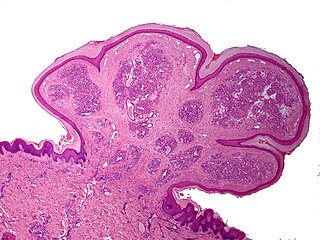
An epidermoid cyst or epidermal inclusion cyst is a benign cyst usually found on the skin. The cyst develops out of ectodermal tissue. Histologically, it is made of a thin layer of squamous epithelium.

Actinic keratosis

Actinic keratosis (AK), sometimes called solar keratosis or senile keratosis, is a pre-cancerous area of thick, scaly, or crusty skin. Actinic keratosis is a disorder (-osis) of epidermal keratinocytes that is induced by ultraviolet (UV) light exposure (actin-). These growths are more common in fair-skinned people and those who are frequently in the sun. They are believed to form when skin gets damaged by UV radiation from the sun or indoor tanning beds, usually over the course of decades. Given their pre-cancerous nature, if left untreated, they may turn into a type of skin cancer called squamous cell carcinoma. Untreated lesions have up to a 20% risk of progression to squamous cell carcinoma, so treatment by a dermatologist is recommended.

Syringoma

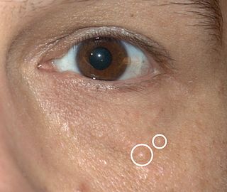
Syringomas are benign eccrine sweat duct tumors, typically found clustered on eyelids, although they may also be found in the armpits, abdomen, chest, neck, scalp or groin area including genitals in a symmetric pattern. They are skin-colored or yellowish firm, rounded bumps, 1–3 mm in diameter, and may be confused with xanthoma, milia, hidrocystoma, trichoepithelioma, and xanthelasma. They are more common in women and are most commonly found in middle-aged Asian women. While they can present at any time in life, they typically present during adolescence. They are usually not associated with any other symptoms although can sometimes cause itchiness or irritation.

Pyogenic granuloma A vascular tumor on both mucosa and skin

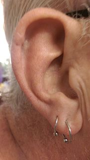
Pyogenic granuloma or pyogenic fibroma is a vascular tumor that occurs on both mucosa and skin, and appears as an overgrowth of tissue due to irritation, physical trauma, or hormonal factors. It is often found to involve the gums, the skin and nasal septum, and has also been found far from the head such as in the thigh.

Mohs surgery Microscopically controlled surgery used to treat common types of skin cancer

Mohs surgery, developed in 1938 by a general surgeon, Frederic E. Mohs, is microscopically controlled surgery used to treat common types of skin cancer. During the surgery, after each removal of tissue and while the patient waits, the tissue is examined for cancer cells. That examination dictates the decision for additional tissue removal. Mohs surgery is the gold standard method for obtaining complete margin control during removal of a skin cancer using frozen section histology. CCPDMA or Mohs surgery allows for the removal of a skin cancer with very narrow surgical margin and a high cure rate.

Verrucous carcinoma

Verrucous carcinoma (VC) is an uncommon variant of squamous cell carcinoma. This form of cancer is often seen in those who chew tobacco or use snuff orally, so much so that it is sometimes referred to as "Snuff dipper's cancer".

Vulvar cancer Cancer involving the vulva

Vulvar cancer is a cancer of the vulva, the outer portion of the female genitals. It most commonly affects the outer vaginal lips. Less often, the inner vaginal lips, clitoris, or vaginal glands. Symptoms include a lump, itchiness, changes in the skin, or bleeding from the vulva.

Skin biopsy

Skin biopsy is a biopsy technique in which a skin lesion is removed to be sent to a pathologist to render a microscopic diagnosis. It is usually done under local anesthetic in a physician's office, and results are often available in 4 to 10 days. It is commonly performed by dermatologists. Skin biopsies are also done by family physicians, internists, surgeons, and other specialties. However, performed incorrectly, and without appropriate clinical information, a pathologist's interpretation of a skin biopsy can be severely limited, and therefore doctors and patients may forgo traditional biopsy techniques and instead choose Mohs surgery. There are four main types of skin biopsies: shave biopsy, punch biopsy, excisional biopsy, and incisional biopsy. The choice of the different skin biopsies is dependent on the suspected diagnosis of the skin lesion. Like most biopsies, patient consent and anesthesia are prerequisites.

Discoid lupus erythematosus Autoimmune skin condition

Discoid lupus erythematosus is the most common type of chronic cutaneous lupus (CCLE), an autoimmune skin condition on the lupus erythematosus spectrum of illnesses. It presents with red, inflamed, coin-shaped patches of skin with a scaling and crusty appearance, most often on the scalp, cheeks, and ears. Hair loss may occur if the lesions are on the scalp. The lesions can then develop severe scarring, and the centre areas may appear lighter in color with a rim darker than the normal skin. These lesions can last for years without treatment.

Cutaneous horn

Cutaneous horns, also known by the Latin name cornu cutaneum, are unusual keratinous skin tumors with the appearance of horns, or sometimes of wood or coral. Formally, this is a clinical diagnosis for a "conical projection above the surface of the skin." They are usually small and localized but can, in very rare cases, be much larger. Although often benign, they can also be malignant or premalignant.

Sebaceous carcinoma

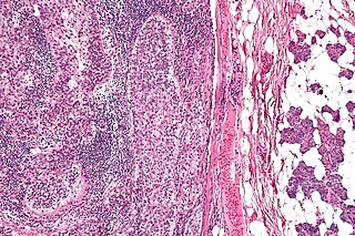
Sebaceous carcinoma, also known as sebaceous gland carcinoma (SGc), sebaceous cell carcinoma, and meibomian gland carcinoma is an uncommon malignant cutaneous tumor. Most are typically about 1.4 cm at presentation. SGc originates from sebaceous glands in the skin and, therefore, may originate anywhere in the body where these glands are found. SGc can be divided into 2 types: periocular and extraocular. The periocular region is rich in sebaceous glands making it a common site of origin. The cause of these lesions in the vast majority of cases is unknown. Occasional cases may be associated with Muir-Torre syndrome. SGc accounts for approximately 0.7% of all skin cancers, and the incidence of SGc is highest in Caucasian, Asian, and Indian populations. Due to the rarity of this tumor and variability in clinical and histological presentation, SGc is often misdiagnosed as an inflammatory condition or a more common neoplasm. SGc is commonly treated with wide local excision or Mohs micrographic surgery, and the relative survival rates at 5 and 10 years are 92.72 and 86.98%, respectively.

Electrodesiccation and curettage is a medical procedure commonly performed by dermatologists, surgeons and general practitioners for the treatment of basal cell cancers and squamous cell cancers of the skin. It provides desiccation, coagulation/cauterization, and curettage to remove lesions from the skin.

Arthur Rook (dermatologist)

Arthur James Rook FRCP was a leading British dermatologist and the principal author of Rook's Textbook of Dermatology (1968), known as "Rook's", which reached its ninth edition in 2016.

References

  1. 1 2 3 Zito, Patrick M.; Scharf, Richard (2018), "Keratoacanthoma", StatPearls, StatPearls Publishing, PMID   29763106 , retrieved 17 September 2018
  2. 1 2 3 4 Joseph A. Regezi; James Sciubba; Richard C. K. Jordan (2012). "6. Neoplasms". Oral Pathology - E-Book: Clinical Pathologic Correlations. Elsevier Saunders. p. 155. ISBN   978-1-4557-0262-6.
  3. Schwartz RA. The Keratoacanthoma: A Review. J Surg Oncol 1979; 12:305–17.
  4. 1 2 "Keratoacanthoma | DermNet New Zealand". www.dermnetnz.org. Retrieved 17 September 2018.
  5. Ko CJ, Keratoacanthoma: facts and controversies. Clin Dermatol. 2010; 28(3):254–61 ( ISSN   1879-1131)
  6. 1 2 "Keratoacanthoma: Background, Pathophysiology, Etiology". Medscape. 14 August 2018.(subscription required)
  7. Kossard S; Tan KB; Choy C; Keratoacanthoma and infundibulocystic squamous cell carcinoma. Am J Dermatopathol. 2008; 30(2):127–34 ( ISSN   1533-0311)
  8. Weedon DD, et al. Squamous cell carcinoma arising in keratoacanthoma: a neglected phenomenon in the elderly. Am J Dermatopathol. 2010; 32(5):423–6
  9. 1 2 3 4 5 6 Freedberg, et al. (2003). Fitzpatrick's Dermatology in General Medicine. (6th ed.). McGraw-Hill. ISBN   0-07-138076-0.
  10. 1 2 3 4 5 James, William; Berger, Timothy; Elston, Dirk (2005). Andrews' Diseases of the Skin: Clinical Dermatology. (10th ed.). Saunders. ISBN   0-7216-2921-0.
  11. "Grzybowski generalized eruptive keratoacanthomas | DermNet New Zealand". www.dermnetnz.org. Retrieved 17 September 2018.
  12. Niebuhr M, et al. Giant keratoacanthoma in an immunocompetent patient with detection of HPV 11. Hautarzt. 2009; 60(3):229–32 ( ISSN   1432-1173)
  13. Niezgoda, A (2015). "Novel Approaches to Treatment of Advanced Melanoma: A Review on Targeted Therapy and Immunotherapy". Biomed Res Int. 2015: 851387. doi:10.1155/2015/851387. PMC   4478296 . PMID   26171394.
  14. 1 2 3 Keratoacanthoma. Désirée Ratner. 2004. http://www.medscape.com/viewarticle/467069 accessed 23 June 2015
  15. Baran, Robert; Berker, David A. R. de; Holzberg, Mark; Thomas, Luc (2012). Baran and Dawber's Diseases of the Nails and their Management. John Wiley & Sons. ISBN   9780470657355.
  16. 1 2 Cerroni, Lorenzo; Kerl, Helmut (2012), Goldsmith, Lowell A.; Katz, Stephen I.; Gilchrest, Barbara A.; Paller, Amy S. (eds.), "Chapter 117. Keratoacanthoma", Fitzpatrick's Dermatology in General Medicine (8 ed.), The McGraw-Hill Companies, retrieved 2018-08-20
  17. Levy, Edwin J. (1954-06-05). "Keratoacanthoma". Journal of the American Medical Association. 155 (6): 562–4. doi:10.1001/jama.1954.03690240028008. ISSN   0002-9955. PMID   13162754.
  18. HJORTH, NIELS (August 1960). "Keratoacanthoma: A Historical Note". British Journal of Dermatology. 72 (8–9): 292–295. doi:10.1111/j.1365-2133.1960.tb13896.x. ISSN   0007-0963. S2CID   71452344.
  19. ROOK, ARTHUR; WHIMSTER, IAN (January 1979). "Keratoacanthoma–a thirty year retrospect". British Journal of Dermatology. 100 (1): 41–47. doi:10.1111/j.1365-2133.1979.tb03568.x. ISSN   0007-0963. PMID   427012. S2CID   27373097.
Classification
D
External resources