Related Research Articles

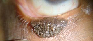
Nevus is a nonspecific medical term for a visible, circumscribed, chronic lesion of the skin or mucosa. The term originates from nævus, which is Latin for "birthmark"; however, a nevus can be either congenital or acquired. Common terms, including mole, birthmark, and beauty mark, are used to describe nevi, but these terms do not distinguish specific types of nevi from one another.

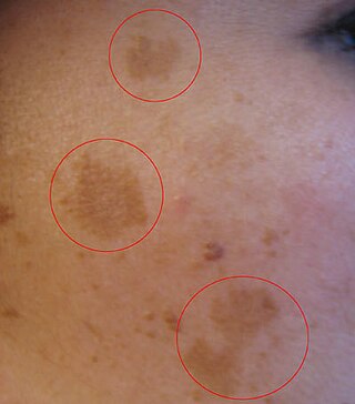
Melasma is a tan or dark skin discoloration. Melasma is thought to be caused by sun exposure, genetic predisposition, hormone changes, and skin irritation. Although it can affect anyone, it is particularly common in women, especially pregnant women and those who are taking oral or patch contraceptives or hormone replacement therapy medications.

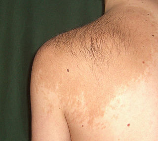
Becker's nevus is a benign skin disorder predominantly affecting males. The nevus can be present at birth, but more often shows up around puberty. It generally first appears as an irregular pigmentation on the torso or upper arm, and gradually enlarges irregularly, becoming thickened and often hairy (hypertrichosis). The nevus is due to an overgrowth of the epidermis, pigment cells (melanocytes), and hair follicles. This form of nevus was first documented in 1948 by American dermatologist Samuel William Becker (1894–1964).

Blaschko's lines, also called the lines of Blaschko, are lines of normal cell development in the skin. These lines are only visible in those with a mosaic skin condition or in chimeras where different cell lines contain different genes. These lines may express different amounts of melanin, or become visible due to a differing susceptibility to disease. In such individuals, they can become apparent as whorls, patches, streaks or lines in a linear or segmental distribution over the skin. They follow a V shape over the back, S-shaped whirls over the chest and sides, and wavy shapes on the head. Not all mosaic skin conditions follow Blaschko's lines.

Nevus of Ota is a hyperpigmentation that occurs on the face, most often appearing on the white of the eye. It also occurs on the forehead, nose, cheek, periorbital region, and temple.
Phakomatosis pigmentovascularis is a rare neurocutanous condition where there is coexistence of a capillary malformation with various melanocytic lesions, including dermal melanocytosis, nevus spilus, and nevus of Ota.

Superficial lymphatic malformation is a congenital vascular anomaly of the superficial lymphatics, presenting as groups of deep-seated, vesicle-like papules resembling frog spawn, at birth or shortly thereafter. Lymphangioma circumscriptum is the most common congenital lymphatic malformation. It is a benign condition, and treatment is not required if the person who has it does not experience symptoms from the condition.

Nevus sebaceus or sebaceous nevus is a congenital, hairless plaque that typically occurs on the face or scalp. Such nevi are classified as epidermal nevi and can be present at birth, or early childhood, and affect males and females of all races equally. The condition is named for an overgrowth of sebaceous glands, a relatively uncommon hamartoma, in the area of the nevus. NSJ is first described by Josef Jadassohn in 1895.

Linear verrucous epidermal nevus is a skin lesion characterized by a verrucous skin-colored, dirty-gray or brown papule. Generally, multiple papules present simultaneously, and coalesce to form a serpiginous plaque. When this nevus covers a diffuse or extensive portion of the body's surface area, it may be referred to as a systematized epidermal nevus, when it involved only one-half of the body it is called a nevus unius lateris.

Epidermal nevus syndrome, also known as Feuerstein and Mims syndrome, and Solomon's syndrome is a rare disease that was first described in 1968 and consists of extensive epidermal nevi with abnormalities of the central nervous system (CNS), skeleton, skin, cardiovascular system, genitourinary system and eyes. However, since the syndrome's first description, a broader concept for the "epidermal nevus" syndrome has been proposed, with at least six types being described:

Schimmelpenning syndrome is a neurocutaneous condition characterized by one or more sebaceous nevi, usually appearing on the face or scalp, associated with anomalies of the central nervous system, ocular system, skeletal system, cardiovascular system and genitourinary system.
Nevus comedonicus syndrome is a skin condition characterized by a nevus comedonicus associated with cataracts, scoliosis, and neurologic abnormalities.
Pigmented hairy epidermal nevus syndrome, also known as Becker's naevus syndrome, is a cutaneous condition characterized by a Becker nevus, ipsilateral hypoplasia of the breast, and skeletal defects such as scoliosis.
Phakomatosis pigmentokeratotica is a rare neurocutanous condition characterized by the combination of an organoid sebaceous nevus and speckled lentiginous nevus. It is an unusual variant of epidermal naevus syndrome. It was first described by Happle et al. It is often associated with neurological or skeletal anomalies such as hemiatrophy, dysaesthesia and hyperhidrosis in a segmental pattern, mild mental retardation, seizures, deafness, ptosis and strabismus.

Inflammatory Linear Verrucous Epidermal Nevus is a rare disease of the skin that presents as multiple, discrete, red papules that tend to coalesce into linear plaques that follow the Lines of Blaschko. The plaques can be slightly warty (psoriaform) or scaly (eczema-like). ILVEN is caused by somatic mutations that result in genetic mosaicism. There is no cure, but different medical treatments can alleviate the symptoms.
Nevus unius lateris is a cutaneous condition, an epidermal nevus in which the skin lesions are distributed on one-half of the body.

Acantholytic dyskeratotic epidermal nevus is a cutaneous condition identical to the generalized form of Darier's disease. "Acantholytic dyskeratotic epidermal nevus" is probably the same disorder.

Supernumerary nipples–uropathies–Becker's nevus syndrome is a skin condition that may be associated with genitourinary tract abnormalities. Supernumerary nipples, also referred to as polythelia or accessory nipples, is a pigmented lesion of the skin that is present at birth. This pigmentation usually occurs along the milk lines, which are the precursors to breast and nipple development. Clinically, this congenital condition is generally considered benign, but some studies have suggested there may be an association with kidney diseases and cancers of the urogenital system.
Porokeratotic eccrine ostial and dermal duct nevus (PEODDN) is a skin lesion that resembles a comedonal nevus, but it occurs on the palms and soles where pilosebaceous follicles are normally absent. It is probably transmitted by paradominant transmission.
References
- ↑ Freedberg, et al. (2003). Fitzpatrick's Dermatology in General Medicine. (6th ed.). McGraw-Hill. ISBN 0-07-138076-0.