Maxillary lateral incisor agenesis

Last updated
Schematic of maxillary lateral incisors in the human mouth for both permanent and primary teeth. Maxillary lateral incisors01-01-06.png
Schematic of maxillary lateral incisors in the human mouth for both permanent and primary teeth.

Maxillary lateral incisor agenesis (MLIA) is lack of development (agenesis) of one or both of the maxillary lateral incisor teeth. In normal human dentition, this would be the second tooth on either side from the center of the top row of teeth. The condition is bilateral if the incisor is absent on both sides or unilateral if only one is missing. It appears to have a genetic component.

Contents

History

Dental anthropology has been a research topic of great interest for investigating tooth development evolution. The variations in number, size, and morphology of teeth among populations have been able to provide insights for genetic basis of odontogenesis. [1] The origins of teeth is believed to have come from dermal structures called "odontodes," which became associated with bones. [1] Phylogenetic changes in teeth has been associated with functional adaptation. [1] The reduction of teeth number has been connected with the decrease size of the jaw in human expansion. [1] Monkeys, apes, great apes, and homo sapiens were studied and it showed that homo sapiens have acquired a shorter maxillo-mandibular skeleton when compared to their ancestors. [1]

The first set of maxillary lateral incisors (primary teeth) develop between the 14th and the 16th week, while being inside the uterus. [2] By the age of 8 or 9, the permanent maxillary lateral incisors erupt as the root continues to mineralize until the age of 11 years old. [2]

Causes

Genetics

The reason for tooth agenesis has not been completely understood, despite the identification of several mutations in MSX1 and PAX9 genes which are related to tooth agenesis and mutations in AXIN2 gene which cause oligodontia. [3] MSX1 and PAX9 are necessary transcription factors that help to form normal teeth. [3] Studies done in mice, have shown that MSX1 and PAX9 encode transcription factors with different DNA binding sequence patterns, a paired domain and homeodomain, which are co-expressed during tooth development. [4]

MSX1

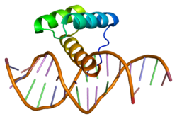
Structure of the MSX1 protein. Based on PyMOL rendering of PDB 1ig7. Protein MSX1 PDB 1ig7.png
Structure of the MSX1 protein. Based on PyMOL rendering of PDB 1ig7.

MSX1 consist of homeobox encoding sequence of 60 amino acid long DNA binding homeodomain. [5] The protein of MSX1 serves as a repressor for gene transcription and interchanges with other homeoproteins. [5] In studies of homozygous mice, it has been found that deletion of the MSX1 gene has resulted in a double cleft palate, deficiency of the alveolar bone, failure of incisor and molar development. [5]

PAX9

PAX9 also plays a key role during embryogenesis. [5] In addition, encoded proteins by PAX9 genes have a 128 amino acid long DNA binding paired domain. [5] Studies suggest that all heterozygous mutations of PAX9 associated with lack of development were due to the loss of function in one of the protein copies, which resulted in haploinsufficiency. [5] Furthermore, mutations of other genes have been identified in syndromes and congenital abnormalities in which tooth agenesis is a regular characteristic. [6]

Endogamy

There have been studies that entail that a possible reason for maxillary lateral incisor agenesis was due to endogamy. A high incidence of the rare anatomical variant of MLIA was found in the population of Basta, southern Jordan. [7] This was believed to be because of close genetic relationships. [7] In anthropology, this would be considered a breed from closely related people or animals. [7]

Applications

Because MLIA can be detected from partial skeletal remains, it is useful in the field of anthropology. Anthropologically-interesting human remains often have relatively well preserved skeletons, but no soft tissues or intact DNA. This makes it hard to determine relationships between the deceased individuals. MLIA is sometimes related to inbreeding, so the presence of MLIA in many members of a large collection of remains can indicate that the population that lived there was relatively inbred. This technique has been used to study a group of Neolithic farmers. [8]

Non-metric morphological traits, also known as epigenetic or anatomical variants, are a great tool for detecting individuals who are genetically related in historic populations if DNA is not well preserved. [7] The method used is non-metric cranial and dental traits, which has already been tested on several burial sites. [7] Identifying individuals who are genetically related from past populations is very important in order to determine if the families share a number of characteristics and phenotypical traits. [7] Traits that are used for kinship analysis must be determine by genetic factors, must be rare in the general population, and the individual traits must be genetically independent from each other. [7] The traits that are closely examined include: anatomical variants of tooth crowns and roots; shape, size, number, structure, and position of the teeth. [7]

See also

Related Research Articles

<span class="mw-page-title-main">Human tooth</span> Calcified whitish structure in humans mouths used to break down food

Human teeth function to mechanically break down items of food by cutting and crushing them in preparation for swallowing and digesting. As such, they are considered part of the human digestive system. Humans have four types of teeth: incisors, canines, premolars, and molars, which each have a specific function. The incisors cut the food, the canines tear the food and the molars and premolars crush the food. The roots of teeth are embedded in the maxilla or the mandible and are covered by gums. Teeth are made of multiple tissues of varying density and hardness.

<span class="mw-page-title-main">Incisor</span> Front teeth of most mammals

Incisors are the front teeth present in most mammals. They are located in the premaxilla above and on the mandible below. Humans have a total of eight. Opossums have 18, whereas armadillos have none.

<span class="mw-page-title-main">Orthodontics</span> Correctional branch of dentistry

Orthodontics is a dentistry specialty that addresses the diagnosis, prevention, management, and correction of mal-positioned teeth and jaws, as well as misaligned bite patterns. It may also address the modification of facial growth, known as dentofacial orthopedics.

<span class="mw-page-title-main">Wisdom tooth</span> Large tooth at the back of the human mouth

The third molar, commonly called wisdom tooth, is the most posterior of the three molars in each quadrant of the human dentition. The age at which wisdom teeth come through (erupt) is variable, but this generally occurs between late teens and early twenties. Most adults have four wisdom teeth, one in each of the four quadrants, but it is possible to have none, fewer, or more, in which case the extras are called supernumerary teeth. Wisdom teeth may become stuck (impacted) against other teeth if there is not enough space for them to come through normally. Impacted wisdom teeth are still sometimes removed for orthodontic treatment, believing that they move the other teeth and cause crowding, though this is not held anymore as true.

<span class="mw-page-title-main">Hyperdontia</span> Condition of having extra teeth beyond the regular number of teeth

Hyperdontia is the condition of having supernumerary teeth, or teeth that appear in addition to the regular number of teeth. They can appear in any area of the dental arch and can affect any dental organ. The opposite of hyperdontia is hypodontia, where there is a congenital lack of teeth, which is a condition seen more commonly than hyperdontia. The scientific definition of hyperdontia is "any tooth or odontogenic structure that is formed from tooth germ in excess of usual number for any given region of the dental arch." The additional teeth, which may be few or many, can occur on any place in the dental arch. Their arrangement may be symmetrical or non-symmetrical.

Hypodontia is defined as the developmental absence of one or more teeth excluding the third molars. It is one of the most common dental anomalies, and can have a negative impact on function, and also appearance. It rarely occurs in primary teeth and the most commonly affected are the adult second premolars and the upper lateral incisors. It usually occurs as part of a syndrome that involves other abnormalities and requires multidisciplinary treatment.

<span class="mw-page-title-main">Human tooth development</span> Process by which teeth form

Tooth development or odontogenesis is the complex process by which teeth form from embryonic cells, grow, and erupt into the mouth. For human teeth to have a healthy oral environment, all parts of the tooth must develop during appropriate stages of fetal development. Primary (baby) teeth start to form between the sixth and eighth week of prenatal development, and permanent teeth begin to form in the twentieth week. If teeth do not start to develop at or near these times, they will not develop at all, resulting in hypodontia or anodontia.

<span class="mw-page-title-main">Malocclusion</span> Medical condition

In orthodontics, a malocclusion is a misalignment or incorrect relation between the teeth of the upper and lower dental arches when they approach each other as the jaws close. The English-language term dates from 1864; Edward Angle (1855-1930), the "father of modern orthodontics", popularised it. The word "malocclusion" derives from occlusion, and refers to the manner in which opposing teeth meet.

<span class="mw-page-title-main">Maxillary central incisor</span> Tooth

The maxillary central incisor is a human tooth in the front upper jaw, or maxilla, and is usually the most visible of all teeth in the mouth. It is located mesial to the maxillary lateral incisor. As with all incisors, their function is for shearing or cutting food during mastication (chewing). There is typically a single cusp on each tooth, called an incisal ridge or incisal edge. Formation of these teeth begins at 14 weeks in utero for the deciduous (baby) set and 3–4 months of age for the permanent set.

Anodontia is a rare genetic disorder characterized by the congenital absence of all primary or permanent teeth. It is divided into two subsections, complete absence of teeth or only some absence of teeth. It is associated with the group of skin and nerve syndromes called the ectodermal dysplasias. Anodontia is usually part of a syndrome and seldom occurs as an isolated entity. There is usually no exact cause for anodontia. The defect results in the dental lamina obstruction during embryogenesis due to local, systemic and genetic factors.

Macrodontia is a type of localized gigantism in which teeth are larger than normal. Macrodontia seen in permanent teeth is thought to affect around 0.03 to 1.9 percent of the worldwide population. Generally, patients with macrodontia have one or two teeth in their mouth that is abnormally large; however, single tooth growth is seen in a number of cases as well.

<span class="mw-page-title-main">Shovel-shaped incisors</span> Possible shape of human incisor teeth

Shovel-shaped incisors are incisors whose lingual surfaces are scooped as a consequence of lingual marginal ridges, crown curvature, or basal tubercles, either alone or in combination.

<span class="mw-page-title-main">Talon cusp</span> Rare dental anomaly resulting in teeth having more than one cusp

Talon cusp is a rare dental anomaly resulting in an extra cusp or cusp-like projection on an anterior tooth, located on the inside surface of the affected tooth. Sometimes it can also be found on the facial surface of the anterior tooth.

Dental anatomy is a field of anatomy dedicated to the study of human tooth structures. The development, appearance, and classification of teeth fall within its purview. Tooth formation begins before birth, and the teeth's eventual morphology is dictated during this time. Dental anatomy is also a taxonomical science: it is concerned with the naming of teeth and the structures of which they are made, this information serving a practical purpose in dental treatment.

<span class="mw-page-title-main">Overjet</span> Medical condition

Overjet is the extent of horizontal (anterior-posterior) overlap of the maxillary central incisors over the mandibular central incisors. In class II malocclusion the overjet is increased as the maxillary central incisors are protruded.

<span class="mw-page-title-main">Homeobox protein MSX-1</span> Protein-coding gene in the species Homo sapiens

Homeobox protein MSX-1, is a protein that in humans is encoded by the MSX1 gene. MSX1 transcripts are not only found in thyrotrope-derived TSH cells, but also in the TtT97 thyrotropic tumor, which is a well differentiated hyperplastic tissue that produces both TSHß- and a-subunits and is responsive to thyroid hormone. MSX1 is also expressed in highly differentiated pituitary cells which until recently was thought to be expressed exclusively during embryogenesis. There is a highly conserved structural organization of the members of the MSX family of genes and their abundant expression at sites of inductive cell–cell interactions in the embryo suggest that they have a pivotal role during early development.

<span class="mw-page-title-main">Crossbite</span> Medical condition

Crossbite is a form of malocclusion where a tooth has a more buccal or lingual position than its corresponding antagonist tooth in the upper or lower dental arch. In other words, crossbite is a lateral misalignment of the dental arches.

<span class="mw-page-title-main">PAX9</span> Protein-coding gene in humans

Paired box gene 9, also known as PAX9, is a protein which in humans is encoded by the PAX9 gene. It is also found in other mammals.

An ectopic maxillary canine is a canine which is following abnormal path of eruption in the maxilla. An impacted tooth is one which is blocked from erupting by a physical barrier in the path of eruption. Ectopic eruption may lead to impaction. Previously, it was assumed that 85% of ectopic canines are displaced palatally, however a recent study suggests the true occurrence is closer to 50%. While maxillary canines can also be displaced buccally, it is thought this arises as a result of a lack of space. Most of these cases resolve themselves with the permanent canine erupting without intervention.

Orthodontic indices are one of the tools that are available for orthodontists to grade and assess malocclusion. Orthodontic indices can be useful for an epidemiologist to analyse prevalence and severity of malocclusion in any population.

References

  1. 1 2 3 4 5 Vastardis H (June 2000). "The genetics of human tooth agenesis: new discoveries for understanding dental anomalies". American Journal of Orthodontics and Dentofacial Orthopedics. 117 (6): 650–6. doi:10.1016/S0889-5406(00)70173-9. PMID   10842107. S2CID   11886845.
  2. 1 2 Nelson SJ, Ash MM, Wheeler RC (2015). Wheeler's dental anatomy, physiology, and occlusion (Tenth ed.). St. Louis, Missouri. ISBN   978-0-323-26323-8. OCLC   879604219.{{cite book}}: CS1 maint: location missing publisher (link)
  3. 1 2 Kondo S, Townsend G, Matsuno M (2014-11-01). "Morphological variation of the maxillary lateral incisor". Japanese Dental Science Review. 50 (4): 100–107. doi: 10.1016/j.jdsr.2014.06.002 . hdl: 2440/95379 .
  4. Nakatomi M, Wang XP, Key D, Lund JJ, Turbe-Doan A, Kist R, et al. (April 2010). "Genetic interactions between Pax9 and Msx1 regulate lip development and several stages of tooth morphogenesis". Developmental Biology. Special Section: Gene Regulatory Networks for Development. 340 (2): 438–49. doi: 10.1016/j.ydbio.2010.01.031 . PMID   20123092.
  5. 1 2 3 4 5 6 Mostowska A, Kobielak A, Trzeciak WH (October 2003). "Molecular basis of non-syndromic tooth agenesis: mutations of MSX1 and PAX9 reflect their role in patterning human dentition". European Journal of Oral Sciences. 111 (5): 365–70. doi:10.1034/j.1600-0722.2003.00069.x. PMID   12974677.
  6. Garib DG, Alencar BM, Lauris JR, Baccetti T (June 2010). "Agenesis of maxillary lateral incisors and associated dental anomalies". American Journal of Orthodontics and Dentofacial Orthopedics. 137 (6): 732.e1-6, discussion 732–3. doi:10.1016/j.ajodo.2009.12.024. PMID   20685523.
  7. 1 2 3 4 5 6 7 8 Alt KW, Benz M, Müller W, Berner ME, Schultz M, Schmidt-Schultz TH, et al. (2013-06-11). "Earliest evidence for social endogamy in the 9,000-year-old-population of Basta, Jordan". PLOS ONE. 8 (6): e65649. Bibcode:2013PLoSO...865649A. doi: 10.1371/journal.pone.0065649 . PMC   3679157 . PMID   23776517.
  8. Balter M (19 June 2013). "First Farmers Were Also Inbred". Archived from the original on 2013-06-22. Retrieved 20 June 2013.