Plasma cell gingivitis

Last updated
Plasma cell gingivitis
Plasma cell gingivitis in adult.jpg
Plasma cell gingivitis in an adult (histologically verified).

Plasma cell gingivitis [1] [2] is a rare condition, [3] appearing as generalized erythema (redness) and edema (swelling) of the attached gingiva, occasionally accompanied by cheilitis (lip swelling) or glossitis (tongue swelling). [4] It is called plasma cell gingivitis where the gingiva (gums) are involved, [5] plasma cell cheilitis, [5] where the lips are involved, and other terms such as plasma cell orifacial mucositis, [5] or plasma cell gingivostomatitis where several sites in the mouth are involved. On the lips, the condition appears as sharply outlined, infiltrated, dark red plaque with a lacquer-like glazing of the surface of the involved oral area. [5]

Contents

Signs and symptoms

Plasma cell gingivitis in 7-year-old child. Histologically verified. Plasma cell gingivitis on 7 yerars old child.jpg
Plasma cell gingivitis in 7-year-old child. Histologically verified.
Plasma cell gingivitis in a 10-year-old child. Histologically verified. Plasma cell gingivitis in a 10 year old child.jpg
Plasma cell gingivitis in a 10-year-old child. Histologically verified.

Plasma cell gingivitis appears as mild gingival enlargement and may extend from the free marginal gingiva on to the attached gingiva. [6] Sometimes it is blended with a marginal, plaque induced gingivitis, or it does not involve the free marginal gingiva. It may also be found as a solitude red area within the attached gingiva (pictures). In some cases the healing of a plaque-induced gingivitis or a periodontitis resolves a plasma cell gingivitis situated a few mm from the earlier plaque-infected marginal gingiva. In case of one or few solitary areas of plasma cell gingivitis, no symptoms are reported from the patient. Most often solitary entities are therefore found by the dentist. [2]

The gums are red, friable, or sometimes granular, and sometimes bleed easily if traumatised. [6] The normal stippling is lost. [7] There is not usually any loss of periodontal attachment. [6] In a few cases a sore mouth can develop, and if so pain is sometimes made worse by toothpastes, or hot or spicy food. [7] The lesions can extend to involve the palate. [7]

Plasma cell cheilitis appears as well defined, infiltrated, dark red plaque with a superficial lacquer-like glazing. [5] Plasma cell cheilitis usually involves the lower lip. [3] The lips appear dry, atrophic and fissured. [7] Angular cheilitis is sometimes present. [7]

Where the condition involves the tongue, there is an erythematous enlargement with furrows, crenation and loss of the normal dorsal tongue coating. [7]

Causes

Plasma cell gingivitis and plasma cell cheilitis are thought to be hypersensitivity reactions to some antigen. [3] [6] Possible sources of antigens include ingredients in toothpastes, chewing gum, mints, pepper, or foods. [7] [6] Specifically, cinnamonaldehyde and cinnamon flavoring are often to blame. [3] However, the exact cause in most is unknown. [3]

Diagnosis

Plasma cells in Plasma cell gingivitis. Light microscopy X450. Histology of plasma cell gingivitis.jpg
Plasma cells in Plasma cell gingivitis. Light microscopy X450.

Histologically plasma cell gingivitis shows mainly plasma cells. [2] The differential diagnosis is with acute leukemia and multiple myeloma. [4] Hence, blood tests are often involved in ruling out other conditions. [3] A biopsy is usually taken, and allergy testing may also be used. The histopathologic appearance is characterized by diffuse, sub-epithelial plasma cell inflammatory infiltration into the connective tissue. [3] The epithelium shows spongiosis. [6] Some consider that plasmoacanthoma (solitary plasma cell tumor) is part of the same spectrum of disease as plasma cell cheilitis. [5]

Classification

Depending upon the site of involvement, this condition could be considered a type of gingivitis (or gingival enlargement); a type of cheilitis; glossitis; or stomatitis. Sometimes the lips, the gums and the tongue can simultaneously be involved, and some authors have described this triad as a syndrome ("plasma-cell gingivostomatitis"). [3] The mucous membranes of the genitals can also be involved by a similar condition, termed "plasma cell balanitis" [2] or "plasma cell vulvitis". [8]

Other synonyms for this condition not previously mentioned include atypical gingivitis, allergic gingivitis, plasmacytosis of the gingiva, idiopathic gingivostomatitis, and atypical gingivostomatitis. [3] [7] Some of these terms are largely historical.

Plasma cell gingivitis has been subclassified into 3 types based upon the cause; namely, allergic, neoplastic and of unknown cause. [3]

Treatment

Preventing exposure to the causative antigen leads to resolution of the condition. [6] Tacrolimus or clobetasol propionate have also been used to treat plasma cell cheilitis. [5]

Epidemiology

Plasma cell gingivitis is rare, and plasma cell cheilitis is very rare. [3] Most people with plasma cell cheilitis have been elderly. [3]

History

Plasma cell gingivitis was first described in the late 1960-early 1970s. A wave of cases occurred during this period, thought to be caused by allergic reactions to a component in chewing gum. Since, the number of cases has decreased, but they are still occasionally reported. [7]

See also

Related Research Articles

<span class="mw-page-title-main">Gums</span> Mouth anatomy

The gums or gingiva consist of the mucosal tissue that lies over the mandible and maxilla inside the mouth. Gum health and disease can have an effect on general health.

<span class="mw-page-title-main">Stomatitis</span> Medical condition

Stomatitis is inflammation of the mouth and lips. It refers to any inflammatory process affecting the mucous membranes of the mouth and lips, with or without oral ulceration.

Periodontology or periodontics is the specialty of dentistry that studies supporting structures of teeth, as well as diseases and conditions that affect them. The supporting tissues are known as the periodontium, which includes the gingiva (gums), alveolar bone, cementum, and the periodontal ligament. A periodontist is a dentist that specializes in the prevention, diagnosis and treatment of periodontal disease and in the placement of dental implants.

<span class="mw-page-title-main">Glossitis</span> Medical condition

Glossitis can mean soreness of the tongue, or more usually inflammation with depapillation of the dorsal surface of the tongue, leaving a smooth and erythematous (reddened) surface,. In a wider sense, glossitis can mean inflammation of the tongue generally. Glossitis is often caused by nutritional deficiencies and may be painless or cause discomfort. Glossitis usually responds well to treatment if the cause is identified and corrected. Tongue soreness caused by glossitis is differentiated from burning mouth syndrome, where there is no identifiable change in the appearance of the tongue, and there are no identifiable causes.

<span class="mw-page-title-main">Angular cheilitis</span> Cheilitis characterized by inflammation of one or both of the corners of the mouth

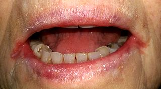
Angular cheilitis (AC) is inflammation of one or both corners of the mouth. Often the corners are red with skin breakdown and crusting. It can also be itchy or painful. The condition can last for days to years. Angular cheilitis is a type of cheilitis.

<span class="mw-page-title-main">Veterinary dentistry</span> Branch of veterinary medicine

Veterinary dentistry is the field of dentistry applied to the care of animals. It is the art and science of prevention, diagnosis, and treatment of conditions, diseases, and disorders of the oral cavity, the maxillofacial region, and its associated structures as it relates to animals.

<span class="mw-page-title-main">Gingival recession</span>

Gingival recession, also known as gum recession and receding gums, is the exposure in the roots of the teeth caused by a loss of gum tissue and/or retraction of the gingival margin from the crown of the teeth. Gum recession is a common problem in adults over the age of 40, but it may also occur starting in adolescence, or around the age of 10. It may exist with or without concomitant decrease in crown-to-root ratio.

<span class="mw-page-title-main">Cheilitis</span> Inflammation of the lips

Cheilitis is a medical condition characterized by inflammation of the lips. The inflammation may include the perioral skin, the vermilion border, or the labial mucosa. The skin and the vermilion border are more commonly involved, as the mucosa is less affected by inflammatory and allergic reactions.

Orofacial granulomatosis (OFG) is a condition characterized by persistent enlargement of the soft tissues of the mouth, lips and the area around the mouth on the face, causing in most cases extreme pain. The mechanism of the enlargement is granulomatous inflammation. The underlying cause of the condition is not completely understood, and there is disagreement as to how it relates to Crohn's disease and sarcoidosis.

Desquamative gingivitis is an erythematous (red), desquamatous (shedding) and ulcerated appearance of the gums. It is a descriptive term and can be caused by several different disorders.

<span class="mw-page-title-main">Scaling and root planing</span> Dental procedure

Scaling and root planing, also known as conventional periodontal therapy, non-surgical periodontal therapy or deep cleaning, is a procedure involving removal of dental plaque and calculus and then smoothing, or planing, of the (exposed) surfaces of the roots, removing cementum or dentine that is impregnated with calculus, toxins, or microorganisms, the agents that cause inflammation. It is a part of non-surgical periodontal therapy. This helps to establish a periodontium that is in remission of periodontal disease. Periodontal scalers and periodontal curettes are some of the tools involved.

<span class="mw-page-title-main">Gingival enlargement</span> Human disease

Gingival enlargement is an increase in the size of the gingiva (gums). It is a common feature of gingival disease. Gingival enlargement can be caused by a number of factors, including inflammatory conditions and the side effects of certain medications. The treatment is based on the cause. A closely related term is epulis, denoting a localized tumor on the gingiva.

Dental pertains to the teeth, including dentistry. Topics related to the dentistry, the human mouth and teeth include:

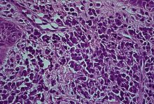
Oral and maxillofacial pathology refers to the diseases of the mouth, jaws and related structures such as salivary glands, temporomandibular joints, facial muscles and perioral skin. The mouth is an important organ with many different functions. It is also prone to a variety of medical and dental disorders.

<span class="mw-page-title-main">Gingivitis</span> Inflammation of the gums

Gingivitis is a non-destructive disease that causes inflammation of the gums; ulitis is an alternative term. The most common form of gingivitis, and the most common form of periodontal disease overall, is in response to bacterial biofilms that are attached to tooth surfaces, termed plaque-induced gingivitis. Most forms of gingivitis are plaque-induced.

Epulis is any tumor like enlargement situated on the gingival or alveolar mucosa. The word literally means "(growth) on the gingiva", and describes only the location of the mass and has no further implications on the nature of the lesion. There are three types: fibromatous, ossifying and acanthomatous. The related term parulis refers to a mass of inflamed granulation tissue at the opening of a draining sinus on the alveolus over the root of an infected tooth. Another closely related term is gingival enlargement, which tends to be used where the enlargement is more generalized over the whole gingiva rather than a localized mass.

<span class="mw-page-title-main">Periodontal abscess</span> Medical condition

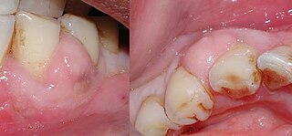
A periodontal abscess, is a localized collection of pus within the tissues of the periodontium. It is a type of dental abscess. A periodontal abscess occurs alongside a tooth, and is different from the more common periapical abscess, which represents the spread of infection from a dead tooth. To reflect this, sometimes the term "lateral (periodontal) abscess" is used. In contrast to a periapical abscess, periodontal abscesses are usually associated with a vital (living) tooth. Abscesses of the periodontium are acute bacterial infections classified primarily by location.

Gingivoplasty is the process by which the gingiva are reshaped to correct deformities. Gingivoplasty is similar to gingivectomy but with a different objective. This is a procedure performed to eliminate periodontal pockets along with the reshaping as part of the technique. This procedure is followed to create physiological gingival contours with the sole purpose of recontouring the gingiva in the absence of the pockets.

Hereditary gingival fibromatosis (HGF), also known as idiopathic gingival hyperplasia, is a rare condition of gingival overgrowth. HGF is characterized as a benign, slowly progressive, nonhemorrhagic, fibrous enlargement of keratinized gingiva. It can cover teeth in various degrees, and can lead to aesthetic disfigurement. Fibrous enlargement is most common in areas of maxillary and mandibular tissues of both arches in the mouth. Phenotype and genotype frequency of HGF is 1:175,000 where males and females are equally affected but the cause is not entirely known. It mainly exists as an isolated abnormality but can also be associated with a multi-system syndrome.

References

  1. Glauser RO, Humpreys PK, Stanley HR, Baer PN: An unusual gingivitis among adolescent navajo Indians. periodontics 1963;1:255-259.
  2. 1 2 3 4 Hedin, CA; Karpe, B; Larsson, A (1994). "Plasma-cell gingivitis in children and adults. A clinical and histological description". Swedish Dental Journal. 18 (4): 117–24. PMID   7825113.
  3. 1 2 3 4 5 6 7 8 9 10 11 12 Janam, P; Nayar, BR; Mohan, R; Suchitra, A (January 2012). "Plasma cell gingivitis associated with cheilitis: A diagnostic dilemma!". Journal of Indian Society of Periodontology . 16 (1): 115–9. doi: 10.4103/0972-124X.94618 . PMC   3357019 . PMID   22628976.
  4. 1 2 Greenberg MS, Glick M (2003). Burket's oral medicine diagnosis & treatment (10th ed.). Hamilton, Ont.: BC Decker. pp.  60. ISBN   978-1550091861.
  5. 1 2 3 4 5 6 7 James, William D.; Berger, Timothy G.; et al. (2006). Andrews' Diseases of the Skin: Clinical Dermatology . Saunders Elsevier. p.  797. ISBN   978-0-7216-2921-6.
  6. 1 2 3 4 5 6 7 Newman MG, Takei HH, Klokkevold PR, Carranza FA, eds. (2012). Carranza's clinical periodontology (11th ed.). St. Louis, Mo.: Elsevier/Saunders. pp.  91–92. ISBN   978-1-4377-0416-7.
  7. 1 2 3 4 5 6 7 8 9 Neville BW, Damm DD, Allen CA, Bouquot JE (2002). Oral & maxillofacial pathology (2nd ed.). Philadelphia: W.B. Saunders. pp.  141, 142. ISBN   978-0721690032.
  8. Hanami, Y; Motoki, Y; Yamamoto, T (Feb 15, 2011). "Successful treatment of plasma cell cheilitis with topical tacrolimus: report of two cases". Dermatology Online Journal. 17 (2): 6. doi:10.5070/D34RD1P1JS. PMID   21382289.