Pulp stone

Last updated
X-ray manual - U.S. Army (1917) (14734336166).jpg

Pulp stones (also denticles or endoliths) [1] are nodular, calcified masses appearing in either or both the coronal and root portion of the pulp organ in teeth. Pulp stones are not painful unless they impinge on nerves.

Contents

They are classified: [2]

A) On the basis of structure
1) True pulp stones: formed of dentin by odontoblasts
2) False pulp stones: formed by mineralization of degenerating pulp cells, often in a concentric pattern
B) On the basis of location
1) Free: entirely surrounded by pulp tissue
2) Adherent: partly fused with dentin
3) Embedded: entirely surrounded by dentin

Introduction

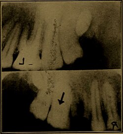
Pulp stones are discrete calcifications found in the pulp chamber of the tooth which may undergo changes to become diffuse pulp calcifications such as dystrophic calcification. [2] They are usually noticed by radiographic examination and appeared as round or ovoid radiopaque lesions. [3] Clinically, a tooth with a pulp stone has normal appearance like any other tooth. [4] The number of pulp stones in a single tooth may vary from 1 to 12 or more, with varying sizes from minute particles to large masses which tend to occlude the pulpal space. It is reported that pulp stones are more commonly found in the coronal region of pulp, albeit also found in the radicular pulp. [2]

It is believed that pulp stones develop around a central nidus of pulp tissue, for instance collagen fibril, ground substance and necrotic cell remnants. Initial calcification occurs around the central nidus and extends outward with regular calcified material in a concentric or radial manner. [5]

Etiology

The pulp calcifications can arise due to:

It is shown that pulp stone occurring in adolescents is significantly associated with carious and/or restored teeth, which suggests a causative relationship of chronic pulp irritation to pulp stone formation. A defence reaction in the pulpodentinal complex may be triggered by caries and microleakage around restorations which lead to pulp calcifications. The formation of pulp stone may have a similar mechanism as the tertiary dentine formation near the irritated odontoblasts. Apart from that, with aging, the pulp decreases in size due to the deposition of secondary or tertiary dentine. This subsequently results in favourable conditions for the formation of pulpal calcifications.

The other reported etiologic factors also include:

Types/classification

Pulp stones can be classified based on different location and structure. [2]

Based on location, they can classified into free, embedded and adherent pulp stones. Free pulp stones are found within the pulp tissue and is the most common encounter. The size vary from 50μm in diameter to several millimetres and may occlude the entire pulp chamber. Embedded pulp stone is fully embedded in dentine and most commonly found in the apical portion of the root. Adherent pulp stones are attached to the wall of pulp space but not fully enclosed by dentine.

Structurally, pulp stones can be classified as true and false pulp stones. True pulp stones are made up of dentine that is lined by odontoblast. True pulp stones are quite rare. On the other hand, false pulp stones are made up of concentric layers of mineralised tissue around blood thrombi, collagen fibres, or dying and dead cells.

Histopathology

Histologically, there are two types of stones: (1) stones with regular calcifications (2) stones with irregular calcifications.  For regular calcification, the pulp stones are smooth, round or ovoid with concentric laminations. It is commonly found in the coronal pulp. As for irregular calcifications without laminations, pulp stones may have the shape of rods or leaves and the surface is rough. It is more common in the radicular pulp. Pulp stones with regular calcification grow in size by addition of collagen fibrils to their surface, whereas the irregular type of pulp stones are formed by calcification of pre-existing collagen fibres.

Pulp stones may also form around epithelial cells such as remnants of Hertwig's epithelial root sheath. It is presumed that epithelial remnants are able to induce adjacent mesenchymal stem cells to differentiate into odontoblasts. [7]

Associations

A pilot study was done with patients with cardiovascular disease (CVD) and it shows increased incidence of pulp stones in teeth with patients with CVD compared to healthy patients without CVD. [8] There are also researchers which suggest the link between pulpal calcification and carotid artery calcification, despite not having a strong proof on this correlation. [5] Besides cardiovascular disease, other disease such as end stage renal disease, Marfan syndrome, Ehlers-Danlos syndrome, Calcinosis universalis, tumoral calcinosis are also discovered to be in association with pulpal calcifications. [9]

Several genetic diseases such as dentin dysplasia and dentinogenesis imperfecta are also accompanied by pulpal calcifications and hence, Marfan syndrome was suspected to be in association with pulp stones due to abnormal dentin formation, leading to the increased frequency of pulpal calcifications in these individuals. [9] Another theory suggests that individuals with Marfan syndrome have connective tissue dysplasia or vascular defects which in the case of tooth pulp, endothelial rupture of the pulp arterioles will lead to hemorrhagic areas in the pulp. It was proposed that these hemorrhagic areas in the pulp will induce mineralization within the pulp. [9]

Prevalence

Pulpal calcifications can be developed throughout the life and prevalence rates from 8–9% in worldwide population had been reported in studies. It was also found that pulpal stones occurred most frequently over the fourth decade, in advancing age. [10] [5]

Generally, pulp stones are more frequent to be found in maxillary teeth compared to mandibular teeth. [11] A study in Australia resulted higher occurrences of pulp stones in molars as opposed to premolars, and first molars as opposed to second molars. [6] First molars which were restored and/or with caries showed a higher incidence of pulp stones as compared to intact, unrestored first molars.

Clinical implications

Pulp stones generally do not have significant clinical implications as they are usually not a source of pain, discomfort or any form of pulpitis. [5] [12] However, when the tooth concerned will undergo endodontic treatment such as root canal treatment, presence of large pulp stones will be clinically significant. [12] [13]

Large pulp stones in the pulp chamber might block the access to canal orifices and prevent the exploring dental instruments from passaging down the canal. [12] In these cases, burs or even ultrasonic instrumentation can be used to remove the blocking pulp stones. [12] During the removal process, sodium hypochlorite which has dissolving action can also be used as a synergistic effect. [14]

Related Research Articles

<span class="mw-page-title-main">Human tooth</span> Calcified whitish structure in humans mouths used to break down food

Human teeth function to mechanically break down items of food by cutting and crushing them in preparation for swallowing and digesting. As such, they are considered part of the human digestive system. Humans have four types of teeth: incisors, canines, premolars, and molars, which each have a specific function. The incisors cut the food, the canines tear the food and the molars and premolars crush the food. The roots of teeth are embedded in the maxilla or the mandible and are covered by gums. Teeth are made of multiple tissues of varying density and hardness.

<span class="mw-page-title-main">Cementum</span> Specialized calcified substance covering the root of a tooth

Cementum is a specialized calcified substance covering the root of a tooth. The cementum is the part of the periodontium that attaches the teeth to the alveolar bone by anchoring the periodontal ligament.

<span class="mw-page-title-main">Tooth decay</span> Deformation of teeth due to acids produced by bacteria

Tooth decay, also known as cavities or caries, is the breakdown of teeth due to acids produced by bacteria. The cavities may be a number of different colors from yellow to black. Symptoms may include pain and difficulty with eating. Complications may include inflammation of the tissue around the tooth, tooth loss and infection or abscess formation.

<span class="mw-page-title-main">Root canal</span> Hollow part of the root of a tooth

A root canal is the naturally occurring anatomic space within the root of a tooth. It consists of the pulp chamber, the main canal(s), and more intricate anatomical branches that may connect the root canals to each other or to the surface of the root.

<span class="mw-page-title-main">Dentin</span> Calcified tissue of the body; one of the four major components of teeth

Dentin or dentine is a calcified tissue of the body and, along with enamel, cementum, and pulp, is one of the four major components of teeth. It is usually covered by enamel on the crown and cementum on the root and surrounds the entire pulp. By volume, 45% of dentin consists of the mineral hydroxyapatite, 33% is organic material, and 22% is water. Yellow in appearance, it greatly affects the color of a tooth due to the translucency of enamel. Dentin, which is less mineralized and less brittle than enamel, is necessary for the support of enamel. Dentin rates approximately 3 on the Mohs scale of mineral hardness. There are two main characteristics which distinguish dentin from enamel: firstly, dentin forms throughout life; secondly, dentin is sensitive and can become hypersensitive to changes in temperature due to the sensory function of odontoblasts, especially when enamel recedes and dentin channels become exposed.

<span class="mw-page-title-main">Toothache</span> Medical condition of the teeth

Toothache, also known as dental pain or tooth pain, is pain in the teeth or their supporting structures, caused by dental diseases or pain referred to the teeth by non-dental diseases. When severe it may impact sleep, eating, and other daily activities.

<span class="mw-page-title-main">Pulp (tooth)</span> Part in the center of a tooth made up of living connective tissue and cells called odontoblasts

The pulp is the connective tissue, nerves, blood vessels, and odontoblasts that comprise the innermost layer of a tooth. The pulp's activity and signalling processes regulate its behaviour.

<span class="mw-page-title-main">Enamel organ</span> Aggregate of cells involved in tooth development

The enamel organ, also known as the dental organ, is a cellular aggregation seen in a developing tooth and it lies above the dental papilla. The enamel organ which is differentiated from the primitive oral epithelium lining the stomodeum. The enamel organ is responsible for the formation of enamel, initiation of dentine formation, establishment of the shape of a tooth's crown, and establishment of the dentoenamel junction.

<span class="mw-page-title-main">Human tooth development</span> Process by which teeth form

Tooth development or odontogenesis is the complex process by which teeth form from embryonic cells, grow, and erupt into the mouth. For human teeth to have a healthy oral environment, all parts of the tooth must develop during appropriate stages of fetal development. Primary (baby) teeth start to form between the sixth and eighth week of prenatal development, and permanent teeth begin to form in the twentieth week. If teeth do not start to develop at or near these times, they will not develop at all, resulting in hypodontia or anodontia.

Dentinogenesis is the formation of dentin, a substance that forms the majority of teeth. Dentinogenesis is performed by odontoblasts, which are a special type of biological cell on the outer wall of dental pulps, and it begins at the late bell stage of a tooth development. The different stages of dentin formation after differentiation of the cell result in different types of dentin: mantle dentin, primary dentin, secondary dentin, and tertiary dentin.

Pulpitis is inflammation of dental pulp tissue. The pulp contains the blood vessels, the nerves, and connective tissue inside a tooth and provides the tooth's blood and nutrients. Pulpitis is mainly caused by bacterial infection which itself is a secondary development of caries. It manifests itself in the form of a toothache.

<span class="mw-page-title-main">Dentinogenesis imperfecta</span> Medical condition

Dentinogenesis imperfecta (DI) is a genetic disorder of tooth development. It is inherited in an autosomal dominant pattern, as a result of mutations on chromosome 4q21, in the dentine sialophosphoprotein gene (DSPP). It is one of the most frequently occurring autosomal dominant features in humans. Dentinogenesis imperfecta affects an estimated 1 in 6,000-8,000 people.

Dens invaginatus (DI), also known as tooth within a tooth, is a rare dental malformation where there is an infolding of enamel into dentine. The prevalence of condition is 0.3 - 10%, affecting more males than females. The condition is presented in two forms, coronal and radicular, with the coronal form being more common.

<span class="mw-page-title-main">Dentin dysplasia</span> Medical condition

Dentin dysplasia (DD) is a rare genetic developmental disorder affecting dentine production of the teeth, commonly exhibiting an autosomal dominant inheritance that causes malformation of the root. It affects both primary and permanent dentitions in approximately 1 in every 100,000 patients. It is characterized by the presence of normal enamel but atypical dentin with abnormal pulpal morphology. Witkop in 1972 classified DD into two types which are Type I (DD-1) is the radicular type, and type II (DD-2) is the coronal type. DD-1 has been further divided into 4 different subtypes (DD-1a,1b,1c,1d) based on the radiographic features.

<span class="mw-page-title-main">Pulpotomy</span>

Pulpotomy is a minimally invasive procedure performed in children on a primary tooth with extensive caries but without evidence of root pathology. The minimally invasive endodontic techniques of vital pulp therapy (VPT) are based on improved understanding of the capacity of pulp (nerve) tissues to heal and regenerate plus the availability of advanced endodontic materials. During the caries removal, this results in a carious or mechanical pulp exposure (bleeding) from the cavity. During pulpotomy, the inflamed/diseased pulp tissue is removed from the coronal pulp chamber of the tooth leaving healthy pulp tissue which is dressed with a long-term clinically successful medicament that maintains the survival of the pulp and promotes repair. There are various types of medicament placed above the vital pulp such as Buckley's Solution of formocresol, ferric sulfate, calcium hydroxide or mineral trioxide aggregate (MTA). MTA is a more recent material used for pulpotomies with a high rate of success, better than formocresol or ferric sulfate. It is also recommended to be the preferred pulpotomy agent in the future. After the coronal pulp chamber is filled, the tooth is restored with a filling material that seals the tooth from microleakage, such as a stainless steel crown which is the most effective long-term restoration. However, if there is sufficient remaining supporting tooth structure, other filling materials such as amalgam or composite resin can provide a functional alternative when the primary tooth has a life span of two years or less. The medium- to long-term treatment outcomes of pulpotomy in symptomatic permanent teeth with caries, especially in young people, indicate that pulpotomy can be a potential alternative to root canal therapy (RCT).

Pulp necrosis is a clinical diagnostic category indicating the death of cells and tissues in the pulp chamber of a tooth with or without bacterial invasion. It is often the result of many cases of dental trauma, caries and irreversible pulpitis.

In dentistry, the smear layer is a layer found on root canal walls after root canal instrumentation. It consists of microcrystalline and organic particle debris. It was first described in 1975 and research has been performed since then to evaluate its importance in bacteria penetration into the dentinal tubules and its effects on endodontic treatment. More broadly, it is the organic layer found over all hard tooth surfaces.

<span class="mw-page-title-main">Regenerative endodontics</span> Dental specialty

Regenerative endodontic procedures is defined as biologically based procedures designed to replace damaged structures such as dentin, root structures, and cells of the pulp-dentin complex. This new treatment modality aims to promote normal function of the pulp. It has become an alternative to heal apical periodontitis. Regenerative endodontics is the extension of root canal therapy. Conventional root canal therapy cleans and fills the pulp chamber with biologically inert material after destruction of the pulp due to dental caries, congenital deformity or trauma. Regenerative endodontics instead seeks to replace live tissue in the pulp chamber. The ultimate goal of regenerative endodontic procedures is to regenerate the tissues and the normal function of the dentin-pulp complex.

<span class="mw-page-title-main">Pulp capping</span>

Pulp capping is a technique used in dental restorations to prevent the dental pulp from necrosis, after being exposed, or nearly exposed during a cavity preparation, from a traumatic injury, or by a deep cavity that reaches the center of the tooth causing the pulp to die. When dental caries is removed from a tooth, all or most of the infected and softened enamel and dentin are removed. This can lead to the pulp of the tooth either being exposed or nearly exposed which causes pulpitis (inflammation). Pulpitis, in turn, can become irreversible, leading to pain and pulp necrosis, and necessitating either root canal treatment or extraction. The ultimate goal of pulp capping or stepwise caries removal is to protect a healthy dental pulp and avoid the need for root canal therapy.

Silver diammine fluoride (SDF), also known as silver diamine fluoride in most of the dental literature, is a topical medication used to treat and prevent dental caries and relieve dentinal hypersensitivity. It is a colorless or blue-tinted, odourless liquid composed of silver, ammonium and fluoride ions at a pH of 10.4 or 13. Ammonia compounds reduce the oxidative potential of SDF, increase its stability and helps to maintain a constant concentration over a period of time, rendering it safe for use in the mouth. Silver and fluoride ions possess antimicrobial properties and are used in the remineralization of enamel and dentin on teeth for preventing and arresting dental caries.

References

  1. Mosby's Medical Dictionary (9th ed.). Elsevier Health Sciences. 2013. p. 507. ISBN   978-0323112581 . Retrieved 10 February 2016.
  2. 1 2 3 4 Goga R, Chandler NP, Oginni AO (June 2008). "Pulp stones: a review" (PDF). International Endodontic Journal. 41 (6): 457–68. doi:10.1111/j.1365-2591.2008.01374.x. PMID   18422587.
  3. Feng XJ, Luo X, Li R, Dong W, Qi MC (August 2015). "[Multiple pulp stones: report of a case and literature review]". Shanghai Kou Qiang Yi Xue = Shanghai Journal of Stomatology. 24 (4): 511–2. PMID   26383583.
  4. 1 2 Berès F, Isaac J, Mouton L, Rouzière S, Berdal A, Simon S, Dessombz A (March 2016). "Comparative Physicochemical Analysis of Pulp Stone and Dentin" (PDF). Journal of Endodontics. 42 (3): 432–8. doi:10.1016/j.joen.2015.11.007. PMID   26794341.
  5. 1 2 3 4 Neville BW, Damm DD, Allen CM, Chi AC (2015-05-13). Oral and maxillofacial pathology (Fourth ed.). St. Louis, MO. ISBN   9781455770526. OCLC   908336985.{{cite book}}: CS1 maint: location missing publisher (link)
  6. 1 2 Kannan S, Kannepady SK, Muthu K, Jeevan MB, Thapasum A (March 2015). "Radiographic assessment of the prevalence of pulp stones in Malaysians". Journal of Endodontics. 41 (3): 333–7. doi:10.1016/j.joen.2014.10.015. PMID   25476972.
  7. Cohen's Pathway of Pulp.
  8. Edds AC, Walden JE, Scheetz JP, Goldsmith LJ, Drisko CL, Eleazer PD (July 2005). "Pilot study of correlation of pulp stones with cardiovascular disease". Journal of Endodontics. 31 (7): 504–6. doi:10.1097/01.don.0000168890.42903.2b. PMID   15980708.
  9. 1 2 3 Bauss O, Neter D, Rahman A (December 2008). "Prevalence of pulp calcifications in patients with Marfan syndrome". Oral Surgery, Oral Medicine, Oral Pathology, Oral Radiology, and Endodontics. 106 (6): e56–61. doi:10.1016/j.tripleo.2008.06.029. PMID   18805711.
  10. Udoye C, Sede M (January 2011). "Prevalence and analysis of factors related to ooccurrence of pulp stone in adult restorative patients". Annals of Medical and Health Sciences Research. 1 (1): 9–14. PMC   3507086 . PMID   23209949.
  11. Hsieh CY, Wu YC, Su CC, Chung MP, Huang RY, Ting PY, Lai CK, Chang KS, Tsai YW, Shieh YS (June 2018). "The prevalence and distribution of radiopaque, calcified pulp stones: A cone-beam computed tomography study in a northern Taiwanese population". Journal of Dental Sciences. 13 (2): 138–144. doi:10.1016/j.jds.2017.06.005. PMC   6388828 . PMID   30895109.
  12. 1 2 3 4 Goga R, Chandler NP, Oginni AO (June 2008). "Pulp stones: a review". International Endodontic Journal. 41 (6): 457–68. doi:10.1111/j.1365-2591.2008.01374.x. PMID   18422587.
  13. Regezi JA, Sciubba J, Jordan RC (2012). "Abnormalities of Teeth". Oral Pathology: Clinical Pathologic Correlations (6th ed.). Elsevier. pp.  373–389. doi:10.1016/b978-1-4557-0262-6.00016-1. ISBN   9781455702626.
  14. Ertas ET, Veli I, Akin M, Ertas H, Atici MY (January 2017). "Dental pulp stone formation during orthodontic treatment: A retrospective clinical follow-up study". Nigerian Journal of Clinical Practice. 20 (1): 37–42. doi: 10.4103/1119-3077.164357 . PMID   27958244. S2CID   4873355.