| Enamel pearl | |
|---|---|
 | |
| Recently pulled wisdom tooth with enamel pearl | |
| Specialty | Dentistry |
Enamel pearls are developmental variations of teeth that present as beads or nodules of enamel in places where they are not normally observed (ectopic enamel).
| Enamel pearl | |
|---|---|
| | |
| Recently pulled wisdom tooth with enamel pearl | |
| Specialty | Dentistry |
Enamel pearls are developmental variations of teeth that present as beads or nodules of enamel in places where they are not normally observed (ectopic enamel).
Enamel pearls most commonly present as spheroid in shape, but can also be conical, cylindrical, oval, teardrop, and irregularly shaped. They vary in size with an average diameter of 1.7mm. [1]
Enamel pearls are most frequently found in the root furcation of molars, and are not commonly seen in teeth with a single root, such as incisors and canines. [1]
Several theories of enamel pearl development exist, although no definitive mechanism has been established. Enamel pearls are composed primarily of enamel, but most also have a core of dentin within them.
The most widely accepted theory suggests enamel pearls are formed from remnants of Hertwig’s epithelial root sheath (HERS), which adhere to the tooth surface after root development. [1] During normal tooth development, after dentin formation is initiated on the root surface, the root sheath disintegrates and moves away from the root allowing the cells of the dental sac to contact predentin. This begins the differentiation of cementoblasts which in turn deposit cementum. However, when HERS remains adherent to the root surface, these fragments have the potential to develop into functional ameloblasts, which then deposit enamel onto the root surface, thereby forming enamel pearls. [1] This theory is considered inconclusive as it does not account for the dentin component seen in some enamel pearls.
Alternative theories propose that enamel pearl formation is akin to the development of supernumerary tubercles or cusps, thus pearl formation must occur during initial dentin formation. [1] Intradental enamel pearls may form during tooth formation, when ameloblasts are invaginated inside the developing dentin. [1] Inner enamel epithelium cells may similarly invade connective tissue of the dental papilla, resulting in an internal enamel pearl. [1]
Enamel pearls can be composed of different dental tissues (enamel, dentin, etc.) and can thus be classified based on this composition. Enamel-dentin pearls make up the largest proportion pearls and consist of a core of tubular dentin surrounded by enamel. Large enamel-dentin pearls may contain pulp within and are termed enamel-dentin-pulp pearls. Pearls that are composed exclusively of enamel are termed true enamel pearls or simple enamel pearls. [1]
Enamel pearls are estimated to occur in 1.1-9.7% of permanent molars, although higher rates are found when pearl detection is performed histologically instead of clinically. [1] The highest prevalence of enamel pearls is found in the maxillary third molar, with an incidence of approximately 75%. [1] Other common sites of occurrence include the mandibular third molar, and the maxillary second molar. The presence of two enamel pearls on the same tooth occurs in 8.7% of affected teeth, and up to four enamel pearls on the same tooth have been documented. [1]

Human teeth function to mechanically break down items of food by cutting and crushing them in preparation for swallowing and digesting. As such, they are considered part of the human digestive system. Humans have four types of teeth: incisors, canines, premolars, and molars, which each have a specific function. The incisors cut the food, the canines tear the food and the molars and premolars crush the food. The roots of teeth are embedded in the maxilla or the mandible and are covered by gums. Teeth are made of multiple tissues of varying density and hardness.
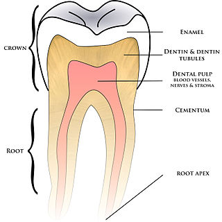
Cementum is a specialized calcified substance covering the root of a tooth. The cementum is the part of the periodontium that attaches the teeth to the alveolar bone by anchoring the periodontal ligament.

Tooth enamel is one of the four major tissues that make up the tooth in humans and many animals, including some species of fish. It makes up the normally visible part of the tooth, covering the crown. The other major tissues are dentin, cementum, and dental pulp. It is a very hard, white to off-white, highly mineralised substance that acts as a barrier to protect the tooth but can become susceptible to degradation, especially by acids from food and drink. In rare circumstances enamel fails to form, leaving the underlying dentin exposed on the surface.

Tooth decay, also known as cavities or caries, is the breakdown of teeth due to acids produced by bacteria. The cavities may be a number of different colors, from yellow to black. Symptoms may include pain and difficulty eating. Complications may include inflammation of the tissue around the tooth, tooth loss and infection or abscess formation. Tooth regeneration is an on-going stem cell based field of study that is trying to reverse the effects of decay, unlike most current methods which only try to make dealing with the effects easier.

Dentin or dentine is a calcified tissue of the body and, along with enamel, cementum, and pulp, is one of the four major components of teeth. It is usually covered by enamel on the crown and cementum on the root and surrounds the entire pulp. By volume, 45% of dentin consists of the mineral hydroxyapatite, 33% is organic material, and 22% is water. Yellow in appearance, it greatly affects the color of a tooth due to the translucency of enamel. Dentin, which is less mineralized and less brittle than enamel, is necessary for the support of enamel. Dentin rates approximately 3 on the Mohs scale of mineral hardness. There are two main characteristics which distinguish dentin from enamel: firstly, dentin forms throughout life; secondly, dentin is sensitive and can become hypersensitive to changes in temperature due to the sensory function of odontoblasts, especially when enamel recedes and dentin channels become exposed.

Ameloblasts are cells present only during tooth development that deposit tooth enamel, which is the hard outermost layer of the tooth forming the surface of the crown.
Cementoenamel junction (CEJ) is defined as the area of the union of cementum and enamel at the cervical region of the tooth. It is a slightly visible anatomical border identified on a tooth. It is the location where the enamel, which covers the anatomical crown of a tooth, and the cementum, which covers the anatomical root of a tooth, meet. Informally it is known as the neck of the tooth. The border created by these two dental tissues has much significance as it is usually the location where the gingiva attaches to a healthy tooth by fibers called the gingival fibers.
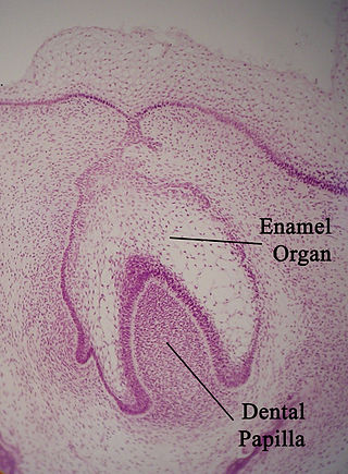
The enamel organ, also known as the dental organ, is a cellular aggregation seen in a developing tooth and it lies above the dental papilla. The enamel organ which is differentiated from the primitive oral epithelium lining the stomodeum. The enamel organ is responsible for the formation of enamel, initiation of dentine formation, establishment of the shape of a tooth's crown, and establishment of the dentoenamel junction.

Tooth development or odontogenesis is the complex process by which teeth form from embryonic cells, grow, and erupt into the mouth. For human teeth to have a healthy oral environment, all parts of the tooth must develop during appropriate stages of fetal development. Primary (baby) teeth start to form between the sixth and eighth week of prenatal development, and permanent teeth begin to form in the twentieth week. If teeth do not start to develop at or near these times, they will not develop at all, resulting in hypodontia or anodontia.

In embryology and prenatal development, the dental papilla is a condensation of ectomesenchymal cells called odontoblasts, seen in histologic sections of a developing tooth. It lies below a cellular aggregation known as the enamel organ. The dental papilla appears after 8–10 weeks of intra uteral life. The dental papilla gives rise to the dentin and pulp of a tooth.
In vertebrates, an odontoblast is a cell of neural crest origin that is part of the outer surface of the dental pulp, and whose biological function is dentinogenesis, which is the formation of dentin, the substance beneath the tooth enamel on the crown and the cementum on the root.
Dentin hypersensitivity is dental pain which is sharp in character and of short duration, arising from exposed dentin surfaces in response to stimuli, typically thermal, evaporative, tactile, osmotic, chemical or electrical; and which cannot be ascribed to any other dental disease.
Concrescence is an uncommon developmental condition of teeth where the cementum overlying the roots of at least two teeth fuse together without the involvement of dentin. Usually, two teeth are involved with the upper second and third molars being most commonly fused together. The prevalence ranges 0.04–0.8% in permanent teeth, with the incidence being highest in the posterior maxilla.
Dens invaginatus (DI), also known as tooth within a tooth, is a rare dental malformation and a developmental anomaly where there is an infolding of enamel into dentin. The prevalence of this condition is 0.3 - 10%, affecting males more frequently than females. The condition presents in two forms, coronal involving tooth crown and radicular involving tooth root, with the former being more common.
Dental anatomy is a field of anatomy dedicated to the study of human tooth structures. The development, appearance, and classification of teeth fall within its purview. Tooth formation begins before birth, and the teeth's eventual morphology is dictated during this time. Dental anatomy is also a taxonomical science: it is concerned with the naming of teeth and the structures of which they are made, this information serving a practical purpose in dental treatment.
Dental pertains to the teeth, including dentistry. Topics related to the dentistry, the human mouth and teeth include:

Cracked tooth syndrome (CTS) is where a tooth has incompletely cracked but no part of the tooth has yet broken off. Sometimes it is described as a greenstick fracture. The symptoms are very variable, making it a notoriously difficult condition to diagnose.

Pulp stones are nodular, calcified masses appearing in either or both the coronal and root portion of the pulp organ in teeth. Pulp stones are not painful unless they impinge on nerves.

Regenerative endodontic procedures is defined as biologically based procedures designed to replace damaged structures such as dentin, root structures, and cells of the pulp-dentin complex. This new treatment modality aims to promote normal function of the pulp. It has become an alternative to heal apical periodontitis. Regenerative endodontics is the extension of root canal therapy. Conventional root canal therapy cleans and fills the pulp chamber with biologically inert material after destruction of the pulp due to dental caries, congenital deformity or trauma. Regenerative endodontics instead seeks to replace live tissue in the pulp chamber. The ultimate goal of regenerative endodontic procedures is to regenerate the tissues and the normal function of the dentin-pulp complex.

Tooth discoloration is abnormal tooth color, hue or translucency. External discoloration is accumulation of stains on the tooth surface. Internal discoloration is due to absorption of pigment particles into tooth structure. Sometimes there are several different co-existent factors responsible for discoloration.