
Human teeth function to mechanically break down items of food by cutting and crushing them in preparation for swallowing and digesting. As such, they are considered part of the human digestive system. Humans have four types of teeth: incisors, canines, premolars, and molars, which each have a specific function. The incisors cut the food, the canines tear the food and the molars and premolars crush the food. The roots of teeth are embedded in the maxilla or the mandible and are covered by gums. Teeth are made of multiple tissues of varying density and hardness.
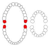
The third molar, commonly called wisdom tooth, is the most posterior of the three molars in each quadrant of the human dentition. The age at which wisdom teeth come through (erupt) is variable, but this generally occurs between late teens and early twenties. Most adults have four wisdom teeth, one in each of the four quadrants, but it is possible to have none, fewer, or more, in which case the extras are called supernumerary teeth. Wisdom teeth may become stuck (impacted) against other teeth if there is not enough space for them to come through normally. Impacted wisdom teeth are still sometimes removed for orthodontic treatment, believing that they move the other teeth and cause crowding, though this is not held anymore as true.

Toothache, also known as dental pain or tooth pain, is pain in the teeth or their supporting structures, caused by dental diseases or pain referred to the teeth by non-dental diseases. When severe it may impact sleep, eating, and other daily activities.

Cementoblastoma, or benign cementoblastoma, is a relatively rare benign neoplasm of the cementum of the teeth. It is derived from ectomesenchyme of odontogenic origin. Cementoblastomas represent less than 0.69–8% of all odontogenic tumors.

The dental follicle, also known as dental sac, is made up of mesenchymal cells and fibres surrounding the enamel organ and dental papilla of a developing tooth. It is a vascular fibrous sac containing the developing tooth and its odontogenic organ. The dental follicle (DF) differentiates into the periodontal ligament. In addition, it may be the precursor of other cells of the periodontium, including osteoblasts, cementoblasts and fibroblasts. They develop into the alveolar bone, the cementum with Sharpey's fibers and the periodontal ligament fibers respectively. Similar to dental papilla, the dental follicle provides nutrition to the enamel organ and dental papilla and also have an extremely rich blood supply.
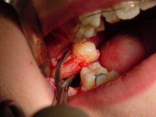
A dental extraction is the removal of teeth from the dental alveolus (socket) in the alveolar bone. Extractions are performed for a wide variety of reasons, but most commonly to remove teeth which have become unrestorable through tooth decay, periodontal disease, or dental trauma, especially when they are associated with toothache. Sometimes impacted wisdom teeth cause recurrent infections of the gum (pericoronitis), and may be removed when other conservative treatments have failed. In orthodontics, if the teeth are crowded, healthy teeth may be extracted to create space so the rest of the teeth can be straightened.
Dilaceration is a developmental disturbance in shape of teeth. It refers to an angulation, or a sharp bend or curve, in the root or crown of a formed tooth. This disturbance is more likely to affect the maxillary incisors and occurs in permanent dentition. Although this may seem more of an aesthetics issue, an impacted maxillary incisor will cause issues related to occlusion, phonetics, mastication, and psychology on young patients.

A dentigerous cyst, also known as a follicular cyst, is an epithelial-lined developmental cyst formed by accumulation of fluid between the reduced enamel epithelium and the crown of an unerupted tooth. It is formed when there is an alteration in the reduced enamel epithelium and encloses the crown of an unerupted tooth at the cemento-enamel junction. Fluid is accumulated between reduced enamel epithelium and the crown of an unerupted tooth.

A buccal exostosis is an exostosis on the buccal surface of the alveolar ridge of the maxilla or mandible. More commonly seen in the maxilla than the mandible, buccal exostoses are considered to be site specific. Existing as asymptomatic bony nodules, buccal exostoses don’t usually present until adult life, and some consider buccal exostoses to be a variation of normal anatomy rather than disease. Bone is thought to become hyperplastic, consisting of mature cortical and trabecular bone with a smooth outer surface. They are less common when compared with mandibular tori.
Dens invaginatus (DI), also known as tooth within a tooth, is a rare dental malformation and a developmental anomaly where there is an infolding of enamel into dentin. The prevalence of this condition is 0.3 - 10%, affecting males more frequently than females. The condition presents in two forms, coronal involving tooth crown and radicular involving tooth root, with the former being more common.

Dentin dysplasia (DD) is a rare genetic developmental disorder affecting dentine production of the teeth, commonly exhibiting an autosomal dominant inheritance that causes malformation of the root. It affects both primary and permanent dentitions in approximately 1 in every 100,000 patients. It is characterized by the presence of normal enamel but atypical dentin with abnormal pulpal morphology. Witkop in 1972 classified DD into two types which are Type I (DD-1) is the radicular type, and type II (DD-2) is the coronal type. DD-1 has been further divided into 4 different subtypes (DD-1a,1b,1c,1d) based on the radiographic features.

Idiopathic osteosclerosis, also known as enostosis or dense bone island, is a condition which may be found around the roots of a tooth, usually a premolar or molar. It is usually painless and found during routine radiographs as an amorphous radiopaque (light) area around a tooth. There is no sign of inflammation of the tooth, and if the island is associated with the root the periodontal ligament space is preserved.
“Lateral periodontal cysts (LPCs) are defined as non-keratinised and non-inflammatory developmental cysts located adjacent or lateral to the root of a vital tooth.” LPCs are a rare form of jaw cysts, with the same histopathological characteristics as gingival cysts of adults (GCA). Hence LPCs are regarded as the intraosseous form of the extraosseous GCA. They are commonly found along the lateral periodontium or within the bone between the roots of vital teeth, around mandibular canines and premolars. Standish and Shafer reported the first well-documented case of LPCs in 1958, followed by Holder and Kunkel in the same year although it was called a periodontal cyst. Since then, there has been more than 270 well-documented cases of LPCs in literature.
Tooth gemination is a dental phenomenon that appears to be two teeth developed from one. There is one main crown with a cleft in it that, within the incisal third of the crown, looks like two teeth, though it is not two teeth. The number of the teeth in the arch will be normal.

Cementoma is an odontogenic tumor of cementum. It is usually observed as a benign spherical mass of hard tissue fused to the root of a tooth. It is found most commonly in the mandible in the region of the lower molar teeth, occurring between the ages of 8 and 30 in both sexes with equal frequency. It causes distortion of surrounding areas but is usually a painless growth, at least initially. Considerable thickening of the cementum can often be observed. A periapical form is also recognized. Cementoma is not exclusive to the mandible as it can infrequently occur in the maxilla and other parts of the body such as the long bones.

In dentistry, a furcation defect is bone loss, usually a result of periodontal disease, affecting the base of the root trunk of a tooth where two or more roots meet. The extent and configuration of the defect are factors in both diagnosis and treatment planning.
Hypercementosis is an idiopathic, non-neoplastic condition characterized by the excessive buildup of normal cementum on the roots of one or more teeth. A thicker layer of cementum can give the tooth an enlarged appearance, which mainly occurs at the apex or apices of the tooth.

Impacted wisdom teeth is a condition where the third molars are prevented from erupting into the mouth. This can be caused by a physical barrier, such as other teeth, or when the tooth is angled away from a vertical position. Completely unerupted wisdom teeth usually result in no symptoms, although they can sometimes develop cysts or neoplasms. Partially erupted wisdom teeth or wisdom teeth that are not erupted but are exposed to oral bacteria through deep periodontal pocket, can develop cavities or pericoronitis. Removal of impacted wisdom teeth is advised for the future prevention of or in the current presence of certain pathologies, such as caries, periodontal disease or cysts. Prophylactic (preventative) extraction of wisdom teeth is preferred to be done at a younger age to take advantage of incomplete root development, which is associated with an easier surgical procedure and less probability of complications.

Tooth mobility is the horizontal or vertical displacement of a tooth beyond its normal physiological boundaries around the gingival (gum) area, i.e. the medical term for a loose tooth.
Tooth ankylosis refers to a fusion between a tooth and underlying bony support tissues. In some species, this is a normal process that occurs during the formation or maintenance of the dentition. By contrast, in humans tooth ankylosis is pathological, whereby a fusion between alveolar bone and the cementum of a tooth occurs.
