The human teeth function to mechanically break down items of food by cutting and crushing them in preparation for swallowing and digesting. Humans have four types of teeth: incisors, canines, premolars, and molars, which each have a specific function. The incisors cut the food, the canines tear the food and the molars and premolars crush the food. The roots of teeth are embedded in the maxilla or the mandible and are covered by gums. Teeth are made of multiple tissues of varying density and hardness.

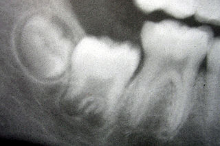
A wisdom tooth or third molar is one of the three molars per quadrant of the human dentition. It is the most posterior of the three. The age at which wisdom teeth come through (erupt) is variable, but generally occurs between late teens and early twenties. Most adults have four wisdom teeth, one in each of the four quadrants, but it is possible to have none, fewer, or more, in which case the extras are called supernumerary teeth. Wisdom teeth may get stuck (impacted) against other teeth if there is not enough space for them to come through normally. While this does not cause movement of other teeth, it can cause tooth decay if the impaction makes oral hygiene difficult. Wisdom teeth which are partially erupted through the gum may also cause inflammation and infection in the surrounding gum tissues, termed pericoronitis. Wisdom teeth are often extracted when or even before these problems occur. However, some, including the National Institute for Health and Care Excellence in the UK, recommend against the prophylactic extraction of disease-free impacted wisdom teeth.

Hyperdontia is the condition of having supernumerary teeth, or teeth that appear in addition to the regular number of teeth. They can appear in any area of the dental arch and can affect any dental organ. The opposite of hyperdontia is hypodontia, where there is a congenital lack of teeth, a condition which is seen more commonly than hyperdontia. The scientific definition of hyperdontia is "any tooth or odontogenic structure that is formed from tooth germ in excess of usual number for any given region of the dental arch." The additional teeth, which may be small in number, or many, can occur on any place in the dental arch. Their arrangement may be symmetrical or non-symmetrical.

Tooth development or odontogenesis is the complex process by which teeth form from embryonic cells, grow, and erupt into the mouth. For human teeth to have a healthy oral environment, all parts of the tooth must develop during appropriate stages of fetal development. Primary (baby) teeth start to form between the sixth and eighth week of prenatal development, and permanent teeth begin to form in the twentieth week. If teeth do not start to develop at or near these times, they will not develop at all, resulting in hypodontia or anodontia.

Pericoronitis is inflammation of the soft tissues surrounding the crown of a partially erupted tooth, including the gingiva (gums) and the dental follicle. The soft tissue covering a partially erupted tooth is known as an operculum, an area which can be difficult to access with normal oral hygiene methods. The synonym operculitis technically refers to inflammation of the operculum alone.

Torus mandibularis is a bony growth in the mandible along the surface nearest to the tongue. Mandibular tori are usually present near the premolars and above the location of the mylohyoid muscle's attachment to the mandible. In 90% of cases, there is a torus on both the left and right sides, making this finding a predominantly bilateral condition.

Microdontia is a condition in which one or more teeth appear smaller than normal. In the generalized form, all teeth are involved. In the localized form, only a few teeth are involved. The most common teeth affected are the upper lateral incisors and third molars.
Macrodontia is a type of localized gigantism in which teeth are larger than normal for the particular type(s) of teeth involved. The three types of macrodontia are true generalized macrodontia, relative generalized macrodontia, and macrodontia of a single tooth. True generalized macrodontia is very rare. Macrodontia of a single tooth is more common. Some kind of macrodontia in the permanent dentition occurs in 1.1% of the total population. It should not be confused with taurodontism, fusion or the jaws being relatively small, giving the appearance of macrodontia.

Dentin dysplasia (DD) is a rare genetic developmental disorder dentine production of the teeth, commonly exhibiting an autosomal dominant inheritance that causes malformation of the root. It affects both primary and permanent dentitions in approximately 1 in every 100,000 patients. It is characterized by presence of normal enamel but atypical dentin with abnormal pulpal morphology. Witkop in 1972 classified DD into two types which are Type I (DD-1) is the radicular type, and type II (DD-2) is the coronal type. DD-1 has been further divided into 4 different subtypes (DD-1a,1b,1c,1d) based on the radiographic features.

Central giant-cell granuloma (CGCG) is a localised benign condition of the jaws. It is twice as common in females and is more likely to occur before age 30. Central giant-cell granulomas are more common in the anterior mandible, often crossing the midline and causing painless swellings.

Talon Cusp is a rare dental anomaly. Generally a person with this develops "cusp-like" projections located on the inside surface of the affected tooth. Talon cusp is an extra cusp on an anterior tooth. Although talon cusp may not appear serious, it can cause clinical, diagnostic, functional problems and alters the aesthetic appeal.

Tooth eruption is a process in tooth development in which the teeth enter the mouth and become visible. It is currently believed that the periodontal ligament plays an important role in tooth eruption. The first human teeth to appear, the deciduous (primary) teeth, erupt into the mouth from around 6 months until 2 years of age, in a process known as "teething". These teeth are the only ones in the mouth until a person is about 6 years old creating the primary dentition stage. At that time, the first permanent tooth erupts and begins a time in which there is a combination of primary and permanent teeth, known as the mixed dentition stage, which lasts until the last primary tooth is lost. Then, the remaining permanent teeth erupt into the mouth during the permanent dentition stage.

An impacted tooth is one that fails to erupt into the dental arch within the expected developmental window. Because impacted teeth do not erupt, they are retained throughout the individual's lifetime unless extracted or exposed surgically. Teeth may become impacted because of adjacent teeth, dense overlying bone, excessive soft tissue or a genetic abnormality. Most often, the cause of impaction is inadequate arch length and space in which to erupt. That is the total length of the alveolar arch is smaller than the tooth arch. The wisdom teeth are frequently impacted because they are the last teeth to erupt in the oral cavity. Mandibular third molars are more commonly impacted than their maxillary counterparts.
Oral and maxillofacial pathology refers to the diseases of the mouth, jaws and related structures such as salivary glands, temporomandibular joints, facial muscles and perioral skin. The mouth is an important organ with many different functions. It is also prone to a variety of medical and dental disorders.

Enamel hypoplasia is a defect of the teeth in which the enamel is deficient in amount, caused by defective enamel matrix formation. Defects are commonly split into one of four categories, pit-form, plane-form, linear-form, and localised enamel hypoplasia. In many cases the enamel crown has pits or a groove on it, and in extreme cases, sections of the tooth have no enamel, exposing the dentin. Enamel hypoplasia varies substantially among populations and can be used to infer health and behaviour in past populations. Defects have also been found in a variety of non-human animals.

Impacted wisdom teeth is a disorder where the third molars are prevented from erupting into the mouth. This can be caused by a physical barrier, such as other teeth, or a disorder of development where the tooth is angled away from a vertical position. Impacted wisdom teeth are usually asymptomatic, until they develop a communication to the mouth, their movement causes damage to adjacent teeth, or they develop pathology around the tooth such as a cyst, at which localized pain and swelling can develop.

Tooth pathology is any condition of the teeth that can be congenital or acquired. Sometimes a congenital tooth diseases are called tooth abnormalities. These are among the most common diseases in humans The prevention, diagnosis, treatment and rehabilitation of these diseases are the base to the dentistry profession, in which are dentists and dental hygienists, and its sub-specialties, such as oral medicine, oral and maxillofacial surgery, and endodontics. Tooth pathology is usually separated from other types of dental issues, including enamel hypoplasia and tooth wear.

Tricho-dento-osseous syndrome (TDO) is a rare, systemic, autosomal dominant genetic disorder that causes defects in hair, teeth, and bones respectively. This disease is present at birth. TDO has been shown to occur in areas of close geographic proximity and within families; most recent documented cases are in Virginia, Tennessee, and North Carolina. The cause of this disease is a mutation in the DLX3 gene, which controls hair follicle differentiation and induction of bone formation. One-hundred percent of patients with TDO suffer from two co-existing conditions called enamel hypoplasia and taurodontism in which the abnormal growth patterns of the teeth result in severe external and internal defects. The hair defects are characterized as being rough, course, with profuse shedding. Hair is curly and kinky at infancy but later straightens. Dental defects are characterized by dark-yellow/brownish colored teeth, thin and/or possibly pitted enamel, that is malformed. The teeth can also look normal in color, but also have a physical impression of extreme fragility and thinness in appearance. Additionally, severe underbites where the top and bottom teeth fail to correctly align may be present; it is common for the affected individual to have a larger, more pronounced lower jaw and longer bones. The physical deformities that TDO causes become more noticeable with age, and emotional support for the family as well as the affected individual is frequently recommended. Adequate treatment for TDO is a team based approach, mostly involving physical therapists, dentists, and oromaxillofacial surgeons. Genetic counseling is also recommended.
Tooth ankylosis is the pathological fusing of cementum or dentine of a tooth root to the alveolar bone. Ankylosis of teeth is uncommon, it more often occurs in deciduous teeth than permanent teeth.

Tooth discoloration is abnormal tooth color, hue or translucency. External discoloration is accumulation of stains on the tooth surface. Internal discoloration is due to absorption of pigment particles into tooth structure. Sometimes there are several different co-existent factors responsible for discoloration.


