Hypercementosis

Last updated
Hypercementosis
Specialty Dentistry   OOjs UI icon edit-ltr-progressive.svg

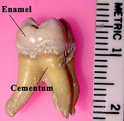
Hypercementosis is an idiopathic, non-neoplastic condition characterized by the excessive buildup of normal cementum (calcified tissue) on the roots of one or more teeth. [1] A thicker layer of cementum can give the tooth an enlarged appearance, mainly occurring at the apex or apices of the tooth. The cellular cementum functions at the bottom half of the tooth roots which contain cementocytes that anchor the tooth into the jaw socket, protect the tooth's pulp, and repair external root resorption [2] .

Contents

Signs and symptoms

Location of where excess cementum may appear Labeledmolar.jpg
Location of where excess cementum may appear

It is experienced as an uncomfortable sensation in the tooth, followed by an aching pain. [3] Excess amounts of cementum may cause pressure on periodontal ligaments and adjacent teeth. The teeth affected may present as asymptomatic [4] . It may be shown on radiographs as a radiopaque (or lighter) mass at each root apex to confirm the diagnosis.

Cause

Trauma and other developmental disorders such as Paget's disease may be more prone to develop hypercementosis in the maxillary region. [5]

Local factors:

Systemic factors:

It may be one of the complications of Paget's disease of bone in the form of generalized hypercementosis.

It may also be a compensatory mechanism in response to attrition to increase occlusal tooth height.

Pathophysiology

Research has suggested that mutations in the ENPP1 and GACI genes may contribute to the development of hypercementosis. [6] Loss of function in ENPP1 caused generalized arterial calcification of infancy (GACI) which was directly associated with individuals with hypercementosis. [7] When ENPP1 is inhibited, there is a deficiency in pyrophosphate (PPi) that regulates the mineralization of bone by stopping hydroxyapatite crystals from forming [8] . PPi naturally inhibits crystal formation in inappropriate areas such as in the sub-gingival area [8] . Loss of control in PPi may result in excessive cementum deposition in the lower third of the tooth. [8]

Diagnosis

Periapical radiographs can locate radiopaque structures in proximity to the root which can appear as dense bone islands or periapical osseous dysplasia in cases of hypercementosis. [9] The majority of affected teeth appear club-shaped due to cemental hyperplasia diffusing in a variety of severities. Most appear in the apical third of the root. [10]

Complications

Such deposits form bulbous enlargements on the roots and may interfere with extraction s, especially if adjacent teeth become fused (concrescence). It may also result in pulpal necrosis by blocking blood supply via the apical foramen. [11] Teeth affected do not necessarily need treatment unless it causes complications to adjacent teeth and structures.

Epidemiology

Hypercementosis occurs more frequently in adults and increases with age [6] . Teeth that are affected are primary mandibular molars followed by secondary premolars in the mandible and maxilla, however any teeth may be affected [6] . While no one race is primarily affected, those with conditions that affect bone hormone levels such as Paget's disease and acromegaly are more likely to develop hypercementosis. [6]

Treatment

While treatment is not necessary if teeth are asymptomatic, it is crucial to monitor the progression of hypercementosis to reduce the chances of surrounding teeth being affected. [6] If pain is associated with teeth affected by hypercementosis, extractions or endodontic treatment may be required. A risk assessment must be considered as excess cementum build-up may make determining the apical limit challenging during a root canal [12] . The prognosis is the same as a regular tooth as long as the root canal is done properly [12] .

Recent Research

A recent study has proposed a "teeth-as-tools" hypothesis that suggests early humans may have used anterior teeth for non-dietary purposes [13] . When excessive mechanical force is used in this way, it stimulates the production of cementum to improve the stability of the tooth. [13] Research focuses on the types of occlusion and contamination that may give insight into how Neanderthals adapted to diverse challenges in the world.

Related Research Articles

<span class="mw-page-title-main">Cementum</span> Specialized calcified substance covering the root of a tooth

Cementum is a specialized calcified substance covering the root of a tooth. The cementum is the part of the periodontium that attaches the teeth to the alveolar bone by anchoring the periodontal ligament.

<span class="mw-page-title-main">Toothache</span> Medical condition of the teeth

Toothaches, also known as dental pain or tooth pain, is pain in the teeth or their supporting structures, caused by dental diseases or pain referred to the teeth by non-dental diseases. When severe it may impact sleep, eating, and other daily activities.

<span class="mw-page-title-main">Periodontal fiber</span> Group of specialized connective tissue fibers

The periodontal ligament, commonly abbreviated as the PDL, are a group of specialized connective tissue fibers that essentially attach a tooth to the alveolar bone within which they sit. It inserts into root cementum on one side and onto alveolar bone on the other.

<span class="mw-page-title-main">Cementoblastoma</span> Benign tumor of the teeth roots

Cementoblastoma, or benign cementoblastoma, is a relatively rare benign neoplasm of the cementum of the teeth. It is derived from ectomesenchyme of odontogenic origin. Cementoblastomas represent less than 0.69–8% of all odontogenic tumors.

<span class="mw-page-title-main">Dentinogenesis imperfecta</span> Genetic disorder impairing tooth development

Dentinogenesis imperfecta (DI) is a genetic disorder of tooth development. It is inherited in an autosomal dominant pattern, as a result of mutations on chromosome 4q21, in the dentine sialophosphoprotein gene (DSPP). It is one of the most frequently occurring autosomal dominant features in humans. Dentinogenesis imperfecta affects an estimated 1 in 6,000-8,000 people.

<span class="mw-page-title-main">Condensing osteitis</span> Hardening of tooth roots due to infection

Condensing osteitis, also known as focal sclerosing osteomyelitis, is a rare periapical inflammatory condition characterized by the formation of sclerotic bone near the roots of premolars and molars. This condition arises as a response to dental infections, such as periapical pulp inflammation or low-intensity trauma. The lesion typically appears as a radiopacity in the periapical area due to the sclerotic reaction. While most commonly associated with non-vital teeth, condensing osteitis can also occur in vital teeth following occlusal trauma. The condition was first described by Dr. Carl Garré in 1893.

Dens invaginatus (DI), also known as tooth within a tooth, is a rare dental malformation and a developmental anomaly where there is an infolding of enamel into dentin. The prevalence of this condition is 0.3 - 10%, affecting males more frequently than females. The condition presents in two forms, coronal involving tooth crown and radicular involving tooth root, with the former being more common.

Dens evaginatus is a rare odontogenic developmental anomaly that is found in teeth where the outer surface appears to form an extra bump or cusp.

<span class="mw-page-title-main">Dentin dysplasia</span> Medical condition

Dentin dysplasia (DD) is a rare genetic developmental disorder affecting dentine production of the teeth, commonly exhibiting an autosomal dominant inheritance that causes malformation of the root. It affects both primary and permanent dentitions in approximately 1 in every 100,000 patients. It is characterized by the presence of normal enamel but atypical dentin with abnormal pulpal morphology. Witkop in 1972 classified DD into two types which are Type I (DD-1) is the radicular type, and type II (DD-2) is the coronal type. DD-1 has been further divided into 4 different subtypes (DD-1a,1b,1c,1d) based on the radiographic features.

<span class="mw-page-title-main">Idiopathic osteosclerosis</span> Hardening of the teeth roots for an unknown reason

Idiopathic osteosclerosis, also known as enostosis or dense bone island, is a condition which may be found around the roots of a tooth, usually a premolar or molar. It is usually painless and found during routine radiographs as an amorphous radiopaque (light) area around a tooth. There is no sign of inflammation of the tooth, and if the island is associated with the root the periodontal ligament space is preserved.

<span class="mw-page-title-main">Dental abscess</span> Collection of pus in or around a tooth

A dental abscess is a localized collection of pus associated with a tooth. The most common type of dental abscess is a periapical abscess, and the second most common is a periodontal abscess. In a periapical abscess, usually the origin is a bacterial infection that has accumulated in the soft, often dead, pulp of the tooth. This can be caused by tooth decay, broken teeth or extensive periodontal disease. A failed root canal treatment may also create a similar abscess.

<span class="mw-page-title-main">Dental radiography</span> X-ray imaging in dentistry

Dental radiographs, commonly known as X-rays, are radiographs used to diagnose hidden dental structures, malignant or benign masses, bone loss, and cavities.

<span class="mw-page-title-main">Root canal treatment</span> Procedure to disinfect and fortify the interior of a tooth

Root canal treatment is a treatment sequence for the infected pulp of a tooth that is intended to result in the elimination of infection and the protection of the decontaminated tooth from future microbial invasion. Root canals, and their associated pulp chamber, are the physical hollows within a tooth that are naturally inhabited by nerve tissue, blood vessels and other cellular entities.

<span class="mw-page-title-main">Cementoma</span> Medical condition

Cementoma is an odontogenic tumor of cementum. It is usually observed as a benign spherical mass of hard tissue fused to the root of a tooth. It is found most commonly in the mandible in the region of the lower molar teeth, occurring between the ages of 8 and 30 in both sexes with equal frequency. It causes distortion of surrounding areas but is usually a painless growth, at least initially. Considerable thickening of the cementum can often be observed. A periapical form is also recognized. Cementoma is not exclusive to the mandible as it can infrequently occur in the maxilla and other parts of the body such as the long bones.

<span class="mw-page-title-main">Tooth resorption</span> Breakdown of the tooth root to be absorbed by the blood

Resorption of the root of the tooth, or root resorption, is the progressive loss of dentin and cementum by the action of odontoclasts. Root resorption is a normal physiological process that occurs in the exfoliation of the primary dentition. However, pathological root resorption occurs in the permanent or secondary dentition and sometimes in the primary dentition.

Pulp necrosis is a clinical diagnostic category indicating the death of cells and tissues in the pulp chamber of a tooth with or without bacterial invasion. It is often the result of many cases of dental trauma, caries and irreversible pulpitis.

A phoenix abscess is an acute exacerbation of a chronic periapical lesion. It is a dental abscess that can occur immediately following root canal treatment. Another cause is due to untreated necrotic pulp. It is also the result of inadequate debridement during the endodontic procedure. Risk of occurrence of a phoenix abscess is minimised by correct identification and instrumentation of the entire root canal, ensuring no missed anatomy.

<span class="mw-page-title-main">Pulp stone</span>

Pulp stones are nodular, calcified masses appearing in either or both the coronal and root portion of the pulp organ in teeth. Pulp stones are not painful unless they impinge on nerves.

<span class="mw-page-title-main">Periapical periodontitis</span> Inflammation at the apex of a tooth root

Periapical periodontitis or apical periodontitis (AP) is an acute or chronic inflammatory lesion around the apex of a tooth root, most commonly caused by bacterial invasion of the pulp of the tooth. It is a likely outcome of untreated dental caries, and in such cases it can be considered a sequela in the natural history of tooth decay, irreversible pulpitis and pulpal necrosis. Other causes can include occlusal trauma due to 'high spots' after restoration work, extrusion from the tooth of root filling material, or bacterial invasion and infection from the gums. Periapical periodontitis may develop into a periapical abscess, where a collection of pus forms at the end of the root, the consequence of spread of infection from the tooth pulp, or into a periapical cyst, where an epithelial lined, fluid-filled structure forms.

In the dental specialty of endodontics, periradicular surgery is surgery to the external root surface. Examples of periradicular surgery include apicoectomy, root resection, repair of root perforation or resorption defects, removal of broken fragments of the tooth or a filling material, and exploratory surgery to look for root fractures.

References

  1. L Napier Souza, S Monteiro Lima Júnior FJ Garcia Santos Pimenta, AC Rodrigues Antunes Souza and R Santiago Gomez. "Atypical hypercementosis versus cementoblastoma". dmfr.birjournals.org. Retrieved 2009-09-08.{{cite web}}: CS1 maint: multiple names: authors list (link)
  2. Massé, Léa; Garot, Elsa; Maureille, Bruno; Le Cabec, Adeline (February 2023). "Insights into the aetiologies of hypercementosis: A systematic review and a scoring system". Archives of Oral Biology. 146: 105599. doi:10.1016/j.archoralbio.2022.105599. ISSN   0003-9969.
  3. "Hypercementosis or Dental Exostosis". chestofbooks.com. Retrieved 2009-09-08.
  4. "Hypercementosis: Causes, Symptoms, and Diagnosis". www.medicoverhospitals.in. Retrieved 2024-11-06.
  5. Rao, V. M.; Karasick, D. (1982). "Hypercementosis--an important clue to Paget disease of the maxilla". Skeletal Radiology. 9 (2): 126–128. doi:10.1007/BF00360497. ISSN   0364-2348. PMID   7163823.
  6. 1 2 3 4 5 "Hypercementosis - an overview | ScienceDirect Topics". www.sciencedirect.com. Retrieved 2024-11-06.
  7. Markley, John L.; Westler, William Milo (August 2017). "Biomolecular NMR: Past and future". Archives of Biochemistry and Biophysics. 628: 3–16. doi:10.1016/j.abb.2017.05.003. ISSN   0003-9861. PMC   5701516 .
  8. 1 2 3 Ralph, Douglas; van de Wetering, Koen; Uitto, Jouni; Li, Qiaoli (May 2022). "Inorganic Pyrophosphate Deficiency Syndromes and Potential Treatments for Pathologic Tissue Calcification". The American Journal of Pathology. 192 (5): 762–770. doi:10.1016/j.ajpath.2022.01.012. ISSN   0002-9440. PMC   9088198 . PMID   35182493.
  9. Lam, Ernest W. N. (2014-01-01), White, Stuart C.; Pharoah, Michael J. (eds.), "Chapter 31 - Dental Anomalies", Oral Radiology (Seventh Edition), St. Louis (MO): Mosby, pp. 582–611, ISBN   978-0-323-09633-1 , retrieved 2024-11-06
  10. Mupparapu, Mel; Shi, Katherine Jie; Ko, Eugene (2020-01-01). "Differential Diagnosis of Periapical Radiopacities and Radiolucencies". Dental Clinics of North America. Oral Diseases for the General Dentist. 64 (1): 163–189. doi:10.1016/j.cden.2019.08.010. ISSN   0011-8532.
  11. Illustrated Dental Embryology, Histology, and Anatomy, Bath-Balogh and Fehrenbach, Elsevier, 2011
  12. 1 2 Pappen, Fernanda Geraldes; Fagonde, Cecilia Definski; Martos, Josué; Silveira, Luiz Fernando Machado (September 2011). "Hypercementosis: a challenge for endodontic therapy". RSBO (Online). 8 (3): 321–334. doi:10.21726/rsbo.v8i3.1078. ISSN   1984-5685.
  13. 1 2 Massé, Léa; d’Incau, Emmanuel; Souron, Antoine; Vanderesse, Nicolas; Santos, Frédéric; Maureille, Bruno; Le Cabec, Adeline (2024-01-13). "Unraveling the Life History of Past Populations through Hypercementosis: Insights into Cementum Apposition Patterns and Possible Etiologies Using Micro-CT and Confocal Microscopy". Biology. 13 (1): 43. doi: 10.3390/biology13010043 . ISSN   2079-7737. PMC   10813066 . PMID   38248474.