Cracked tooth syndrome

Last updated
Cracked tooth syndrome
Other namesCracked cusp syndrome, [1] split tooth syndrome, [1] incomplete fracture of posterior teeth [1]
Tooth Section.svg
Cross-section of a posterior tooth.
Specialty Dentistry

Cracked tooth syndrome (CTS) [2] is where a tooth has incompletely cracked but no part of the tooth has yet broken off. Sometimes it is described as a greenstick fracture. [1] The symptoms are very variable, making it a notoriously difficult condition to diagnose.

Contents

Classification and definition

Cracked tooth syndrome could be considered a type of dental trauma and also one of the possible causes of dental pain. One definition of cracked tooth syndrome is "a fracture plane of unknown depth and direction passing through tooth structure that, if not already involving, may progress to communicate with the pulp and/or periodontal ligament." [1]

Signs and symptoms

Tooth crack in the upper first molar tooth in a patient who suffers from bruxism. Cracked tooth.jpg
Tooth crack in the upper first molar tooth in a patient who suffers from bruxism.

The reported symptoms are very variable, [2] and frequently have been present for many months before the condition is diagnosed. [1] Reported symptoms may include some of the following:

If the crack propagates into the pulp, irreversible pulpitis, pulpal necrosis and periapical periodontitis may develop, with the respective associated symptoms. [1]

Pathophysiology

Crack (vertical fracture) of tooth and root #1.4 (green arrows) splitting it in two even pieces which has caused a lateral periodontal abscess (blue arrows). Cracked tooth lateral periodontal abscess.jpg
Crack (vertical fracture) of tooth and root #1.4 (green arrows) splitting it in two even pieces which has caused a lateral periodontal abscess (blue arrows).

CTS is typically characterized by pain when releasing biting pressure on an object. This is because when biting down the segments are usually moving apart and thereby reducing the pressure in the nerves in the dentin of the tooth. When the bite is released the "segments" snap back together sharply increasing the pressure in the intradentin nerves causing pain. The pain is often inconsistent, and frequently hard to reproduce. Pain associated with CTS has been reported to occur more commonly on biting, rather than on release of pressure after biting. If untreated, CTS can lead to severe pain, possible pulpal death, abscess, and even the loss of the tooth.

If the fracture propagates into the pulp, this is termed a complete fracture, and pulpitis and pulp death may occur. If the crack propagates further into the root, a periodontal defect may develop, or even a vertical root fracture. [1]

According to one theory, the pain on biting is caused by the 2 fractured sections of the tooth moving independently of each other, triggering sudden movement of fluid within the dentinal tubules. [1] This activates A-type nociceptors in the dentin-pulp complex, reported by the pulp-dentin complex as pain. Another theory is that the pain upon cold stimuli results from leak of noxious substances via the crack, irritating the pulp. [1]

Diagnosis

Cracked tooth syndrome (CTS) was defined as 'an incomplete fracture of a vital posterior tooth that involves the dentine and occasionally extends to the pulp' by Cameron in 1964 and more recently has included 'a fracture plane of unknown depth and direction passing through tooth structure that, if not already involving, may progress to communicate with the pulp and/or periodontal ligament'. [4] The diagnosis of cracked tooth syndrome is notoriously difficult even for experienced clinicians. [2] The features are highly variable and may mimic sinusitis, temporomandibular disorders, headaches, ear pain, or atypical facial pain/atypical odontalgia (persistent idiopathic facial pain). [2] When diagnosing cracked tooth syndrome, a dentist takes many factors into consideration. Effective management and good prognosis of cracked teeth is linked to prompt diagnosis. A detailed history may reveal pain on release of pressure when eating or sharp pain when consuming cold food and drink. There are a variety of habits which predispose patients to CTS including chewing ice, pens and hard sweets etc. Recurrent occlusal adjustment of restorations due to discomfort may also be indicative of CTS, alongside a history of extensive dental treatment. Below different techniques used for diagnosing CTS are discussed.

Clinical examination

Cracks are difficult to see during a clinical exam which may limit diagnosis. However other clinical signs which may lead to the diagnosis of CTS includes wear faceting indicating excessive forces perhaps from clenching or grinding or the presence of an isolated deep periodontal pocket which may symbolise a split tooth. Removing restorations may help to visualise fracture lines but should only be carried out after gaining informed consent from the patient, as removing a restoration may prove to be of little diagnostic benefit. Tactile examination with a sharp probe may also aid diagnosis.

Gentian violet or methylene blue stains

Dyes may be used to aid visualisation of fractures. The technique requires 2–5 days to be effective and a temporary restoration may be required. The structural integrity can be weakened by this method, leading to crack propagation.

Transillumination

Transillumination of tooth 2.1 showing vertical fracture (blue arrows) and inflammation in marginal gingiva at fracture site (green arrow) Transillumination of tooth marked.jpg
Transillumination of tooth 2.1 showing vertical fracture (blue arrows) and inflammation in marginal gingiva at fracture site (green arrow)

Transillumination is best performed by placing a fibre optic light source directly onto the tooth and optimal results can be achieved with the aid of magnification. Cracks involving dentine interrupt the light transmission. However, transillumination may cause cracks to appear enlarged as well as causing colour changes to become invisible.

Radiographs

Radiographs offer little benefit in visualising cracks. This is due to the fact that cracks propagate in a direction which is parallel to the plane of the film (Mesiodistal) however radiographs can be useful when examining the periodontal and pulpal status.

Bite test

Different tools can be used when carrying out a bite test which produce symptoms associated with cracked tooth syndrome. Patients bite down followed by sudden release of pressure. CTS diagnosis is confirmed by pain on release of pressure. The involved cusp can be determined by biting on individual cusps separately. Tooth Slooth II (Professional Results Inc., Laguna Niguel, CA, USA) and Fractfinder (Denbur, Oak Brook, IL, USA) are commercially available tools.

Epidemiology

Aetiology of CTS is multifactorial, the causative factors include: [5]

Most commonly involved teeth are mandibular molars followed by maxillary premolars, maxillary molars and maxillary premolars. in a recent audit, mandibular first molar thought to be most affected by CTS possibly due to the wedging effect of opposing pointy, protruding maxillary mesio-palatal cusp onto the mandibular molar central fissure. Studies have also found signs of cracked teeth following the cementation of porcelain inlays; it is suggested that the debonding of intracoronal restorations may be caused by unrecognized cracks in the tooth. [6]

Treatments

Fractured tooth (blue arrows) viewed in the mouth (left) and after extraction (right). Cracked tooth.png
Fractured tooth (blue arrows) viewed in the mouth (left) and after extraction (right).

There is no universally accepted treatment strategy in the treatment of a cracked tooth, as judging the extent of a fracture is challenging. Minor fractures without deep propagation may require no more than observation, as tertiary dentin formation can reinforce and stabilize some fractures not impinging on pulp. Generally, treatments for more severe fractures aim to prevent movement of the segments of the involved tooth so they do not move or flex independently during biting and grinding (stabilization of the crack) to prevent propagation of the crack. [7] Provisionally, a band may be placed around the tooth or a direct composite splint can be placed in supra-occlusion to minimize flexing. Definitive options include: [8]

Teeth originally presenting with CTS may subsequently require root canal therapy (if pain persists after above) or extraction.

History

The term "cuspal fracture odontalgia" was suggested in 1954 by Gibbs. [1] Subsequently, the term "cracked tooth syndrome" was coined in 1964 by Cameron, [2] who defined the condition as "an incomplete fracture of a vital posterior tooth that involves the dentin and occasionally extends into the pulp." [1]

Related Research Articles

<span class="mw-page-title-main">Human tooth</span> Calcified whitish structure in humans mouths used to break down food

Human teeth function to mechanically break down items of food by cutting and crushing them in preparation for swallowing and digesting. As such, they are considered part of the human digestive system. Humans have four types of teeth: incisors, canines, premolars, and molars, which each have a specific function. The incisors cut the food, the canines tear the food and the molars and premolars crush the food. The roots of teeth are embedded in the maxilla or the mandible and are covered by gums. Teeth are made of multiple tissues of varying density and hardness.

<span class="mw-page-title-main">Toothache</span> Medical condition of the teeth

Toothache, also known as dental pain or tooth pain, is pain in the teeth or their supporting structures, caused by dental diseases or pain referred to the teeth by non-dental diseases. When severe it may impact sleep, eating, and other daily activities.

<span class="mw-page-title-main">Pulp (tooth)</span> Part in the center of a tooth made up of living connective tissue and cells called odontoblasts

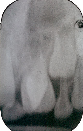
The pulp is the connective tissue, nerves, blood vessels, and odontoblasts that comprise the innermost layer of a tooth. The pulp's activity and signalling processes regulate its behaviour.

<span class="mw-page-title-main">Human tooth development</span> Process by which teeth form

Tooth development or odontogenesis is the complex process by which teeth form from embryonic cells, grow, and erupt into the mouth. For human teeth to have a healthy oral environment, all parts of the tooth must develop during appropriate stages of fetal development. Primary (baby) teeth start to form between the sixth and eighth week of prenatal development, and permanent teeth begin to form in the twentieth week. If teeth do not start to develop at or near these times, they will not develop at all, resulting in hypodontia or anodontia.

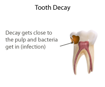
Pulpitis is inflammation of dental pulp tissue. The pulp contains the blood vessels, the nerves, and connective tissue inside a tooth and provides the tooth's blood and nutrients. Pulpitis is mainly caused by bacterial infection which itself is a secondary development of caries. It manifests itself in the form of a toothache.

A dental emergency is an issue involving the teeth and supporting tissues that are of high importance to be treated by the relevant professional. Dental emergencies do not always involve pain, although this is a common signal that something needs to be looked at. Pain can originate from the tooth, surrounding tissues or can have the sensation of originating in the teeth but be caused by an independent source. Depending on the type of pain experienced an experienced clinician can determine the likely cause and can treat the issue as each tissue type gives different messages in a dental emergency.

<span class="mw-page-title-main">Veterinary dentistry</span> Branch of veterinary medicine

Veterinary dentistry is the field of dentistry applied to the care of animals. It is the art and science of prevention, diagnosis, and treatment of conditions, diseases, and disorders of the oral cavity, the maxillofacial region, and its associated structures as it relates to animals.

Dentin hypersensitivity is dental pain which is sharp in character and of short duration, arising from exposed dentin surfaces in response to stimuli, typically thermal, evaporative, tactile, osmotic, chemical or electrical; and which cannot be ascribed to any other dental disease.

Concrescence is an uncommon developmental condition of teeth where the cementum overlying the roots of at least two teeth fuse together without the involvement of dentin. Usually, two teeth are involved with the upper second and third molars being most commonly fused together. The prevalence ranges 0.04–0.8% in permanent teeth, with the incidence being highest in the posterior maxilla.

Dens invaginatus (DI), also known as tooth within a tooth, is a rare dental malformation and a developmental anomaly where there is an infolding of enamel into dentin. The prevalence of this condition is 0.3 - 10%, affecting males more frequently than females. The condition presents in two forms, coronal involving tooth crown and radicular involving tooth root, with the former being more common.

Dens evaginatus is a rare odontogenic developmental anomaly that is found in teeth where the outer surface appears to form an extra bump or cusp.

<span class="mw-page-title-main">Talon cusp</span> Rare dental anomaly resulting in teeth having more than one cusp

Talon cusp is a rare dental anomaly resulting in an extra cusp or cusp-like projection on an anterior tooth, located on the inside surface of the affected tooth. Sometimes it can also be found on the facial surface of the anterior tooth.

<span class="mw-page-title-main">Dental attrition</span>

Dental attrition is a type of tooth wear caused by tooth-to-tooth contact, resulting in loss of tooth tissue, usually starting at the incisal or occlusal surfaces. Tooth wear is a physiological process and is commonly seen as a normal part of aging. Advanced and excessive wear and tooth surface loss can be defined as pathological in nature, requiring intervention by a dental practitioner. The pathological wear of the tooth surface can be caused by bruxism, which is clenching and grinding of the teeth. If the attrition is severe, the enamel can be completely worn away leaving underlying dentin exposed, resulting in an increased risk of dental caries and dentin hypersensitivity. It is best to identify pathological attrition at an early stage to prevent unnecessary loss of tooth structure as enamel does not regenerate.

Dental anatomy is a field of anatomy dedicated to the study of human tooth structures. The development, appearance, and classification of teeth fall within its purview. Tooth formation begins before birth, and the teeth's eventual morphology is dictated during this time. Dental anatomy is also a taxonomical science: it is concerned with the naming of teeth and the structures of which they are made, this information serving a practical purpose in dental treatment.

Occlusion, in a dental context, means simply the contact between teeth. More technically, it is the relationship between the maxillary (upper) and mandibular (lower) teeth when they approach each other, as occurs during chewing or at rest.

<span class="mw-page-title-main">Root canal treatment</span> Dental treatment

Root canal treatment is a treatment sequence for the infected pulp of a tooth which is intended to result in the elimination of infection and the protection of the decontaminated tooth from future microbial invasion. Root canals, and their associated pulp chamber, are the physical hollows within a tooth that are naturally inhabited by nerve tissue, blood vessels and other cellular entities. Together, these items constitute the dental pulp.

Dental pertains to the teeth, including dentistry. Topics related to the dentistry, the human mouth and teeth include:

<span class="mw-page-title-main">Barodontalgia</span> Tooth pain caused by ambient pressure change

Barodontalgia, commonly known as tooth squeeze, is a pain in a tooth caused by a change in ambient pressure. The pain usually ceases at ground level. Dental barotrauma is a condition in which such changes in barometric pressure changes cause damage to the dentition.

<span class="mw-page-title-main">Pulpotomy</span>

Pulpotomy is a minimally invasive procedure performed in children on a primary tooth with extensive caries but without evidence of root pathology. The minimally invasive endodontic techniques of vital pulp therapy (VPT) are based on improved understanding of the capacity of pulp (nerve) tissues to heal and regenerate plus the availability of advanced endodontic materials. During the caries removal, this results in a carious or mechanical pulp exposure (bleeding) from the cavity. During pulpotomy, the inflamed/diseased pulp tissue is removed from the coronal pulp chamber of the tooth leaving healthy pulp tissue which is dressed with a long-term clinically successful medicament that maintains the survival of the pulp and promotes repair. There are various types of medicament placed above the vital pulp such as Buckley's Solution of formocresol, ferric sulfate, calcium hydroxide or mineral trioxide aggregate (MTA). MTA is a more recent material used for pulpotomies with a high rate of success, better than formocresol or ferric sulfate. It is also recommended to be the preferred pulpotomy agent in the future. After the coronal pulp chamber is filled, the tooth is restored with a filling material that seals the tooth from microleakage, such as a stainless steel crown which is the most effective long-term restoration. However, if there is sufficient remaining supporting tooth structure, other filling materials such as amalgam or composite resin can provide a functional alternative when the primary tooth has a life span of two years or less. The medium- to long-term treatment outcomes of pulpotomy in symptomatic permanent teeth with caries, especially in young people, indicate that pulpotomy can be a potential alternative to root canal therapy (RCT).

<span class="mw-page-title-main">Pulp stone</span>

Pulp stones are nodular, calcified masses appearing in either or both the coronal and root portion of the pulp organ in teeth. Pulp stones are not painful unless they impinge on nerves.

References

  1. 1 2 3 4 5 6 7 8 9 10 11 12 13 14 15 16 17 18 19 Banerji, S; Mehta, SB; Millar, BJ (May 22, 2010). "Cracked tooth syndrome. Part 1: aetiology and diagnosis". British Dental Journal. 208 (10): 459–63. doi: 10.1038/sj.bdj.2010.449 . PMID   20489766.
  2. 1 2 3 4 5 6 7 Mathew, S; Thangavel, B; Mathew, CA; Kailasam, S; Kumaravadivel, K; Das, A (Aug 2012). "Diagnosis of cracked tooth syndrome". Journal of Pharmacy & Bioallied Sciences. 4 (Suppl 2): S242–4. doi: 10.4103/0975-7406.100219 . PMC   3467890 . PMID   23066261.
  3. Bailey, O; Whitworth, J (2020). "Cracked tooth syndrome diagnosis part 1: integrating the old with the new". Dental Update. 47 (6): 494–499. doi:10.12968/denu.2020.47.6.494.
  4. Millar, B. J.; Mehta, S. B.; Banerji, S. (May 2010). "Cracked tooth syndrome. Part 1: aetiology and diagnosis". British Dental Journal. 208 (10): 459–463. doi: 10.1038/sj.bdj.2010.449 . ISSN   1476-5373. PMID   20489766.
  5. Banerji, S. (May 2017). "The management of Cracked Tooth Syndrome in Dental Practice" (PDF). British Dental Journal. 222 (9): 659–666. doi:10.1038/sj.bdj.2017.398. PMID   28496251.
  6. Mathew, Sebeena; Thangavel, Boopathi; Mathew, Chalakuzhiyil Abraham; Kailasam, SivaKumar; Kumaravadivel, Karthick; Das, Arjun (August 2012). "Diagnosis of cracked tooth syndrome". Journal of Pharmacy & Bioallied Sciences. 4 (Suppl 2): S242–S244. doi: 10.4103/0975-7406.100219 . ISSN   0976-4879. PMC   3467890 . PMID   23066261.
  7. Banerji, S.; Mehta, S. B.; Millar, B. J. (12 June 2010). "Cracked tooth syndrome. Part 2: restorative options for the management of cracked tooth syndrome". BDJ. 208 (11): 503–514. doi: 10.1038/sj.bdj.2010.496 . PMID   20543791.
  8. Bailey, O (2020). "Cracked tooth syndrome management part 2: integrating the old with the new" (PDF). Dental Update. 47 (7): 570–582. doi:10.12968/denu.2020.47.7.570.