Osteomyelitis of the jaws

Last updated

Osteomyelitis of the jaws is osteomyelitis (which is infection and inflammation of the bone marrow, sometimes abbreviated to OM) which occurs in the bones of the jaws (i.e. maxilla or the mandible). Historically, osteomyelitis of the jaws was a common complication of odontogenic infection (infections of the teeth). Before the antibiotic era, it was frequently a fatal condition. [1]

Contents

Former and colloquial names include Osteonecrosis of the jaws (ONJ), cavitations, dry or wet socket, and NICO (Neuralgia-Inducing Cavitational osteonecrosis). The current, more correct, term, osteomyelitis of the jaws, differentiates the condition from the relatively recent and better known phenomenon of bisphosphonate-caused osteonecrosis of the jaws. The latter is found primarily in post-menopausal women given bisphosphonate medications, usually against osteoporosis.

Classification

The classification is similar to the classification of OM generally, according to the length of time the inflammation has been present and whether there is suppuration (the formation of pus). Acute osteomyelitis is loosely defined as OM which has been present for less than one month and chronic osteomyelitis is the term used for when the condition lasts for more than one month. Suppurative osteomyelitis of the jaws is uncommon in developed regions, and more common in developing countries. In Europe and the United States, most cases follow dental infections, oral surgery or mandibular fractures. There have been many reported cases occurring in Africa which are coexistent with acute necrotizing ulcerative gingivitis or cancrum oris. [2]

In the pre-antibiotic era, acute OM of the jaws was more extensive. Massive, diffuse infections commonly involved the whole side of the mandible, or the whole of one side and the opposite side as far as the mental foramen. Localized osteomyelitis tended to be described as either vertical, where a short segment of the body of the mandible from the alveolar crest to the lower border was involved, and alveolar, where a segment of alveolar bone down to the level of the inferior alveolar canal would sequestrate, including the sockets of several teeth. Treatment with antibiotics has significantly altered the natural history of OM of the jaws. Today, however, the condition is often a hidden infection, due in part to not being visible on most dental X-rays unless there is a substantial loss of bone density. In addition, some schools of dentistry do not recognize "silent" OM of the jaws—occurrence of the condition without visually obvious manifestations—in their curriculum. In addition, as circulation is intrinsically diminished in jawbones, antibiotics are frequently ineffective.

Signs and symptoms

The signs and symptoms depend upon the type of OM, and may include:

Unlike acute OM in the long bones, acute OM in the jaws gives only a moderate systemic reaction and systemic inflammatory markers, such as blood tests, usually remain normal. Acute OM of the jaws may give a similar appearance to a typical odontogenic infection or dry socket, but cellulitis does not tend to spread from the periosteal envelope of the involved bone. If the infection is not controlled, the process becomes chronic and visible signs may be present, including draining fistulas, loosening of teeth and sequestra formation. Untreated chronic osteomyelitis tends to feature occasional acute exacerbations.

Cause

OM is usually a polymicrobial, opportunistic infection, caused primarily by a mixture of alpha hemolytic streptococci and anaerobic bacteria from the oral cavity such as Peptostreptococcus , Fusobacterium and Prevotella , (in contrast to OM of the long bones, usually caused by isolated Staphylococcus aureus infection). These are the same as the common causative organisms in odotonogenic infections. However, when OM in the jaws follows trauma, is the likely cause is still staphylococcal (usually Staphylococcus epidermis . [3]

Other risk factors can be any familial hypercoagulation tendency, including for example, Factor V (Five) Leiden heterozygosity.

Pathogenesis

OM may occur by direct inoculation of pathogens into the bone (through surgery or injury), by spread of an adjacent area of infection or by seeding of the infection from a non adjacent site via the blood supply (hematogenous spread). Unlike OM of the long bones, hematogenous OM in the bones of the jaws is rare. OM of the jaws is mainly caused by spread of adjacent odontogenic infection. The second most common cause is trauma, including traumatic fracture and usually following a compound fracture (i.e. one that communicates with the mouth or the external environment). In OM of the long bones, a single invading pathogenic micro-organism is usually found (commonly staphylococci spp.). [3]

The mandible is affected more commonly than the maxilla. This is thought to be related to the differences in blood supply between the mandible and the maxilla. The maxilla has a better blood supply, and has thin cortical plates and less medullary spaces. These factors mean that infections of the maxilla are not readily confined to the bone, and readily dissipate edema and pus into the surrounding soft tissues and the paranasal air sinuses. OM of the maxilla may rarely occur during an uncontrolled infection of the middle ear or in infants who have sustained birth injury due to forceps. The mandible in contrast has a relatively poor blood supply, which deteriorates with increasing age. The cortical plates are thick and there is a medullary cavity. The sites of the mandible most commonly affected by OM are (decreasing order of frequency) the body, the symphysis, the angle, the ramus and finally the condyle. The mandible's blood supply is primarily via the inferior alveolar artery, and secondarily via the periosteum. Compromise of this supply is a critical factor in the development of OM in the mandible. [3]

Most periapical and periodontal infections are isolated by the body which produces a protective pyogenic membrane or abscess wall to keep the area of infection localized. Micro-organisms which are sufficiently virulent may destroy this barrier. Factors which may contribute to this are decreased host resistance, surgery or repeated movement of fracture segments, as may occur with an untreated fracture. Mechanical trauma burnishes the bone, causing ischemia by crushing blood vessels and seeds micro-organisms into the tissues. [3]

The events preceding OM are acute inflammatory changes such as hyperemia, increased capillary permeability and infiltration of granulocytes. Proteolytic enzymes are released, and thrombus formation in the blood vessels and tissue necrosis occur. Pus accumulates in the medullary spaces of the bone, which increases the pressure and leads to collapse of the blood vessels, venous stasis and ischemia. Pus may also spread to the sub-periosteal layer, dissecting it away from the surface of the bone and further reducing the blood supply. The inferior alveolar neurovascular bundle is compressed within the mandible, causing anesthesia or paresthesia in the distribution of the mental nerve. Pus may drain via sinuses on the skin and in the mouth, and these may in time become lined with epithelium, when they are termed fistulas. [3]

Chronic OM is characterized by a degree of healing which takes place as a lesser degree of inflammation is present. Granulation tissue and new blood vessels form, and fragments of necrotic bone (sequestra) are separated from vital bone. Small sections of necrotic bone may be resorbed completely, and larger segments may become surrounded by granulation tissue and new bone (an involucrum). Sequestra may also be revascularized by new blood vessels, cause no symptoms or become chronically infected. Sometimes the involucrum is penetrated by channels (cloacae) through which pus drains to the skin or mouth. [3]

OM of the jaws often occurs in the presence of one or more predisposing factors. These factors are related to compromised vascular perfusion locally, regionally or systemically, causes of immunocompromise and poor wound healing. Specific examples include familial hypercoagulation, diabetes, autoimmune diseases, Agranulocytosis, leukemia, severe anemia, syphilis, chemotherapy, corticosteroid therapy, sickle cell disease, acquired immunodeficiency syndrome, old age, malnutrition, smoking and alcohol consumption, radiotherapy, osteoporosis, Paget's disease of bone, fibrous dysplasia, bone malignancy and causes of bone necrosis such as Bismuth, Mercury or arsenic. Poor compliance or access to health care is also a risk factor. [3]

Rarely, OM of the jaws may be a complication of trigeminal herpes zoster.

Prevention

Regular dental and periodontal assessment and care.

Treatment

Culture and sensitivity of the wound site determines the choice of antibiotic; however positive cultures rates are generally low for OM, leading to the need for empirical treatment and an increased risk of antibiotic failure. PCR testing may also be done to identify microbe DNA. Repeated culture and sensitivity testing is often carried out in OM since the treatment is prolonged and antibiotic resistance may occur, when a change in the drug may be required. [3]

Prognosis

Pathologic fracture of the mandible is a possible complication of OM where the bone has been weakened significantly.

Epidemiology

OM of the jaws can occur in all genders, races and age groups. The mandible is affected more commonly than the maxilla. Globally, the most common cause of OM of the jaws is the spread of adjacent odontogenic infection, followed by trauma, including fracture and surgery.

Related Research Articles

<span class="mw-page-title-main">Sinusitis</span> An inflammation of the mucous membrane that lines the sinuses resulting in symptoms

Sinusitis, also known as rhinosinusitis, is an inflammation of the mucous membranes that line the sinuses resulting in symptoms that may include thick nasal mucus, a plugged nose, and facial pain. Other signs and symptoms may include fever, headaches, a poor sense of smell, sore throat, a feeling that phlegm is oozing out from the back of the nose to the throat along with a necessity to clear the throat frequently and frequent attacks of cough.

<span class="mw-page-title-main">Trismus</span> Condition of limited jaw mobility

Trismus is a condition of restricted opening of the mouth. The term was initially used in the setting of tetanus. Trismus may be caused by spasm of the muscles of mastication or a variety of other causes. Temporary trismus occurs much more frequently than permanent trismus. It is known to interfere with eating, speaking, and maintaining proper oral hygiene. This interference, specifically with an inability to swallow properly, results in an increased risk of aspiration. In some instances, trismus presents with altered facial appearance. The condition may be distressing and painful. Examination and treatments requiring access to the oral cavity can be limited, or in some cases impossible, due to the nature of the condition itself.

<span class="mw-page-title-main">Septic arthritis</span> Medical condition

Acute septic arthritis, infectious arthritis, suppurative arthritis, pyogenic arthritis, osteomyelitis, or joint infection is the invasion of a joint by an infectious agent resulting in joint inflammation. Generally speaking, symptoms typically include redness, heat and pain in a single joint associated with a decreased ability to move the joint. Onset is usually rapid. Other symptoms may include fever, weakness and headache. Occasionally, more than one joint may be involved, especially in neonates, younger children and immunocompromised individuals. In neonates, infants during the first year of life, and toddlers, the signs and symptoms of septic arthritis can be deceptive and mimic other infectious and non-infectious disorders.

<span class="mw-page-title-main">Osteomyelitis</span> Infection of the bones

Osteomyelitis (OM) is an infection of bone. Symptoms may include pain in a specific bone with overlying redness, fever, and weakness. The long bones of the arms and legs are most commonly involved in children e.g. the femur and humerus, while the feet, spine, and hips are most commonly involved in adults.

<span class="mw-page-title-main">Toothache</span> Medical condition of the teeth

Toothache, also known as dental pain or tooth pain, is pain in the teeth or their supporting structures, caused by dental diseases or pain referred to the teeth by non-dental diseases. When severe it may impact sleep, eating, and other daily activities.

<span class="mw-page-title-main">Alveolar osteitis</span> Medical condition

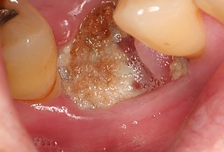
Alveolar osteitis, also known as dry socket, is inflammation of the alveolar bone. Classically, this occurs as a postoperative complication of tooth extraction.

<span class="mw-page-title-main">Maxillary sinus</span> Largest of the paranasal sinuses, and drains into the middle meatus of the nose

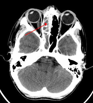
The pyramid-shaped maxillary sinus is the largest of the paranasal sinuses, located in the maxilla. It drains into the middle meatus of the nose through the semilunar hiatus. It is located to the side of the nasal cavity, and below the orbit.

<span class="mw-page-title-main">Dental extraction</span> Operation to remove a tooth

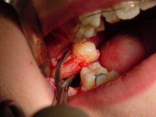
A dental extraction is the removal of teeth from the dental alveolus (socket) in the alveolar bone. Extractions are performed for a wide variety of reasons, but most commonly to remove teeth which have become unrestorable through tooth decay, periodontal disease, or dental trauma, especially when they are associated with toothache. Sometimes impacted wisdom teeth cause recurrent infections of the gum (pericoronitis), and may be removed when other conservative treatments have failed. In orthodontics, if the teeth are crowded, healthy teeth may be extracted to create space so the rest of the teeth can be straightened.

<span class="mw-page-title-main">Pericoronitis</span> Inflammation of the soft tissues surrounding the crown of a partially erupted tooth

Pericoronitis is inflammation of the soft tissues surrounding the crown of a partially erupted tooth, including the gingiva (gums) and the dental follicle. The soft tissue covering a partially erupted tooth is known as an operculum, an area which can be difficult to access with normal oral hygiene methods. The hyponym operculitis technically refers to inflammation of the operculum alone.

<span class="mw-page-title-main">Veterinary dentistry</span> Branch of veterinary medicine

Veterinary dentistry is the field of dentistry applied to the care of animals. It is the art and science of prevention, diagnosis, and treatment of conditions, diseases, and disorders of the oral cavity, the maxillofacial region, and its associated structures as it relates to animals.

A Brodie abscess is a subacute osteomyelitis, which may persist for years before progressing to a chronic, frank osteomyelitis. Classically, this may present after progression to a draining abscess extending from the tibia out through the skin. Occasionally acute osteomyelitis may be contained to a localized area and walled off by fibrous and granulation tissue.

<span class="mw-page-title-main">Osteonecrosis of the jaw</span> Medical condition

Osteonecrosis of the jaw (ONJ) is a severe bone disease (osteonecrosis) that affects the jaws. Various forms of ONJ have been described since 1861, and a number of causes have been suggested in the literature.

<span class="mw-page-title-main">Periapical cyst</span> Medical condition

Commonly known as a dental cyst, the periapical cyst is the most common odontogenic cyst. It may develop rapidly from a periapical granuloma, as a consequence of untreated chronic periapical periodontitis.

<span class="mw-page-title-main">Dental abscess</span> Medical condition

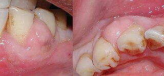
A dental abscess is a localized collection of pus associated with a tooth. The most common type of dental abscess is a periapical abscess, and the second most common is a periodontal abscess. In a periapical abscess, usually the origin is a bacterial infection that has accumulated in the soft, often dead, pulp of the tooth. This can be caused by tooth decay, broken teeth or extensive periodontal disease. A failed root canal treatment may also create a similar abscess.

Mouth infections, also known as oral infections, are a group of infections that occur around the oral cavity. They include dental infection, dental abscess, and Ludwig's angina. Mouth infections typically originate from dental caries at the root of molars and premolars that spread to adjacent structures. In otherwise healthy patients, removing the offending tooth to allow drainage will usually resolve the infection. In cases that spread to adjacent structures or in immunocompromised patients, surgical drainage and systemic antibiotics may be required in addition to tooth extraction. Since bacteria that normally reside in the oral cavity cause mouth infections, proper dental hygiene can prevent most cases of infection. As such, mouth infections are more common in populations with poor access to dental care or populations with health-related behaviors that damage one's teeth and oral mucosa. This is a common problem, representing nearly 36% of all encounters within the emergency department related to dental conditions.

<span class="mw-page-title-main">Medication-related osteonecrosis of the jaw</span> Medical condition

Medication-related osteonecrosis of the jaw is progressive death of the jawbone in a person exposed to a medication known to increase the risk of disease, in the absence of a previous radiation treatment. It may lead to surgical complication in the form of impaired wound healing following oral and maxillofacial surgery, periodontal surgery, or endodontic therapy.

<span class="mw-page-title-main">Mandibular fracture</span> Medical condition

Mandibular fracture, also known as fracture of the jaw, is a break through the mandibular bone. In about 60% of cases the break occurs in two places. It may result in a decreased ability to fully open the mouth. Often the teeth will not feel properly aligned or there may be bleeding of the gums. Mandibular fractures occur most commonly among males in their 30s.

<span class="mw-page-title-main">Periodontal abscess</span> Medical condition

A periodontal abscess, is a localized collection of pus within the tissues of the periodontium. It is a type of dental abscess. A periodontal abscess occurs alongside a tooth, and is different from the more common periapical abscess, which represents the spread of infection from a dead tooth. To reflect this, sometimes the term "lateral (periodontal) abscess" is used. In contrast to a periapical abscess, periodontal abscesses are usually associated with a vital (living) tooth. Abscesses of the periodontium are acute bacterial infections classified primarily by location.
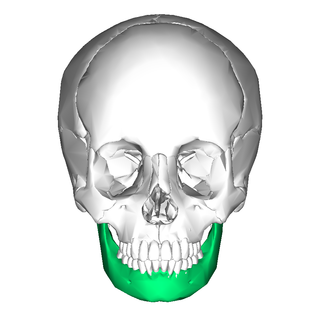
A cyst is a pathological epithelial lined cavity that fills with fluid or soft material and usually grows from internal pressure generated by fluid being drawn into the cavity from osmosis. The bones of the jaws, the mandible and maxilla, are the bones with the highest prevalence of cysts in the human body. This is due to the abundant amount of epithelial remnants that can be left in the bones of the jaws. The enamel of teeth is formed from ectoderm, and so remnants of epithelium can be left in the bone during odontogenesis. The bones of the jaws develop from embryologic processes which fuse, and ectodermal tissue may be trapped along the lines of this fusion. This "resting" epithelium is usually dormant or undergoes atrophy, but, when stimulated, may form a cyst. The reasons why resting epithelium may proliferate and undergo cystic transformation are generally unknown, but inflammation is thought to be a major factor. The high prevalence of tooth impactions and dental infections that occur in the bones of the jaws is also significant to explain why cysts are more common at these sites.

<span class="mw-page-title-main">Osteoradionecrosis</span> Osteoradionecrosis is an Acute form of Osteomyelitis

Osteoradionecrosis (ORN) is a serious complication of radiation therapy in cancer treatment where radiated bone becomes necrotic and exposed. ORN occurs most commonly in the mouth during the treatment of head and neck cancer, and can arise over 5 years after radiation. Common signs and symptoms include pain, difficulty chewing, trismus, mouth-to-skin fistulas and non-healing ulcers.

References

  1. Peravali RK, Jayade B, Joshi A, Shirganvi M, Bhasker Rao C, Gopalkrishnan K (1 October 2011). "Osteomyelitis of Maxilla in Poorly Controlled Diabetics in a Rural Indian Population". Journal of Maxillofacial and Oral Surgery. 11 (1): 57–66. doi:10.1007/s12663-011-0283-0. PMC   3319832 . PMID   23449555.
  2. Neville BW, Damm DD, Allen CA, Bouquot JE (2002). Oral & maxillofacial pathology (2nd ed.). Philadelphia: W.B. Saunders. pp. 126–132. ISBN   0721690033.
  3. 1 2 3 4 5 6 7 8 Topazian RG, Goldberg MH, Hupp JR (2002). Oral and maxillofacial infections (4th ed.). Philadelphia: W.B. Saunders. pp. 214–235. ISBN   978-0721692715.