Fusobacterium

Last updated

Fusobacterium
Fusobacterium novum 01.jpg
Fusobacterium novum in liquid culture
Scientific classification OOjs UI icon edit-ltr.svg
Domain: Bacteria
Phylum: Fusobacteriota
Class: Fusobacteriia
Order: Fusobacteriales
Family: Fusobacteriaceae
Genus: Fusobacterium
Knorr 1923
Type species
Fusobacterium nucleatum [1]
Knorr 1923
Species

See text

Synonyms
  • "Distasoa" Pribram 1929
  • "Necrobacterium" Lahelle & Thjotta 1945
  • "Sphaerocillus" corrig. Prévot 1938
  • "Sphaerophorus" Prévot 1938 non Persoon 1794 non Gray 1864 non Waltl 1835

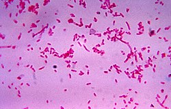
Fusobacterium is a genus of obligate anaerobic, Gram-negative, [2] non-sporeforming bacteria [3] belonging to Gracilicutes. Individual cells are slender, rod-shaped bacilli with pointed ends. [4] [5] Fusobacterium was discovered in 1900 by Courmont and Cade and is common in the flora of humans. [6] [7]

Contents

Strains of Fusobacterium can cause several human diseases and infections, including periodontal diseases, Lemierre's syndrome, [8] oral, head, and neck infections, as well as colorectal cancer and topical skin ulcers. [9]

It has been tied[ clarification needed ] to HIV infection and suboptimal immune recovery. [10] Detection of Fusobacterium is typically through surgical retrieval of tissue, fecal tests, or blood tests in patients showing symptoms. [2] Early detection is preferred and helps to prevent further disease progression. [6]

Although older sources state that Fusobacterium is part of the normal flora of the human oropharynx, the current consensus is that Fusobacterium should always be treated as a pathogen. [11] There are thirteen described Fusobacterium strains; the predominant one affecting humans is F. nucleatum , [12] followed by F. necrophorum , which also affects animals, mainly cattle. [13]

Background

History

Although the genus was not discovered until 1900 by Courmont and Cade, [6] the first documented Fusobacterium infection was reported in 1898 by Veillon and Zuber, who described a case of systemic infection in a young child. [14] The genus was proposed by Knorr in 1923. [15] Fusobacterium has been classically considered a normal part of the human oral, gastrointestinal, and female genital flora, which is why infections are not commonly seen. [7]

Clinical relevance

Fusobacterium is often associated with ulcerative colitis. [16] Research of colon cancer has also shown an overrepresentation of Fusobacterium, both in feces of patients [17] and tumor issue itself. [18] Fusobacterium has also been seen increased in individuals infected with HIV as well as in individuals with suboptimal immune recovery as compared to patients who were not infected and had optimal responses. [10]

Prevalent pathogenic species

F. nucleatum is found in humans more so than any other species of Fusobacterium. [12] It is commonly found in the oral cavity as well as in the intestinal tract. [9] Some of its pathogenic ties include its extraction from amniotic fluid sourced from spontaneous premature labor without reason/a given source. [12] A few additional sources of its pathogenic nature inclue its association with oral inflammation diseases, cancers such as pancreatic, oral, and colorectal, as well as infections of the head and neck. This association is due to the high increase in the prevalence of F. nucleatum in those infected areas. F. nucleatum can worsen or initiate colorectal cancer by stimulating other bacteria such as Streptococcus, Campylobacter spp. and Leptotrichia as well as cancerous gene expression from Beta-catenin signaling. F. nucleatum can be detected in tissues, genomic DNA, and feces using methods such as (FQ, q, and dd) polymerase chain reaction and fluorescence in situ hybridization. However, these are limited because tissues can only be tested after surgery and fecal matter can return false positive results. [9]

F. necrophorum has been found as a common pathogen in the diagnostic of peritonsillar abscess and is more prevalent than other bacteria regarding this infection. It is also the most frequent leading cause associated with Lemierre Syndrome and is not proven to be a normal part of the human oral bacterium population. [8] F. necrophorum commonly infects animals, causing liver abscesses and necrodic diseases, and can combine with other pathogenic bacteria to cause various infections such as foot rot [13] and uterine infections. [19]

Sources of other species of Fusobacterium

Source: [12]

Symptoms and treatment

Fusobacterium infections often cause clinical symptoms such as a fever, inflammation, and a diseased appearance. Further diagnosis can confirm suspicions of Fusobacterium infection through blood testing or culturing the tissue. Upon diagnosing the infection, action to treat it involves the application of antibiotics over a 2-week period which could be in the form of penicillin or other variants as well as using anaerobic antibiotics like clindamycin and metronidazole which work when the Fusobacterium can break down the Beta-lactams. Leaving Fusobacterium untreated could lead to more severe developments of the infection and early testing is recommended. [2] By testing early, fatal diseases such as Lemierre syndrome can be avoided. However, this requires the family physician to be conscious of the danger as infections such as Lemierre syndrome affects younger populations and especially those of male gender. [6] The bacterium is a big anchor for biofilms. [20] [21] It is usually susceptible to clindamycin, [22] while approximately 20% of the clinical strains are resistant to penicillin. [23] In contrast to Bacteroides spp., Fusobacterium has a potent lipopolysaccharide.

Taxonomy

Current species

Fusobacterium is divided into 13 different species, two of which each have their own set of subspecies ( F. nucleatum and F. necrophorum ). [12]

Reclassified species

Other previously declared species of Fusobacterium such as F. symbiosum, F. praecutum, F. plauti,F. alocis, F. sulci, and F. prausnitzii have since been reclassified due to containing different characteristics from the other Fusobacterium members. F. alocis has been reclassified into Filifactor alocis while F. sulci has been reclassified as Eubacterium sulci. F. prausnitzii is a part of the Clostridium leptum subgroup under Eubacterium-like organisms. [12] A few strains F. prausnitzii, a gut commensal associated with healthy patients, was completely reclassified as Faecalibacterium (Clostridiales:Ruminococcaceae) in 2002.

Phylogenic tree

16S rRNA based LTP_08_2023 [24] [25] [26] 120 marker proteins based GTDB 08-RS214 [27] [28] [29]
Fusobacterium

F. perfoetens

F. gonidiaformans

F. equinumDorsch et al. 2001

F. necrophorum

F. n. funduliformeHallé 1898 ex Shinjo et al. 1991

F. n. necrophorum(Flügge 1886) Shinjo et al. 1991

F. ulcerans

F. varium

F. hominisLiu et al. 2022

F. mortiferum

F. necrogenes

F. gastrosuis

F. russii

F. periodonticum

F. polymorphum

F. naviforme(Jungano 1909) Moore & Holdeman 1970

F. vincentii

F. canifelinum

F. nucleatum

F. simiaeSlots and Potts 1982

Filifactor alocis (Cato et al. 1985) Jalava and Eerola 1999

F. animalis

F. watanabeiTomida et al. 2021

Fusobacterium

F. perfoetens(Tissier 1905) Moore and Holdeman 1973

F. ulceransAdriaans and Shah 1988

F. varium(Eggerth and Gagnon 1933) Moore and Holdeman 1969

"Ca. F. pullicola" Gilroy et al. 2021

F. mortiferum (Harris 1901) Moore and Holdeman 1970

F. necrogenes(Weinberg et al. 1937) Moore and Holdeman 1970

F. gonidiaformans(Tunnicliff and Jackson 1925) Moore and Holdeman 1970

F. necrophorum (Flügge 1886) Shinjo et al. 1991

F. gastrosuisDe Witte et al. 2017

F. russii(Hauduroy et al. 1937) Moore and Holdeman 1970

"F. massiliense" Mailhe et al. 2017

F. periodonticumSlots et al. 1984

"F. pseudoperiodonticum" Park et al. 2019

F. animalis(Gharbia & Shah 1992) Kook et al. 2022

F. vincentii(Dzink, Sheenan & Socransky 1990) Kook et al. 2022

F. nucleatum Knorr 1922 (type sp.)

F. polymorphum (Dzink, Sheenan & Socransky 1990) Kook et al. 2022

F. canifelinumcorrig. Conrads et al. 2004

"F. hwasookii" Cho et al. 2014

See also

Related Research Articles

<span class="mw-page-title-main">Lemierre's syndrome</span> Medical condition

Lemierre's syndrome is infectious thrombophlebitis of the internal jugular vein. It most often develops as a complication of a bacterial sore throat infection in young, otherwise healthy adults. The thrombophlebitis is a serious condition and may lead to further systemic complications such as bacteria in the blood or septic emboli.

<span class="mw-page-title-main">Fusobacteriota</span> Phylum of Gram-negative bacteria

Fusobacteriota are obligately anaerobic non-sporeforming Gram-negative bacilli. Since the first reports in the late nineteenth century, various names have been applied to these organisms, sometimes with the same name being applied to different species. More recently, not only have there been changes to the nomenclature, but also attempts to differentiate between species which are believed to be either pathogenic or commensal or both. Because of their asaccharolytic nature, and a general paucity of positive results in routine biochemical tests, laboratory identification of the Fusobacteriota has been difficult. However, the application of novel molecular biological techniques to taxonomy has established a number of new species, together with the subspeciation of Fusobacterium necrophorum and F. nucleatum, and provided new methods for identification. The involvement of Fusobacteriota in a wide spectrum of human infections causing tissue necrosis and septicaemia has long been recognised, and, more recently, their importance in intra-amniotic infections, premature labour and tropical ulcers has been reported.

<span class="mw-page-title-main">Clindamycin</span> Antibiotic

Clindamycin is an antibiotic medication used for the treatment of a number of bacterial infections, including osteomyelitis (bone) or joint infections, pelvic inflammatory disease, strep throat, pneumonia, acute otitis media, and endocarditis. It can also be used to treat acne, and some cases of methicillin-resistant Staphylococcus aureus (MRSA). In combination with quinine, it can be used to treat malaria. It is available by mouth, by injection into a vein, and as a cream or a gel to be applied to the skin or in the vagina.

<i>Actinomyces</i> Genus of bacteria

Actinomyces is a genus of the Actinomycetia class of bacteria. They all are gram-positive. Actinomyces species are facultatively anaerobic and they grow best under anaerobic conditions. Actinomyces species may form endospores, and while individual bacteria are rod-shaped, Actinomyces colonies form fungus-like branched networks of hyphae. The aspect of these colonies initially led to the incorrect assumption that the organism was a fungus and to the name Actinomyces, "ray fungus".

<span class="mw-page-title-main">Peritonsillar abscess</span> Pus behind the tonsil due to an infection

Peritonsillar abscess (PTA), also known as quinsy, is an accumulation of pus due to an infection behind the tonsil. Symptoms include fever, throat pain, trouble opening the mouth, and a change to the voice. Pain is usually worse on one side. Complications may include blockage of the airway or aspiration pneumonitis.

<i>Peptostreptococcus</i> Genus of bacteria

Peptostreptococcus is a genus of anaerobic, Gram-positive, non-spore forming bacteria. The cells are small, spherical, and can occur in short chains, in pairs or individually. They typically move using cilia. Peptostreptococcus are slow-growing bacteria with increasing resistance to antimicrobial drugs. Peptostreptococcus is a normal inhabitant of the healthy lower reproductive tract of women.

<i>Bacteroides</i> Genus of bacteria

Bacteroides is a genus of Gram-negative, obligate anaerobic bacteria. Bacteroides species are non endospore-forming bacilli, and may be either motile or nonmotile, depending on the species. The DNA base composition is 40–48% GC. Unusual in bacterial organisms, Bacteroides membranes contain sphingolipids. They also contain meso-diaminopimelic acid in their peptidoglycan layer.

<i>Bacteroides fragilis</i> Species of bacterium

Bacteroides fragilis is an anaerobic, Gram-negative, pleomorphic to rod-shaped bacterium. It is part of the normal microbiota of the human colon and is generally commensal, but can cause infection if displaced into the bloodstream or surrounding tissue following surgery, disease, or trauma.

Dysbiosis is characterized by a disruption to the microbiome resulting in an imbalance in the microbiota, changes in their functional composition and metabolic activities, or a shift in their local distribution. For example, a part of the human microbiota such as the skin flora, gut flora, or vaginal flora, can become deranged, with normally dominating species underrepresented and normally outcompeted or contained species increasing to fill the void. Dysbiosis is most commonly reported as a condition in the gastrointestinal tract.

<i>Eikenella corrodens</i> Species of bacterium

Eikenella corrodens is a Gram-negative facultative anaerobic bacillus that can cause severe invasive disease in humans. It was first identified by M. Eiken in 1958, who called it Bacteroides corrodens. E. corrodens is a rare pericarditis associated pathogen. It is a fastidious, slow growing, human commensal bacillus, capable of acting as an opportunistic pathogen and causing abscesses in several anatomical sites, including the liver, lung, spleen, and submandibular region. E. corrodens could independently cause serious infection in both immunocompetent and immunocompromised hosts.

Fusobacterium necrophorum is a species of bacteria responsible for Lemierre's syndrome. It has also been known to cause sinusitis, mastoiditis, and odontogenic infections.

Dichelobacter nodosus, formerly Bacteroides nodosus, is a Gram-negative, obligate anaerobe of the family Cardiobacteriaceae. It has polar fimbriae and is the causative agent of ovine foot rot as well as interdigital dermatitis. It is the lone species in the genus Dichelobacter.

Fusobacterium nucleatum is a Gram-negative, anaerobic oral bacterium, commensal to the human oral cavity, that plays a role in periodontal disease. This organism is commonly recovered from different monocultured microbial and mixed infections in humans and animals. In health and disease, it is a key component of periodontal plaque due to its abundance and its ability to coaggregate with other bacteria species in the oral cavity.

Capnocytophaga is a genus of Gram-negative bacteria. Normally found in the oropharyngeal tract of mammals, they are involved in the pathogenesis of some animal bite wounds and periodontal diseases.

A septic embolism is a type of embolism that is infected with bacteria, resulting in the formation of pus. These may become dangerous if dislodged from their original location. Like other emboli, a septic embolism may be fatal.

<i>Veillonella parvula</i> Species of bacterium

Veillonella parvula is a strictly anaerobic, Gram-negative, coccus-shaped bacterium in the genus Veillonella. It is a normal part of the oral flora but can be associated with diseases such as periodontitis and dental caries as well as various systemic infections, including meningitis and osteomyelitis. It has also been isolated from women with bacterial vaginosis and has been associated with hypertension together with Campylobacter rectus and Prevotella melaninogenica.

<i>Porphyromonas</i> Genus of bacteria

Porphyromonas is a Gram-negative, non-spore-forming, obligately anaerobic and non-motile genus from the family Porphyromonadaceae. There were 16 different Porphyromonas species documented as of 2015, which reside in both animal and human reservoirs. It was discovered more recently that Porphyromonas also exist in the environment, albeit to a lesser extent. This genus is notably implicated in the modulation of oral cavity, respiratory tract, and gastrointestinal tract disease states. It is suggested that Porphyromonas either operate as benign bacteria pertinent to host immunity or are potential pathobionts that opportunistically provoke diseased states when homeostasis is disrupted. Despite its characterization not being fully elucidated due to sparse research, various studies report the prevalence of this genus at 58.7% in healthy states compared with 41.3% in diseased states.

Anaerobic infections are caused by anaerobic bacteria. Obligately anaerobic bacteria do not grow on solid media in room air ; facultatively anaerobic bacteria can grow in the presence or absence of air. Microaerophilic bacteria do not grow at all aerobically or grow poorly, but grow better under 10% carbon dioxide or anaerobically. Anaerobic bacteria can be divided into strict anaerobes that can not grow in the presence of more than 0.5% oxygen and moderate anaerobic bacteria that are able of growing between 2 and 8% oxygen. Anaerobic bacteria usually do not possess catalase, but some can generate superoxide dismutase which protects them from oxygen.

<i>Clostridium tertium</i> Species of bacterium

Clostridium tertium is an anaerobic, motile, gram-positive bacterium. Although it can be considered an uncommon pathogen in humans, there has been substantial evidence of septic episodes in human beings. C. tertium is easily decolorized in Gram-stained smears and can be mistaken for a Gram-negative organism. However, C.tertium does not grow on selective media for Gram-negative organisms.

Anaerococcus is a genus of bacteria. Its type species is Anaerococcus prevotii. These bacteria are Gram-positive and strictly anaerobic. The genus Anaerococcus was proposed in 2001. Its genome was sequenced in August 2009. The genus Anaerococcus is one of six genera classified within the group GPAC. These six genera are found in the human body as part of the commensal human microbiota.

References

  1. Parte, A.C. "Fusobacterium". LPSN .
  2. 1 2 3 Arane, Karen; Goldman, Ran (October 2016). "Fusobacterium infections in children". Canadian Family Physician. 62 (10): 813–814. PMC   5063768 . PMID   27737977.
  3. Broadley, Marissa; Schweon, Steven J. (May 2017). "Get the facts about Fusobacterium". Nursing2023. 47 (5): 64–65. doi:10.1097/01.NURSE.0000515524.23032.d5. ISSN   0360-4039. PMID   28445341. S2CID   41693034.
  4. Madigan M; Martinko J, eds. (2005). Brock Biology of Microorganisms (11th ed.). Prentice Hall. ISBN   978-0-13-144329-7.
  5. Taber's Cyclopedic Medical Dictionary, 22nd Edition, ISBN   9780803629790, (2009)n p.983
  6. 1 2 3 4 Cheung, Winson (September 2007). "Fusobacterium". Canadian Family Physician. 53 (9): 1451–1453. PMC   2234623 . PMID   17872873.
  7. 1 2 Garcia-Carretero, Rafael; Lopez-Lomba, Marta; Carrasco-Fernandez, Blanca; Duran-Valle, Maria Teresa (October 2017). "Clinical Features and Outcomes of Fusobacterium Species Infections in a Ten-Year Follow-up". Journal of Critical Care Medicine (Universitatea de Medicina Si Farmacie Din Targu-Mures). 3 (4): 141–147. doi:10.1515/jccm-2017-0029. PMC   5769905 . PMID   29967887.
  8. 1 2 Ehlers Klug, Tejs; Rusan, Maria; Fuursted, Kurt; Ovesen, Therese (15 November 2009). "Fusobacterium necrophorum: Most Prevalent Pathogen in Peritonsillar Abscess in Denmark". Clinical Infectious Diseases. 49 (10): 1467–1472. doi: 10.1086/644616 . PMID   19842975.
  9. 1 2 3 Shang, Fu-Mei; Liu, Hong-Li (15 March 2018). "Fusobacterium nucleatum and colorectal cancer: A review". World Journal of Gastrointestinal Oncology. 10 (3): 71–81. doi: 10.4251/wjgo.v10.i3.71 . PMC   5852398 . PMID   29564037.
  10. 1 2 Lee, Soo Ching; Chua, Ling Ling; Yap, Siew Hwei; Khang, Tsung Fei; Leng, Chan Yoon; Raja Azwa, Raja Iskandar; Lewin, Sharon R.; Kamarulzaman, Adeeba; Woo, Yin Ling; Lim, Yvonne Ai Lian; Loke, P’ng; Rajasuriar, Reena (2018-09-24). "Enrichment of gut-derived Fusobacterium is associated with suboptimal immune recovery in HIV-infected individuals". Scientific Reports. 8 (1): 14277. Bibcode:2018NatSR...814277L. doi:10.1038/s41598-018-32585-x. ISSN   2045-2322. PMC   6155144 . PMID   30250162.
  11. Aliyu SH, Marriott RK, Curran MD, et al. (October 2004). "Real-time PCR investigation into the importance of Fusobacterium necrophorum as a cause of acute pharyngitis in general practice". J Med Microbiol. 53 (Pt 10): 1029–35. doi: 10.1099/jmm.0.45648-0 . PMID   15358827.
  12. 1 2 3 4 5 6 Citron, Diane (September 2002). "Update on the Taxonomy and Clinical Aspects of the Genus Fusobacterium". Clinical Infectious Diseases. 35 (s1): S22–S27. doi:10.1086/341916. PMID   12173104 . Retrieved 5 May 2023.
  13. 1 2 Tadepalli, S.; Narayanan, S. K.; Stewart, G. C.; Chengappa, M. M.; Nagaraja, T. G. (2009-02-01). "Fusobacterium necrophorum: A ruminal bacterium that invades liver to cause abscesses in cattle". Anaerobe. Foodborne and Gastrointestinal Pathogen Ecology and Control in the Intestinal Tract. 15 (1): 36–43. doi:10.1016/j.anaerobe.2008.05.005. ISSN   1075-9964. PMID   18595747.
  14. Creemers-Schild, D; Grounthoud, F; Spanjaard, L; Visser, L G; Brouwer, C N M; Kuijper, E J (May 2014). "Fusobacterium necrophorum, an emerging pathogen of otogenic and paranasal infections?". New Microbes and New Infections. 2 (3): 52–57. doi:10.1002/nmi2.39. PMC   4184658 . PMID   25356344.
  15. Bennett, K. W.; Eley, A.YR 1993 (1993). "Fusobacteria: New taxonomy and related diseases". Journal of Medical Microbiology. 39 (4): 246–254. doi: 10.1099/00222615-39-4-246 . ISSN   1473-5644. PMID   8411084.{{cite journal}}: CS1 maint: numeric names: authors list (link)
  16. Su, Wenhao; Chen, Yongyu; Cao, Pan; Chen, Yan; Guo, Yuanmei; Wang, Siwei; Dong, Weiguo (2020-11-27). "Fusobacterium nucleatum Promotes the Development of Ulcerative Colitis by Inducing the Autophagic Cell Death of Intestinal Epithelial". Frontiers in Cellular and Infection Microbiology. 10: 594806. doi: 10.3389/fcimb.2020.594806 . PMC   7728699 . PMID   33330137.
  17. Ahn, J.; Sinha, R.; Pei, Z.; Dominianni, C.; Wu, J.; Shi, J.; Goedert, J. J.; Hayes, R. B.; Yang, L. (18 December 2013). "Human Gut Microbiome and Risk for Colorectal Cancer". J Natl Cancer Inst. 105 (24): 1907–1911. doi:10.1093/jnci/djt300. PMC   3866154 . PMID   24316595.
  18. Alice Park (18 October 2011). "A Surprising Link Between Bacteria and Colon Cancer". Time.com. Retrieved 18 October 2011.
  19. Bicalho, M. L. S.; Machado, V. S.; Oikonomou, G.; Gilbert, R. O.; Bicalho, R. C. (2012-05-25). "Association between virulence factors of Escherichia coli, Fusobacterium necrophorum, and Arcanobacterium pyogenes and uterine diseases of dairy cows". Veterinary Microbiology. 157 (1): 125–131. doi:10.1016/j.vetmic.2011.11.034. ISSN   0378-1135. PMID   22186615.
  20. Saito, Y.; Fujii, R.; Nakagawa, K.-I.; Kuramitsu, H. K.; Okuda, K.; Ishihara, K. (February 2008). "Stimulation of Fusobacterium nucleatum biofilm formation by Porphyromonas gingivalis". Oral Microbiology and Immunology. 23 (1): 1–6. doi:10.1111/j.1399-302X.2007.00380.x. hdl: 10130/821 . PMID   18173791. S2CID   9675789.
  21. Okuda, Tamaki; Kokubu, Eitoyo; Kawana, Tomoko; Saito, Atsushi; Okuda, Katsuji; Ishihara, Kazuyuki (Feb 2012). "Synergy in biofilm formation between Fusobacterium nucleatum and Prevotella species" (PDF). Anaerobe. 18 (1): 110–116. doi:10.1016/j.anaerobe.2011.09.003. hdl: 10130/3922 . ISSN   1095-8274. PMID   21945879.
  22. "Clindamycin" (PDF). Davis. 2017. Archived from the original (PDF) on November 14, 2017. Retrieved November 14, 2017.
  23. Di Bella, Stefano; Antonello, Roberta Maria; Sanson, Gianfranco; Maraolo, Alberto Enrico; Giacobbe, Daniele Roberto; Sepulcri, Chiara; Ambretti, Simone; Aschbacher, Richard; Bartolini, Laura; Bernardo, Mariano; Bielli, Alessandra (June 2022). "Anaerobic bloodstream infections in Italy (ITANAEROBY): A 5-year retrospective nationwide survey". Anaerobe. 75: 102583. doi:10.1016/j.anaerobe.2022.102583. hdl: 11368/3020691 . PMID   35568274. S2CID   248736289.
  24. "The LTP" . Retrieved 20 November 2023.
  25. "LTP_all tree in newick format" . Retrieved 20 November 2023.
  26. "LTP_08_2023 Release Notes" (PDF). Retrieved 20 November 2023.
  27. "GTDB release 08-RS214". Genome Taxonomy Database . Retrieved 10 May 2023.
  28. "bac120_r214.sp_label". Genome Taxonomy Database . Retrieved 10 May 2023.
  29. "Taxon History". Genome Taxonomy Database . Retrieved 10 May 2023.