Related Research Articles

The salivary glands in many vertebrates including mammals are exocrine glands that produce saliva through a system of ducts. Humans have three paired major salivary glands, as well as hundreds of minor salivary glands. Salivary glands can be classified as serous, mucous, or seromucous (mixed).

The parotid gland is a major salivary gland in many animals. In humans, the two parotid glands are present on either side of the mouth and in front of both ears. They are the largest of the salivary glands. Each parotid is wrapped around the mandibular ramus, and secretes serous saliva through the parotid duct into the mouth, to facilitate mastication and swallowing and to begin the digestion of starches. There are also two other types of salivary glands; they are submandibular and sublingual glands. Sometimes accessory parotid glands are found close to the main parotid glands.
The paired submandibular glands are major salivary glands located beneath the floor of the mouth. In adult humans, they each weigh about 15 grams and contribute some 60–67% of unstimulated saliva secretion; on stimulation their contribution decreases in proportion as parotid gland secretion rises to 50%. The average length of the normal adult human submandibular salivary gland is approximately 27 mm, while the average width is approximately 14.3 mm.

Xerostomia, also known as dry mouth, is a subjective complaint of dryness in the mouth, which may be associated with a change in the composition of saliva, or reduced salivary flow, or have no identifiable cause.
Parotitis is an inflammation of one or both parotid glands, the major salivary glands located on either side of the face, in humans. The parotid gland is the salivary gland most commonly affected by inflammation.

Stomatitis is inflammation of the mouth and lips. It refers to any inflammatory process affecting the mucous membranes of the mouth and lips, with or without oral ulceration.
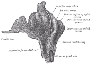
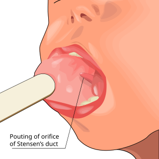
The parotid duct or Stensen duct is a salivary duct. It is the route that saliva takes from the major salivary gland, the parotid gland, into the mouth. It opens into the mouth opposite the second upper molar tooth.

A ranula is a mucus extravasation cyst involving a sublingual gland and is a type of mucocele found on the floor of the mouth. Ranulae present as a swelling of connective tissue consisting of collected mucin from a ruptured salivary gland caused by local trauma. If small and asymptomatic further treatment may not be needed, otherwise minor oral surgery may be indicated.
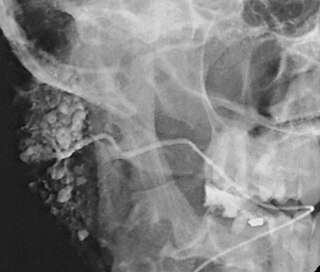
Sialography is the radiographic examination of the salivary glands. It usually involves the injection of a small amount of contrast medium into the salivary duct of a single gland, followed by routine X-ray projections.

Sialadenitis (sialoadenitis) is inflammation of salivary glands, usually the major ones, the most common being the parotid gland, followed by submandibular and sublingual glands. It should not be confused with sialadenosis (sialosis) which is a non-inflammatory enlargement of the major salivary glands.

Sialolithiasis is a crystallopathy where a calcified mass or sialolith forms within a salivary gland, usually in the duct of the submandibular gland. Less commonly the parotid gland or rarely the sublingual gland or a minor salivary gland may develop salivary stones.

Cheilitis is a medical condition characterized by inflammation of the lips. The inflammation may include the perioral skin, the vermilion border, or the labial mucosa. The skin and the vermilion border are more commonly involved, as the mucosa is less affected by inflammatory and allergic reactions.

The Stafne defect is a depression of the mandible, most commonly located on the lingual surface. The Stafne defect is thought to be a normal anatomical variant, as the depression is created by ectopic salivary gland tissue associated with the submandibular gland and does not represent a pathologic lesion as such. This cavity is commonly observed on panoramic radiograph.

Salivary gland tumours, also known as mucous gland adenomas or neoplasms, are tumours that form in the tissues of salivary glands. The salivary glands are classified as major or minor. The major salivary glands consist of the parotid, submandibular, and sublingual glands. The minor salivary glands consist of 800 to 1000 small mucus-secreting glands located throughout the lining of the oral cavity. Patients with these types of tumours may be asymptomatic.
Sialoendoscopy is a minimally invasive technique that allows for salivary gland surgery for the safe and effective treatment of obstructive salivary gland disorders and other conditions of the salivary glands. During sialoendoscopy a small endoscope is placed into the salivary glands through the salivary ducts that empty into the mouth. The procedure is not exclusively diagnostic, but is interventional; thus, it can be used for the extraction of salivary stones, salivary duct lavage, dilatation of stenotic segments, or instillation of various medications such as corticosteroids or antibiotics. Thus, sialoendoscopy is an efficient yet simple mode of treatment for major salivary gland obstructions, strictures and sialoliths. Depending on the obstruction, sialoendoscopy can be conducted under local anesthesia in an outpatient office or in the operating room under general anesthesia.

Salivary gland diseases (SGDs) are multiple and varied in cause. There are three paired major salivary glands in humans: the parotid glands, the submandibular glands, and the sublingual glands. There are also about 800–1,000 minor salivary glands in the mucosa of the mouth. The parotid glands are in front of the ears, one on side, and secrete mostly serous saliva, via the parotid ducts, into the mouth, usually opening roughly opposite the second upper molars. The submandibular gland is medial to the angle of the mandible, and it drains its mixture of serous and mucous saliva via the submandibular duct into the mouth, usually opening in a punctum in the floor of mouth. The sublingual gland is below the tongue, on the floor of the mouth; it drains its mostly mucous saliva into the mouth via about 8–20 ducts, which open along the plica sublingualis, a fold of tissue under the tongue.
Chronic sclerosing sialadenitis is a chronic (long-lasting) inflammatory condition affecting the salivary gland. Relatively rare in occurrence, this condition is benign, but presents as hard, indurated and enlarged masses that are clinically indistinguishable from salivary gland neoplasms or tumors. It is now regarded as a manifestation of IgG4-related disease.
Salivary duct stricture is narrowing of the duct of a major salivary gland.
Ductal papilloma is a group of rare and benign papillary salivary gland tumors arising from the duct system:
Sialectasis is cystic dilation of the ducts of salivary glands. It may be caused by salivary duct strictures or stones (sialolithiasis). It can also rarely be congenital.
References
- 1 2 3 4 White SC; Pharoah MJ (25 September 2008). Oral Radiology: Principles and Interpretation. Elsevier Health Sciences. pp. 1420–1421. ISBN 0-323-07592-4.
- ↑ Yousem DM; Grossman RI (2010). Neuroradiology: The Requisites. Elsevier Health Sciences. p. 480. ISBN 0-323-04521-9.
- 1 2 Mafee M; Becker M (21 November 2012). Imaging of the Head and Neck. Thieme. pp. 10–11. ISBN 978-3-13-150531-6.
- 1 2 Whaites E; Drage N (2013). Essentials of Dental Radiography and Radiology. Elsevier Health Sciences. p. 453. ISBN 978-0-7020-4599-8.
- ↑ Michael Glick (1 September 2014). Burket's oral medicine: 12th edition. coco. pp. 242–. ISBN 978-1-60795-188-9.