Stomatitis

Last updated
Stomatitis
Kwashiorkor 6180.jpg
An infant with stomatitis due to kwashiorkor and an accompanying Vitamin B deficiency.
Specialty Dermatology

Stomatitis is inflammation of the mouth and lips. [1] It refers to any inflammatory process affecting the mucous membranes of the mouth and lips, with or without oral ulceration. [2]

Contents

In its widest meaning, stomatitis can have a multitude of different causes and appearances. Common causes include infections, nutritional deficiencies, allergic reactions, radiotherapy, and many others.

When inflammation of the gums and the mouth generally presents itself, sometimes the term gingivostomatitis is used, though this is also sometimes used as a synonym for herpetic gingivostomatitis.

The term is derived from the Greek stoma (στόμα), meaning "mouth", and the suffix -itis (-ῖτις), meaning "inflammation".

Causes

Nutritional deficiency

Malnutrition (improper dietary intake) or malabsorption (poor absorption of nutrients into the body) can lead to nutritional deficiency states, several of which can lead to stomatitis. For example, deficiencies of iron, vitamin B2 (riboflavin), [3] :490 vitamin B3 (niacin), vitamin B6 (pyridoxine), vitamin B9 (folic acid) or vitamin B12 (cobalamine) may all manifest as stomatitis. Iron is necessary for the upregulation of transcriptional elements for cell replication and repair. Lack of iron can cause genetic downregulation of these elements, leading to ineffective repair and regeneration of epithelial cells, especially in the mouth and lips. Many disorders which cause malabsorption can cause deficiencies, which in turn causes stomatitis. Examples include tropical sprue. [3] :49

Aphthous stomatitis

Aphthous stomatitis (canker sores) is the recurrent appearance of mouth ulcers in otherwise healthy individuals. The cause is not completely understood, but it is thought that the condition represents a T cell mediated immune response which is triggered by a variety of factors. The individual ulcers (aphthae) recur periodically and heal completely, although in the more severe forms, new ulcers may appear in other parts of the mouth before the old ones have finished healing. Aphthous stomatitis is one of the most common diseases of the oral mucosa, and is thought to affect about 20% of the general population to some degree. [4] The symptoms range from a minor nuisance to being disabling in their impact on eating, swallowing, and talking, and the severe forms can cause people to lose weight. There is no cure for aphthous stomatitis, [5] and therapies are aimed at alleviating the pain, reducing the inflammation and promoting healing of the ulcers, but there is little evidence of efficacy for any treatment that has been used.

Angular stomatitis

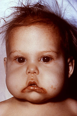
Inflammation of the corners (angles) of the lips is termed angular stomatitis or angular cheilitis. In children a frequent cause is repeated lip-licking, and in adults it may be a sign of underlying iron deficiency anemia, or vitamin B deficiencies (e.g., B2-riboflavin, B9-folate, or B12-cobalamin, which in turn may be evidence of poor diets or malnutrition such as celiac disease).

Also, angular cheilitis can be caused by a patient's jaws at rest being 'overclosed' due to edentulousness or tooth wear, causing the jaws to come to rest closer together than if the complete/unaffected dentition were present. This causes skin folds around the angle of the mouth which are kept moist by saliva, which in turn favours infection; mostly by Candida albicans or similar species. Treatment usually involves the administration of topical nystatin or similar antifungal agents. Another treatment can be to correct the jaw relationship with dental treatment (e.g., dentures or occlusal adjustment).

This is a common condition present in denture wearers. It appears as reddened but painless mucosa beneath the denture. 90% of cases are associated with Candida species, and it is the most common form of oral candidiasis. Treatment is by antifungal medication and improved dental hygiene, such as not wearing the denture during sleep.

Allergic contact stomatitis

Allergic contact stomatitis (also termed "allergic gingivostomatitis" or "allergic contact gingivostomatitis") [6] is a type IV (delayed) hypersensitivity reaction that occurs in susceptible atopic individuals when allergens penetrate the skin or mucosa. [7]

Allergens, which may be different for different individuals, combine with epithelial-derived proteins, forming haptens which bind with Langerhans cells in the mucosa, which in turn present the antigen on their surface to T lymphocytes, sensitizing them to that antigen and causing them to produce many specific clones. The second time that specific antigen is encountered, an inflammatory reaction is triggered at the site of exposure. [7] Allergic contact stomatitis is less common than allergic contact dermatitis because the mouth is coated in saliva, which washes away antigens and acts as a barrier. [7] The oral mucosa is also more vascular (has a better blood supply) than skin, meaning that any antigens are more quickly removed from the area by the circulation. [7] Finally, there is substantially less keratin in oral mucosa, meaning that there is less likelihood that haptens will form. [7]

Allergic contact stomatitis appears as non-specific inflammation, so it may be mistaken for chronic physical irritation. [7] There may be burning or soreness of the mouth and ulceration. [7] Chronic exposure to the allergen may result in a lichenoid lesion. [7] Plasma cell gingivitis may also occur, which may be accompanied by glossitis and cheilitis. [7]

Patch test Epikutanni-test.jpg
Patch test

Allergens that may cause allergic contact stomatitis in some individuals include cinnamaldehyde, Balsam of Peru, peppermint, mercury, gold, pyrophosphates, zinc citrate, free acrylic monomer, nickel, fluoride, and sodium lauryl sulfate. [7] [8] [9] [10] [11] [12] [13] These allergens may originate from many sources, including various foods and drink, chewing gum, toothpaste, mouthwash, dental floss, dental fillings, dentures, orthodontic bands or wires, and many other sources. [7] If the substance containing the allergen comes into contact with the lips, allergic contact cheilitis can occur, together with allergic contact stomatitis.

The diagnosis is confirmed by patch test, and management is by avoidance of exposure to the allergen. [7]

Migratory stomatitis

Migratory stomatitis (or geographic stomatitis) is an atypical presentation of a condition which normally presents on the tongue, termed geographic tongue. Geographic tongue is so named because there are atrophic, erythematous areas of depapillation that migrate over time, giving a map-like appearance.

In migratory stomatitis, other mucosal sites in the mouth, such as the ventral surface (undersurface) of the tongue, buccal mucosa, labial mucosa, soft palate, or floor of mouth may be afflicted with identical lesions, usually in addition to the tongue. [14] Apart from not being restricted to the tongue, migratory stomatitis is an identical condition in every regard to geographic tongue. Another synonym for geographic tongue which uses the term stomatitis is "stomatitis areata migrans".

Herpetic gingivostomatitis

Herpetic stomatitis (herpetic gingivostomatitis) Stomatitis herpetica.jpg
Herpetic stomatitis (herpetic gingivostomatitis)

This is inflammation of the mouth caused by herpes simplex virus.

Irradiation and chemotherapy

Stomatitis may also be caused by chemotherapy, or radiation therapy of the oropharyngeal area. [15] The term mucositis is sometimes used synonymously with stomatitis, however the former usually refers to mucosal reactions to radiotherapy or chemotherapy, and may occur anywhere in the gastrointestinal tract and not just in the mouth. [16]

Necrotizing ulcerative gingivostomatitis

The term necrotizing ulcerative gingivostomatitis is sometimes used as a synonym of the necrotizing periodontal disease more commonly termed necrotizing ulcerative gingivitis, or a more severe form (also termed necrotizing stomatitis). The term necrotizing gingivostomatitis is also sometimes used. [17]

Stomatitis nicotina

Also called smoker's palatal keratosis, [18] :176 this condition may occur in smokers, especially pipe smokers. The palate appears dry and cracked, and white from keratosis. The minor salivary glands appear as small, red and swollen bumps. It is not a premalignant condition, and the appearance reverses if the smoking is stopped. [18] :176

Chronic ulcerative stomatitis

Chronic ulcerative stomatitis is a condition with specific immunopathologic features, which was first described in 1990. [19] It is characterized by erosions and ulcerations which relapse and remit. Lesions are located on the buccal mucosa (inside of the cheeks) or on the gingiva (gums). [20] [21] The condition resembles oral lichen planus when biopsied.

The diagnosis is made by microscopic examination of biopsy tissue: direct immunofluorescence can reveal the presence of antinuclear antibodies specifically directed against the ΔNp63α form of the p63 protein, which is normally expressed within the basal layer of stratified epithelium. [19] Treatment with hydroxychloroquine can be effective. [19]

Plasma cell gingivostomatitis

Terms such as plasma cell gingivostomatitis, [22] atypical gingivostomatitis and idiopathic gingivostomatitis [23] [24] are sometimes a synonym for plasma cell gingivitis, or specifically to refer to a severe form of plasma cell gingivitis.

Other forms of stomatitis

Related Research Articles

<span class="mw-page-title-main">Mouthwash</span> Liquid rinse for oral hygiene

Mouthwash, mouth rinse, oral rinse, or mouth bath is a liquid which is held in the mouth passively or swirled around the mouth by contraction of the perioral muscles and/or movement of the head, and may be gargled, where the head is tilted back and the liquid bubbled at the back of the mouth.

<span class="mw-page-title-main">Mouth ulcer</span> Sore on the mucous membrane of the oral cavity

A mouth ulcer (aphtha) is an ulcer that occurs on the mucous membrane of the oral cavity. Mouth ulcers are very common, occurring in association with many diseases and by many different mechanisms, but usually there is no serious underlying cause. Rarely, a mouth ulcer that does not heal may be a sign of oral cancer. These ulcers may form individually or multiple ulcers may appear at once. Once formed, an ulcer may be maintained by inflammation and/or secondary infection.

<span class="mw-page-title-main">Necrotizing gingivitis</span> Non-contagious, painful bacterial infection of the gums

Necrotizing gingivitis (NG) is a common, non-contagious infection of the gums with sudden onset. The main features are painful, bleeding gums, and ulceration of inter-dental papillae. This disease, along with necrotizing periodontitis (NP) and necrotizing stomatitis, is classified as a necrotizing periodontal disease, one of the three general types of gum disease caused by inflammation of the gums (periodontitis).

<span class="mw-page-title-main">Oral candidiasis</span> Fungal infection

Oral candidiasis, also known as oral thrush among other names, is candidiasis that occurs in the mouth. That is, oral candidiasis is a mycosis of Candida species on the mucous membranes of the mouth.

<span class="mw-page-title-main">Aphthous stomatitis</span> Common condition of mouth ulcers

Aphthous stomatitis, or recurrent aphthous stomatitis (RAS), is a common condition characterized by the repeated formation of benign and non-contagious mouth ulcers (aphthae) in otherwise healthy individuals. The informal term canker sore is also used, mainly in North America, although it may also refer to other types of mouth ulcers. The cause is not completely understood but involves a T cell-mediated immune response triggered by a variety of factors which may include nutritional deficiencies, local trauma, stress, hormonal influences, allergies, genetic predisposition, certain foods, dehydration, some food additives, or some hygienic chemical additives like SDS.

<span class="mw-page-title-main">Glossitis</span> Medical condition

Glossitis can mean soreness of the tongue, or more usually inflammation with depapillation of the dorsal surface of the tongue, leaving a smooth and erythematous (reddened) surface,. In a wider sense, glossitis can mean inflammation of the tongue generally. Glossitis is often caused by nutritional deficiencies and may be painless or cause discomfort. Glossitis usually responds well to treatment if the cause is identified and corrected. Tongue soreness caused by glossitis is differentiated from burning mouth syndrome, where there is no identifiable change in the appearance of the tongue, and there are no identifiable causes.

The oral mucosa is the mucous membrane lining the inside of the mouth. It comprises stratified squamous epithelium, termed "oral epithelium", and an underlying connective tissue termed lamina propria. The oral cavity has sometimes been described as a mirror that reflects the health of the individual. Changes indicative of disease are seen as alterations in the oral mucosa lining the mouth, which can reveal systemic conditions, such as diabetes or vitamin deficiency, or the local effects of chronic tobacco or alcohol use. The oral mucosa tends to heal faster and with less scar formation compared to the skin. The underlying mechanism remains unknown, but research suggests that extracellular vesicles might be involved.

<span class="mw-page-title-main">Noma (disease)</span> Medical condition

Noma is a rapidly progressive and often fatal infection of the mouth and face. This disease predominantly affects children between the ages of two and six years old in the least developed countries around the world, primarily in the "noma belt" of sub-Saharan Africa. Noma has also been seen in severely immunocompromised people in the developed world.

<span class="mw-page-title-main">Herpetic gingivostomatitis</span> Medical condition

Gingivostomatitis is a combination of gingivitis and stomatitis, or an inflammation of the oral mucosa and gingiva. Herpetic gingivostomatitis is often the initial presentation during the first ("primary") herpes simplex infection. It is of greater severity than herpes labialis which is often the subsequent presentations. Primary herpetic gingivostomatitis is the most common viral infection of the mouth.

<span class="mw-page-title-main">Angular cheilitis</span> Cheilitis characterized by inflammation of one or both of the corners of the mouth

Angular cheilitis (AC) is inflammation of one or both corners of the mouth. Often the corners are red with skin breakdown and crusting. It can also be itchy or painful. The condition can last for days to years. Angular cheilitis is a type of cheilitis.

An oral medicine or stomatology doctor/dentist has received additional specialized training and experience in the diagnosis and management of oral mucosal abnormalities including oral cancer, salivary gland disorders, temporomandibular disorders and facial pain, taste and smell disorders; and recognition of the oral manifestations of systemic and infectious diseases. It lies at the interface between medicine and dentistry. An oral medicine doctor is trained to diagnose and manage patients with disorders of the orofacial region, essentially as a "physician of the mouth".

Burning mouth syndrome (BMS) is a burning, tingling or scalding sensation in the mouth, lasting for at least four to six months, with no underlying known dental or medical cause. No related signs of disease are found in the mouth. People with burning mouth syndrome may also have a subjective xerostomia, paraesthesia, or an altered sense of taste or smell.

<span class="mw-page-title-main">Cheilitis</span> Inflammation of the lips

Cheilitis is a medical condition characterized by inflammation of the lips. The inflammation may include the perioral skin, the vermilion border, or the labial mucosa. The skin and the vermilion border are more commonly involved, as the mucosa is less affected by inflammatory and allergic reactions.

Orofacial granulomatosis (OFG) is a condition characterized by persistent enlargement of the soft tissues of the mouth, lips and the area around the mouth on the face, causing in most cases extreme pain. The mechanism of the enlargement is granulomatous inflammation. The underlying cause of the condition is not completely understood, and there is disagreement as to how it relates to Crohn's disease and sarcoidosis.

Oral and maxillofacial pathology refers to the diseases of the mouth, jaws and related structures such as salivary glands, temporomandibular joints, facial muscles and perioral skin. The mouth is an important organ with many different functions. It is also prone to a variety of medical and dental disorders.

<span class="mw-page-title-main">Plasma cell gingivitis</span> Medical condition

Plasma cell gingivitis is a rare condition, appearing as generalized erythema (redness) and edema (swelling) of the attached gingiva, occasionally accompanied by cheilitis or glossitis. It is called plasma cell gingivitis where the gingiva (gums) are involved, plasma cell cheilitis, where the lips are involved, and other terms such as plasma cell orifacial mucositis, or plasma cell gingivostomatitis where several sites in the mouth are involved. On the lips, the condition appears as sharply outlined, infiltrated, dark red plaque with a lacquer-like glazing of the surface of the involved oral area.

<span class="mw-page-title-main">Mouth assessment</span> Part of patient assessment performed on the mouth

A mouth assessment is performed as part of a patient's health assessment. The mouth is the beginning of the digestive system and a substantial part of the respiratory tract. Before an assessment of the mouth, patient is sometimes advised to remove any dentures. The assessment begins with a dental-health questionnaire, including questions about toothache, hoarseness, dysphagia, altered taste or a frequent sore throat, current and previous tobacco use and alcohol consumption and any sores, lesions or bleeding of the gums.

Denture-related stomatitis is a common condition where mild inflammation and redness of the oral mucous membrane occurs beneath a denture. In about 90% of cases, Candida species are involved, which are normally a harmless component of the oral microbiota in many people. Denture-related stomatitis is the most common form of oral candidiasis. It is more common in elderly people, and in those who wear a complete upper denture. Denture-related stomatitis is more likely to develop when the denture is left constantly in the mouth, rather than removing it during sleep, and when the denture is not cleaned regularly.

Oral manifestations of systematic disease are signs and symptoms of disease occurring elsewhere in the body detected in the oral cavity and oral secretions. High blood sugar can be detected by sampling saliva. Saliva sampling may be a non-invasive way to detect changes in the gut microbiome and changes in systemic disease. Another example is tertiary syphilis, where changes to teeth can occur. Syphilis infection can be associated with longitudinal furrows of the tongue.

References

  1. Zaoutis, [edited by] Jeffrey M. Bergelson, Samir S. Shah, Theoklis E. (2008). Pediatric infectious diseases. Philadelphia: Mosby/Elsevier. ISBN   9780323076333.{{cite book}}: |first= has generic name (help)CS1 maint: multiple names: authors list (link)
  2. Michael G. Stewart, Samuel Selesnick (editors) (2010-10-04). "35". Differential diagnosis in otolaryngology – head and neck surgery. New York: Thieme. ISBN   9781604062793.{{cite book}}: |last= has generic name (help)
  3. 1 2 Yamada T, Alpers DH, et al. (2009). Textbook of gastroenterology (5th ed.). Chichester, West Sussex: Blackwell Pub. ISBN   978-1-4051-6911-0.
  4. Neville BW, Damm DD, Allen CM, Bouquot JE (2002). Oral & maxillofacial pathology (2nd ed.). Philadelphia: W.B. Saunders. pp.  253–284. ISBN   978-0721690032.
  5. Brocklehurst, P; Tickle, M; Glenny, AM; Lewis, MA; Pemberton, MN; Taylor, J; Walsh, T; Riley, P; Yates, JM (Sep 12, 2012). Brocklehurst, Paul (ed.). "Systemic interventions for recurrent aphthous stomatitis (mouth ulcers)". The Cochrane Database of Systematic Reviews. 9 (9): CD005411. doi:10.1002/14651858.CD005411.pub2. PMID   22972085.
  6. Kanerva, L.; Alanko, K.; Estlander, T. (1 December 1999). "Allergic contact gingivostomatitis from a temporary crown made of methacrylates and epoxy diacrylates". Allergy. 54 (12): 1316–1321. doi:10.1034/j.1398-9995.1999.00074.x. PMID   10688437. S2CID   11805635.
  7. 1 2 3 4 5 6 7 8 9 10 11 12 Greenberg MS, Glick M (2003). Burket's oral medicine diagnosis & treatment (10th ed.). Hamilton, Ont.: BC Decker. pp.  60, 61. ISBN   978-1550091861.
  8. Gottfried Schmalz; Dorthe Arenholt Bindslev (2008). Biocompatibility of Dental Materials. Springer. ISBN   9783540777823 . Retrieved March 5, 2014.
  9. Thomas P. Habif (2009). Clinical Dermatology. Elsevier Health Sciences. ISBN   978-0323080378 . Retrieved March 6, 2014.
  10. Edward T. Bope; Rick D. Kellerman (2013). Conn's Current Therapy 2014: Expert Consult. Elsevier Health Sciences. ISBN   9780323225724 . Retrieved March 6, 2014.
  11. "Balsam of Peru contact allergy". Dermnetnz.org. December 28, 2013. Retrieved March 5, 2014.
  12. James, William D.; Berger, Timothy G.; et al. (2006). Andrews' Diseases of the Skin: Clinical Dermatology. Saunders Elsevier. p. 63. ISBN   0-7216-2921-0.
  13. J. J. Shea, M.D., F.A.C.A., S. M. Gillespie, M.D., G. L. Waldbott, M.D. Allergy to Fluoride. Annals of Allergy, Volume 25, July, 1967
  14. Treister NS, Bruch JM (2010). Clinical oral medicine and pathology . New York: Humana Press. pp.  20, 21. ISBN   978-1-60327-519-4.
  15. Berger, Ann M.; Shuster, John L.; von Roenn, Jamie H. (2007). Principles and Practice of Palliative Care and Supportive Oncology. Lippincott Williams & Wilkins. ISBN   9780781795951 . Retrieved 2014-03-20.
  16. Zeppetella, Giovambattista (2012-06-14). Palliative care in clinical practice. London: Springer. p. 132. ISBN   9781447128434.
  17. Horning, GM (October 1996). "Necotizing gingivostomatitis: NUG to noma". Compendium of Continuing Education in Dentistry. 17 (10): 951–4, 956, 957–8 passim, quiz 964. PMID   9533316.
  18. 1 2 Coulthard P, Horner K, Sloan P, Theaker E (2008). Master dentistry volume 1, oral and maxillofacial surgery, radiology, pathology and oral medicine (2nd ed.). Edinburgh: Churchill Livingstone/Elsevier. ISBN   9780443068966.
  19. 1 2 3 Azzi L, Cerati M, Lombardo M, et al. (2019). "Chronic ulcerative stomatitis: A comprehensive review and proposal for diagnostic criteria" (PDF). Oral Diseases. 25 (6): 1465–1491. doi:10.1111/odi.13001. PMID   30457193. S2CID   53872327.
  20. Scully, Crispian (2008). Oral and maxillofacial medicine: the basis of diagnosis and treatment (2nd ed.). Edinburgh: Churchill Livingstone. ISBN   9780443068188.
  21. Fourie J, van Heerden WF, McEachen SC, van Zyl A (April 2011). "Chronic ulcerative stomatitis: a distinct clinical entity?". South African Dental Journal. 66 (3): 119–21. PMID   21874892.
  22. Neville BW, Damm DD, Allen CA, Bouquot JE (2002). Oral & maxillofacial pathology (2nd ed.). Philadelphia: W.B. Saunders. pp.  141, 142. ISBN   978-0721690032.
  23. Janam, P; Nayar, BR; Mohan, R; Suchitra, A (January 2012). "Plasma cell gingivitis associated with cheilitis: A diagnostic dilemma!". Journal of Indian Society of Periodontology . 16 (1): 115–9. doi: 10.4103/0972-124X.94618 . PMC   3357019 . PMID   22628976.
  24. Kerr, DA; McClatchey, KD; Regezi, JA (September 1971). "Idiopathic gingivostomatitis. Cheilitis, glossitis, gingivitis syndrome; atypical gingivostomatitis, plasma-cell gingivitis, plasmacytosis of gingiva". Oral Surgery, Oral Medicine, and Oral Pathology. 32 (3): 402–23. doi:10.1016/0030-4220(71)90201-5. PMID   5285187.
  25. Laskaris, George (2006). Pocket atlas of oral diseases (2nd ed.). Stuttgart: Thieme. p.  12. ISBN   9781588902498.