Pyogenic granuloma

Last updated
Pyogenic granuloma
Other namesEruptive hemangioma, granulation tissue-type hemangioma, granuloma gravidarum, lobular capillary hemangioma, pyogenic fibroma, pregnancy tumor, and tumor of pregnancy [1] [2]
SkinTumors-PB061062.JPG
Specialty Dermatology   OOjs UI icon edit-ltr-progressive.svg

A pyogenic granuloma or lobular capillary hemangioma [3] is a vascular tumor that occurs on both mucosa and skin, and appears as an overgrowth of tissue due to irritation, physical trauma, or hormonal factors. [4] [5] It is often found to involve the gums, skin, or nasal septum, and has also been found far from the head, such as in the thigh. [6]

Contents

Pyogenic granulomas may be seen at any age, and are more common in females than males. In pregnant women, lesions may occur in the first trimester with an increasing incidence until the seventh month, and are often seen on the gums. [7]

Signs and symptoms

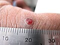
The appearance of pyogenic granuloma is usually a color ranging from red/pink to purple, grows rapidly, and can be smooth or mushroom-shaped. Younger lesions are more likely to be red because of their high number of blood vessels. Older lesions begin to change into a pink color. Size commonly ranges from a few millimeters to centimeters, though smaller or larger lesions may occur.[ medical citation needed ] A pyogenic granuloma can be painful, especially if located in an area of the body where it is constantly disturbed. Pyogenic granulomas can grow rapidly and often bleed profusely with little or no trauma. They may exude an oil-like substance, causing the surface to be damp. This is especially true if the granuloma is located on the scalp.[ medical citation needed ]

Epulis granulomatosum is a variant of pyogenic granuloma that forms only on gingiva, and is often seen forming in a recent extraction socket. Pyogenic granulomas appear on the gingiva in 75% of cases, more often in the maxillary than mandibular jaw. Anterior areas are more often affected than posterior areas. It can also be found on the lips, tongue, and inner cheek. Poor oral hygiene or trauma are usually precipitating factors.[ citation needed ]

One study has suggested a correlation between pyogenic granulomas and Bartonella seropositivity. [8] However, this association has been questioned by others. [9] The microscopic appearance of a pyogenic granuloma consists of highly vascular granulation tissue. Inflammation is present. The lesion may have a fibrous character if it is older, and the surface may have ulcerations. Pyogenic granulomas rarely occur in the conjunctiva, cornea, or connective tissue of the eye following minor local trauma. Grossly, these mass lesions resemble those occurring at more common sites. The relationship of these lesions to lobular capillary hemangiomas of skin and oropharyngeal mucosa commonly referred to as pyogenic granuloma is uncertain.[ citation needed ]

Associated conditions

Due to its overwhelming incidence on the gingiva, the condition is often associated with two other diseases, though not because they occur together. Instead, the three are associated with each other because they appear frequently on gingiva—peripheral giant cell granuloma and peripheral ossifying fibroma. Detailed analysis can be used to distinguish these conditions. [10]

Cause

Pyogenic granulomas are caused by proliferation of capillaries and are not caused by infection or cancer.[ citation needed ]

Diagnosis

Characteristics of pyogenic granuloma on microscopy Histopathology of pyogenic granuloma, high magnification, annotated.jpg
Characteristics of pyogenic granuloma on microscopy

A doctor likely can diagnose a pyogenic granuloma based on its appearance, and might perform a biopsy to make a more accurate diagnosis. A biopsy also helps rule out malignant (cancerous) medical conditions that can cause a similar kind of growth. These conditions include squamous-cell carcinoma, basal-cell carcinoma, and melanoma.[ citation needed ]

Histopathological examination shows multiple capillaries (due to the vascular nature of the tumor), neutrophils (pyogenic), and necrotic tissue.[ citation needed ]

Management

Although pyogenic granulomas are not infectious or cancerous, treatment may be considered because of bleeding or ulceration. Frequently, they are treated with electrodesiccation (cauterization) and curettage (excision), though laser treatment using pulsed dye laser or CO2 laser is often effective. [11] [12]

Several reports have demonstrated the efficacy of topical application of the beta-adrenergic antagonist timolol in the treatment of pediatric pyogenic granuloma. [13]

Usually, no treatment is used if the pyogenic granuloma occurs during pregnancy, since the lesion may heal spontaneously. Recurrent bleeding in either oral or nasal lesions may necessitate excision and cauterization sooner, however. If aesthetics are a concern, then treatment may be prioritized. Usually, only minor surgery may be needed, along with a dental cleaning for oral lesions to remove any calculus or other source of irritation.

History

Pyogenic granulomas were first described in 1897 by two French surgeons, Antonin Poncet and Dor, who named these lesions botryomycosis hominis. [14]

Terminology

The name "pyogenic granuloma" is misleading, as it is neither pyogenic or a true granuloma. Rather, it is a capillary hemangioma of lobular subtype, which is why such a lesion is prone to bleeding. [5] It is also not truly pyogenic (pus-producing), as the cause is hormonal or traumatic and has no association with infection or pus production.

See also

Related Research Articles

<span class="mw-page-title-main">Granuloma inguinale</span> Medical condition

Granuloma inguinale is a bacterial disease caused by Klebsiella granulomatis characterized by genital ulcers. It is endemic in many less-developed regions. It is also known as donovanosis, granuloma genitoinguinale, granuloma inguinale tropicum, granuloma venereum, granuloma venereum genitoinguinale, lupoid form of groin ulceration, serpiginous ulceration of the groin, ulcerating granuloma of the pudendum, and ulcerating sclerosing granuloma. Oral manifestations are also notably seen. The lesions of oral cavity are usually secondary to active genital lesions.

<span class="mw-page-title-main">Infantile hemangioma</span> Raised red skin lesion that affects infants caused by benign vascular tumor

An infantile hemangioma (IH), sometimes called a strawberry mark due to appearance, is a type of benign vascular tumor or anomaly that affects babies. Other names include capillary hemangioma, strawberry hemangioma, strawberry birthmark and strawberry nevus. and formerly known as a cavernous hemangioma. They appear as a red or blue raised lesion on the skin. Typically, they begin during the first four weeks of life, growing until about five months of life, and then shrinking in size and disappearing over the next few years. Often skin changes remain after they shrink. Complications may include pain, bleeding, ulcer formation, disfigurement, or heart failure. It is the most common tumor of orbit and periorbital areas in childhood. It may occur in the skin, subcutaneous tissues and mucous membranes of oral cavities and lips as well as in extracutaneous locations including the liver and gastrointestinal tract.

<span class="mw-page-title-main">Cryosurgery</span> Cauterization by freezing tissue

Cryosurgery is the use of extreme cold in surgery to destroy abnormal or diseased tissue; thus, it is the surgical application of cryoablation. The term comes from the Greek words cryo (κρύο) and surgery meaning "hand work" or "handiwork". Cryosurgery has been historically used to treat a number of diseases and disorders, especially a variety of benign and malignant skin conditions.

<span class="mw-page-title-main">Cherry angioma</span> Small bright red dome-shaped bump on the skin

Cherry angioma, also called cherry hemangioma, is a small bright red dome-shaped bump on the skin. It ranges between 0.5 – 6 mm in diameter and usually several are present, typically on the chest and arms, and increasing in number with age. If scratched, they may bleed.

The oral mucosa is the mucous membrane lining the inside of the mouth. It comprises stratified squamous epithelium, termed "oral epithelium", and an underlying connective tissue termed lamina propria. The oral cavity has sometimes been described as a mirror that reflects the health of the individual. Changes indicative of disease are seen as alterations in the oral mucosa lining the mouth, which can reveal systemic conditions, such as diabetes or vitamin deficiency, or the local effects of chronic tobacco or alcohol use. The oral mucosa tends to heal faster and with less scar formation compared to the skin. The underlying mechanism remains unknown, but research suggests that extracellular vesicles might be involved.

<span class="mw-page-title-main">Lymphangioma</span> Malformations of the lymphatic system characterized by lesions that are thin-walled cysts

Lymphangiomas are malformations of the lymphatic system characterized by lesions that are thin-walled cysts; these cysts can be macroscopic, as in a cystic hygroma, or microscopic. The lymphatic system is the network of vessels responsible for returning to the venous system excess fluid from tissues as well as the lymph nodes that filter this fluid for signs of pathogens. These malformations can occur at any age and may involve any part of the body, but 90% occur in children less than 2 years of age and involve the head and neck. These malformations are either congenital or acquired. Congenital lymphangiomas are often associated with chromosomal abnormalities such as Turner syndrome, although they can also exist in isolation. Lymphangiomas are commonly diagnosed before birth using fetal ultrasonography. Acquired lymphangiomas may result from trauma, inflammation, or lymphatic obstruction.

Epulis fissuratum is a benign hyperplasia of fibrous connective tissue which develops as a reactive lesion to chronic mechanical irritation produced by the flange of a poorly fitting denture. More simply, epulis fissuratum is where excess folds of firm tissue form inside the mouth, as a result of rubbing on the edge of dentures that do not fit well. It is a harmless condition and does not represent oral cancer. Treatment is by simple surgical removal of the lesion, and also by adjustment of the denture or provision of a new denture.

Peripheral giant-cell granuloma (PGCG) is an oral pathologic condition that appears in the mouth as an overgrowth of tissue due to irritation or trauma. Because of its overwhelming incidence on the gingiva, the condition is associated with two other diseases, pyogenic granuloma and peripheral ossifying fibroma. These three diseases are associated because they appear frequently on gingiva. Due to its similar microscopic appearance, peripheral giant-cell granuloma is considered to be the soft tissue equivalent of central giant-cell granuloma.

A peripheral ossifying fibroma, also known as ossifying fibrous epulis, is “a gingival nodule which is composed of a cellular fibroblastic connective tissue stroma which is associated with the formation of randomly dispersed foci of mineralised products, which consists of bone, cementum-like tissue, or a dystrophic calcification. The lesion is considered part of an ossifying fibroma, but that is usually considered to be a jaw tumor. Because of its overwhelming incidence on the gingiva, the condition is associated with two other diseases, though not because they occur together. Instead, the three are associated with each other because they appear frequently on gingiva: pyogenic granuloma and peripheral giant cell granuloma. Some researchers believe peripheral ossifying fibromas to be related to pyogenic fibromas and, in some instances, are the result of a pyogenic granuloma which has undergone fibrosis and calcification.

<span class="mw-page-title-main">Granuloma annulare</span> Medical condition

Granuloma annulare (GA) is a common, sometimes chronic skin condition which presents as reddish bumps on the skin arranged in a circle or ring. It can initially occur at any age, though two-thirds of patients are under 30 years old, and it is seen most often in children and young adults. Females are two times as likely to have it as males.

<span class="mw-page-title-main">Vascular tumor</span> Medical condition

A vascular tumor is a tumor of vascular origin; a soft tissue growth that can be either benign or malignant, formed from blood vessels or lymph vessels. Examples of vascular tumors include hemangiomas, lymphangiomas, hemangioendotheliomas, Kaposi's sarcomas, angiosarcomas, and hemangioblastomas. An angioma refers to any type of benign vascular tumor.

Giant-cell fibroma is a benign localized fibrous mass. It often mimics other fibroepithelial growths and can be distinguished by its histopathology. The exact cause of giant-cell fibromas is unknown however there is no evidence to show that it can be caused by irritation. Giant-cell fibromas can be removed by surgical incision, electrosurgery, or laser excision.

<span class="mw-page-title-main">Periapical cyst</span> Medical condition

Commonly known as a dental cyst, the periapical cyst is the most common odontogenic cyst. It may develop rapidly from a periapical granuloma, as a consequence of untreated chronic periapical periodontitis.

<span class="mw-page-title-main">Gingival enlargement</span> Human disease

Gingival enlargement is an increase in the size of the gingiva (gums). It is a common feature of gingival disease. Gingival enlargement can be caused by a number of factors, including inflammatory conditions and the side effects of certain medications. The treatment is based on the cause. A closely related term is epulis, denoting a localized tumor on the gingiva.

<span class="mw-page-title-main">Eosinophilic ulcer of the oral mucosa</span> Medical condition

Eosinophilic ulcer of the oral mucosa is a condition characterized by an ulcer with an indurated and elevated border. The lesion might be tender, fast-growing and the patient often not be aware of any trauma in the area.

Oral pigmentation is asymptomatic and does not usually cause any alteration to the texture or thickness of the affected area. The colour can be uniform or speckled and can appear solitary or as multiple lesions. Depending on the site, depth, and quantity of pigment, the appearance can vary considerably.

Epulis is any tumor like enlargement situated on the gingival or alveolar mucosa. The word literally means "(growth) on the gingiva", and describes only the location of the mass and has no further implications on the nature of the lesion. There are three types: fibromatous, ossifying and acanthomatous. The related term parulis refers to a mass of inflamed granulation tissue at the opening of a draining sinus on the alveolus over the root of an infected tooth. Another closely related term is gingival enlargement, which tends to be used where the enlargement is more generalized over the whole gingiva rather than a localized mass.

A vascular anomaly is any of a range of lesions from a simple birthmark to a large tumor that may be disfiguring. They are caused by a disorder of the vascular system. A vascular anomaly is a localized defect in blood or lymph vessels. These defects are characterized by an increased number of vessels, and vessels that are both enlarged and sinuous. Some vascular anomalies are congenital, others appear within weeks to years after birth, and others are acquired by trauma or during pregnancy. Inherited vascular anomalies are also described and often present with a number of lesions that increase with age. Vascular anomalies can also be a part of a syndrome.

Plasma cell granulomas (PCGs) are uncommon, non-neoplastic lesions of unknown etiology and are considered an entity of IgG4-related diseases.

References

  1. James, William D.; Berger, Timothy G.; et al. (2006). Andrews' Diseases of the Skin: Clinical Dermatology. Saunders Elsevier. ISBN   978-0-7216-2921-6.
  2. Rapini, Ronald P.; Bolognia, Jean L.; Jorizzo, Joseph L. (2007). Dermatology: 2-Volume Set. St. Louis: Mosby. ISBN   978-1-4160-2999-1.
  3. Nat Pernick. "Oral cavity & oropharynx - Other nonneoplastic - Pyogenic granuloma". PathologyOutlines. Topic Completed: 1 November 2013. Minor changes: 12 October 2020
  4. Freedberg, et al. (2003). Fitzpatrick's Dermatology in General Medicine. (6th ed.). McGraw-Hill. ISBN   0-07-138076-0.
  5. 1 2 Jafarzadeh, Hamid; Sanatkhani, Majid; Mohtasham, Nooshin (2006). "Oral pyogenic granuloma: a review". Journal of Oral Science. 48 (4): 167–175. doi:10.2334/josnusd.48.167. ISSN   1343-4934. PMID   17220613.
  6. Nthumba, Peter M. (31 March 2008). "Giant pyogenic granuloma of the thigh: a case report". Journal of Medical Case Reports. 2 (1): 95. doi: 10.1186/1752-1947-2-95 . ISSN   1752-1947. PMC   2329656 . PMID   18377654.
  7. Rockafellow, Andrew; Florin, Whitney; Philipone, Elizabeth; Koslovsky, David (2015). "Pregnancy Tumor in a 31-Year-Old Female with a Facial Port-Wine Stain". Case Reports in Dentistry. 2015: 472605. doi: 10.1155/2015/472605 . PMC   4698533 . PMID   26798522.
  8. Lee, Joyce; Lynde, Charles (1 November 2001). "Pyogenic Granuloma: Pyogenic Again? Association betweenPyogenic Granuloma and Bartonella". Journal of Cutaneous Medicine and Surgery: IncorporatingMedical and Surgical Dermatology. 5 (6): 467–470. doi:10.1007/s10227-001-0022-0. ISSN   1203-4754. PMID   11907853. S2CID   7408146.
  9. Levy, Itzhak; Rolain, Jean-Marc; Lepidi, Hubert; Raoult, Didler; Feinmesser, Meora; Lapidoth, Moshe; Ben-Amitai, Dan (December 2005). "Is pyogenic granuloma associated with Bartonella infection?". Journal of the American Academy of Dermatology. 53 (6): 1065–1066. doi:10.1016/j.jaad.2005.08.046. ISSN   0190-9622. PMID   16310070.
  10. Salum, F. G.; Yurgel, L. S.; Cherubini, K.; De Figueiredo, M. a. Z.; Medeiros, I. C.; Nicola, F. S. (May 2008). "Pyogenic granuloma, peripheral giant cell granuloma and peripheral ossifying fibroma: retrospective analysis of 138 cases". Minerva Stomatologica. 57 (5): 227–232. ISSN   0026-4970. PMID   18496485.
  11. Pagliai, Kelley A.; Cohen, Bernard A. (2004). "Pyogenic Granuloma in Children". Pediatric Dermatology. 21 (1): 10–13. doi:10.1111/j.0736-8046.2004.21102.x. ISSN   1525-1470. PMID   14871318. S2CID   8958995.
  12. Tay, Yong-Kwang; Weston, William L.; Morelli, Joseph G. (1 March 1997). "Treatment of Pyogenic Granuloma in Children With the Flashlamp-pumped Pulsed Dye Laser". Pediatrics. 99 (3): 368–370. doi:10.1542/peds.99.3.368. ISSN   0031-4005. PMID   9041290.
  13. Lee, Lara Wine; Goff, Kiera L.; Lam, Joseph M.; Low, David W.; Yan, Albert C.; Castelo‐Soccio, Leslie (2014). "Treatment of Pediatric Pyogenic Granulomas Using β-Adrenergic Receptor Antagonists". Pediatric Dermatology. 31 (2): 203–207. doi:10.1111/pde.12217. ISSN   1525-1470. PMID   24138457. S2CID   38987169.
  14. Ferry, Andrew P.; Zimmerman, Lorenz E. (1 August 1965). "Granuloma Pyogenicum of Limbus: Simulating Recurrent Squamous Cell Carcinoma". Archives of Ophthalmology. 74 (2): 229–230. doi:10.1001/archopht.1965.00970040231019. ISSN   0003-9950.