Von Hippel–Lindau disease (VHL), also known as VonHippel–Lindau syndrome, is a rare genetic disorder with multisystem involvement. It is characterized by visceral cysts and benign tumors with potential for subsequent malignant transformation. It is a type of phakomatosis that results from a mutation in the Von Hippel–Lindau tumor suppressor gene on chromosome 3p25.3.
Spinal tumors are neoplasms located in either the vertebral column or the spinal cord. There are three main types of spinal tumors classified based on their location: extradural and intradural. Extradural tumors are located outside the dura mater lining and are most commonly metastatic. Intradural tumors are located inside the dura mater lining and are further subdivided into intramedullary and extramedullary tumors. Intradural-intramedullary tumors are located within the dura and spinal cord parenchyma, while intradural-extramedullary tumors are located within the dura but outside the spinal cord parenchyma. The most common presenting symptom of spinal tumors is nocturnal back pain. Other common symptoms include muscle weakness, sensory loss, and difficulty walking. Loss of bowel and bladder control may occur during the later stages of the disease.

A benign tumor is a mass of cells (tumor) that does not invade neighboring tissue or metastasize. Compared to malignant (cancerous) tumors, benign tumors generally have a slower growth rate. Benign tumors have relatively well differentiated cells. They are often surrounded by an outer surface or stay contained within the epithelium. Common examples of benign tumors include moles and uterine fibroids.
Hypoxia-inducible factors (HIFs) are transcription factors that respond to decreases in available oxygen in the cellular environment, or hypoxia. They are only present in parahoxozoan animals.
Phakomatoses, also known neurocutaneous syndromes, are a group of multisystemic diseases that most prominently affect structures primarily derived from the ectoderm such as the central nervous system, skin and eyes. The majority of phakomatoses are single-gene disorders that may be inherited in an autosomal dominant, autosomal recessive or X-linked pattern. Presentations may vary dramatically between patients with the same particular syndrome due to mosaicism, variable expressivity, and penetrance.
Angiomatosis is a non-neoplastic condition characterised by nests of proliferating capillaries arranged in a lobular pattern, displacing adjacent muscle and fat. It consists of many angiomas.

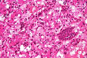
The Von Hippel–Lindau tumor suppressor also known as pVHL is a protein that, in humans, is encoded by the VHL gene. Mutations of the VHL gene are associated with Von Hippel–Lindau disease, which is characterized by hemangioblastomas of the brain, spinal cord and retina. It is also associated with kidney and pancreatic lesions.

Eugen Adolf Arthur von Hippel was a German ophthalmologist born in Königsberg.

Arvid Vilhelm Lindau was a Swedish pathologist and bacteriologist born in Malmö.

Hypoxia-inducible factor 1-alpha, also known as HIF-1-alpha, is a subunit of a heterodimeric transcription factor hypoxia-inducible factor 1 (HIF-1) that is encoded by the HIF1A gene. The Nobel Prize in Physiology or Medicine 2019 was awarded for the discovery of HIF.

Hypoxia-inducible factor prolyl hydroxylase 2 (HIF-PH2), or prolyl hydroxylase domain-containing protein 2 (PHD2), is an enzyme encoded by the EGLN1 gene. It is also known as Egl nine homolog 1. PHD2 is a α-ketoglutarate/2-oxoglutarate-dependent hydroxylase, a superfamily non-haem iron-containing proteins. In humans, PHD2 is one of the three isoforms of hypoxia-inducible factor-proline dioxygenase, which is also known as HIF prolyl-hydroxylase.

Leptomeningeal cancer is a rare complication of cancer in which the disease spreads from the original tumor site to the meninges surrounding the brain and spinal cord. This leads to an inflammatory response, hence the alternative names neoplastic meningitis (NM), malignant meningitis, or carcinomatous meningitis. The term leptomeningeal describes the thin meninges, the arachnoid and the pia mater, between which the cerebrospinal fluid is located. The disorder was originally reported by Eberth in 1870.

Ubiquitin carboxyl-terminal hydrolase 20 is an enzyme that in humans is encoded by the USP20 gene.

The spinal cord is a long, thin, tubular structure made up of nervous tissue, which extends from the medulla oblongata in the brainstem to the lumbar region of the vertebral column (backbone). The backbone encloses the central canal of the spinal cord, which contains cerebrospinal fluid. The brain and spinal cord together make up the central nervous system (CNS). In humans, the spinal cord begins at the occipital bone, passing through the foramen magnum and then enters the spinal canal at the beginning of the cervical vertebrae. The spinal cord extends down to between the first and second lumbar vertebrae, where it ends. The enclosing bony vertebral column protects the relatively shorter spinal cord. It is around 45 cm (18 in) long in adult men and around 43 cm (17 in) long in adult women. The diameter of the spinal cord ranges from 13 mm in the cervical and lumbar regions to 6.4 mm in the thoracic area.

Spinal disease refers to a condition impairing the backbone. These include various diseases of the back or spine ("dorso-"), such as kyphosis. Dorsalgia refers to back pain. Some other spinal diseases include spinal muscular atrophy, ankylosing spondylitis, lumbar spinal stenosis, spina bifida, spinal tumors, osteoporosis and cauda equina syndrome.

An endolymphatic sac tumor (ELST) is a very uncommon papillary epithelial neoplasm arising within the endolymphatic sac or endolymphatic duct. This tumor shows a very high association with Von Hippel–Lindau syndrome (VHL).
The Pacak-Zhuang syndrome is a recently described disease manifestation in females that includes multiple paragangliomas or pheochromocytomas and somatostatinomas, both neuroendocrine tumors, and secondary polycythemia associated with high erythropoietin levels. Paragangliomas in these patients are mainly localized to the abdomen whereas somatostatinomas are found in the second portion of the duodenum, as shown by imaging or biochemistry. This syndrome is of special interest as finding more than one type of neuroendocrine tumor in one individual is unusual. Such co-occurrences are usually seen in patients carrying hereditary syndromes like multiple endocrine neoplasia (MEN), neurofibromatosis 1 (NF1), or von Hippel-Lindau (VHL) disease.

William G. Kaelin Jr. is an American Nobel laureate physician-scientist. He is a professor of medicine at Harvard University and the Dana–Farber Cancer Institute. His laboratory studies tumor suppressor proteins. In 2016, Kaelin received the Albert Lasker Award for Basic Medical Research and the AACR Princess Takamatsu Award. He also won the Nobel Prize in Physiology or Medicine in 2019 along with Peter J. Ratcliffe and Gregg L. Semenza.
A central nervous system tumor is an abnormal growth of cells from the tissues of the brain or spinal cord. CNS tumor is a generic term encompassing over 120 distinct tumor types. Common symptoms of CNS tumors include vomiting, headache, changes in vision, nausea, and seizures. A CNS tumor can be detected and classified via neurological examination, medical imaging, such as x-ray imaging, magnetic resonance imaging (MRI) or computed tomography (CT), or after analysis of a biopsy.

Belzutifan, sold under the brand name Welireg, is an anti-cancer medication used for the treatment of von Hippel–Lindau disease-associated renal cell carcinoma. It is taken by mouth. Belzutifan is an hypoxia-inducible factor-2 alpha (HIF-2α) inhibitor.