Vascular tumor

Last updated
Vascular tumor
Hemangioma.JPG
A hemangioma, a benign type of vascular tumor
Specialty Oncology   OOjs UI icon edit-ltr-progressive.svg

A vascular tumor is a tumor of vascular origin; a soft tissue growth that can be either benign or malignant, formed from blood vessels or lymph vessels. [1] Examples of vascular tumors include hemangiomas, lymphangiomas, hemangioendotheliomas, Kaposi's sarcomas, angiosarcomas, and hemangioblastomas. An angioma refers to any type of benign vascular tumor. [2]

Contents

Some vascular tumors can be associated with serious blood-clotting disorders, making correct diagnosis critical. [3]

A vascular tumor may be described in terms of being highly vascularized, or poorly vascularized, referring to the degree of blood supply to the tumor.

Classification

Vascular tumors make up one of the classifications of vascular anomalies. The other grouping is vascular malformations. Vascular tumors can be further subclassified as being benign, borderline or aggressive, and malignant. Vascular tumors are described as proliferative, and vascular malformations as nonproliferative. [4]

Types

A pyogenic granuloma on a thumb. Pyogenic Granuloma Thumb 1.JPG
A pyogenic granuloma on a thumb.

A vascular tumor typically grows quickly by the proliferation of endothelial cells. Most are not birth defects. [4]

Benign

The most common type of benign vascular tumors are hemangiomas, most commonly infantile hemangiomas, and less commonly congenital hemangiomas.

Infantile hemangioma

Infantile hemangiomas are the most common type of vascular tumor to affect babies, [4] accounting for 90% of hemangiomas. [5] They are characterised by the abnormal proliferation of endothelial cells and of deviant blood vessel formation or architecture. [6] Hypoxic stress seems to be a major trigger for this. [7] Infantile hemangiomas are easily diagnosed, and little if any aggressive treatment is needed. [7] They are characterised by rapid growth in the first few months, followed by spontaneous regression in early childhood. [5]

Congenital hemangioma

Congenital hemangiomas are present and fully formed at birth, [5] and only account for 2% of the hemangiomas. They do not have the postnatal phase of proliferation common to infantile hemangiomas. [6] There are two main variants of congenital hemangioma: non-involuting, and rapidly involuting (beginning in the first year of life). [6] A third variant is also recognised as partially involuting. [8] Congenital hemangiomas can also be distinguished from infantile hemangiomas in that neither variant of congenital hemangioma expresses the glucose transporter GLUT 1. [6] [9]

Some cases have been associated with a mild form of thrombocytopenia. Rare cases have been associated with heart failure. [6]

Hemangioblastoma

Hemangioblastomas are vascular tumors of the central nervous system.

Pyogenic granuloma

A range of benign vascular tumors are described as reactive proliferative lesions that grow in response to a stimulus, such as trauma, or a local thrombosis. They can also form infrequently during pregnancy as a hormonal reaction affecting the gums. [10]

The most common type of reactive proliferative tumors are pyogenic granulomas also known as lobular capillary hemangiomas, that are more often found in children and young adults. [4] These granulomas are well defined growths of less than a centimetre across. They are bright red due to being highly vascularised, and bleed and ulcerate easily. [10] Their colouring fades with age. [4]

Tufted angioma

Tufted angiomas are hereditary hemangiomas found in infants from birth to five years of age, however they may occur in adults. They are found on the neck, shoulders, and trunk as rounded nodules. [11] Tufted angiomas are usually poorly defined lesions of purple colouration. [12] The tumors are of tufts of capillary-sized vessels in lobules that are scattered in the skin, and that sometimes reach into the subcutaneous tissue, and have lymph vessels on the periphery. Their growth is slow to begin with, and progresses to a stable size. They show a high rate of spontaneous regression, particularly in congenital and early-onset cases. [12] They typically have a deep nodular component sometimes extending into the subcutaneous tissue, fascia, and muscle, and can sometimes be painful. [12] Tufted angiomas are associated with arteriovenous malformations. [12]

The origin of tufted angiomas is not clear but markers on the cells suggest a possible derivation from the endothelial cells of lymph vessels. [11] They are also associated with the local secretion of growth factors that affect angiogenesis and promote the development of vascular lobules. [12]

Borderline

Kaposiform hemangioendothelioma Kaposiform hemangioendothelioma.JPG
Kaposiform hemangioendothelioma

Kaposiform hemangioendotheliomas (KHEs) are borderline, locally destructive vascular tumors. [4] They are named after their resemblance to the lesions of Kaposi's sarcoma. [13] KHEs are described as locally destructive because they can infiltrate underlying muscle and fat. [4] They are often seen to overlap with tufted angiomas (TAs) but TAs may be a milder, benign counterpart. [14]

KHEs show as a red or purple expanding mass of soft tissue, [14] found mostly in infants. Under the microscope KHE is characterised by nodules of tumor-like spindled endothelial cells. [14] Unlike infantile hemangiomas, KHEs have a high mortality rate. [14] Both KHEs and TAs are unique in that they carry the risk of the development of Kasabach–Merritt syndrome. [15]

Malignant

Malignant vascular tumors are rare, [7] and include angiosarcomas, and epithelioid hemangioendotheliomas. [4] Other types are hemangiopericytomas, [16] and lymphangiosarcomas.

Related Research Articles

<span class="mw-page-title-main">Birthmark</span> Medical condition

A birthmark is a congenital, benign irregularity on the skin which is present at birth or appears shortly after birth—usually in the first month. Birthmarks can occur anywhere on the skin. They are caused by overgrowth of blood vessels, melanocytes, smooth muscle, fat, fibroblasts, or keratinocytes.

<span class="mw-page-title-main">Central nervous system cavernous hemangioma</span> Medical condition

Cerebral cavernous malformation (CCM) is a cavernous hemangioma that arises in the central nervous system. It can be considered to be a variant of hemangioma, and is characterized by grossly large dilated blood vessels and large vascular channels, less well circumscribed, and more involved with deep structures, with a single layer of endothelium and an absence of neuronal tissue within the lesions. These thinly walled vessels resemble sinusoidal cavities filled with stagnant blood. Blood vessels in patients with cerebral cavernous malformations (CCM) can range from a few millimeters to several centimeters in diameter. Most lesions occur in the brain, but any organ may be involved.

<span class="mw-page-title-main">Infantile hemangioma</span> Raised red skin lesion that affects infants caused by benign vascular tumor

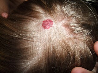
An infantile hemangioma (IH), sometimes called a strawberry mark due to appearance, is a type of benign vascular tumor or anomaly that affects babies. Other names include capillary hemangioma, strawberry hemangioma, strawberry birthmark and strawberry nevus. and formerly known as a cavernous hemangioma. They appear as a red or blue raised lesion on the skin. Typically, they begin during the first four weeks of life, growing until about five months of life, and then shrinking in size and disappearing over the next few years. Often skin changes remain after they shrink. Complications may include pain, bleeding, ulcer formation, disfigurement, or heart failure. It is the most common tumor of orbit and periorbital areas in childhood. It may occur in the skin, subcutaneous tissues and mucous membranes of oral cavities and lips as well as in extracutaneous locations including the liver and gastrointestinal tract.

<span class="mw-page-title-main">Angioma</span> Medical condition

Angiomas are benign tumors derived from cells of the vascular or lymphatic vessel walls (endothelium) or derived from cells of the tissues surrounding these vessels.

<span class="mw-page-title-main">Cherry angioma</span> Small bright red dome-shaped bump on the skin

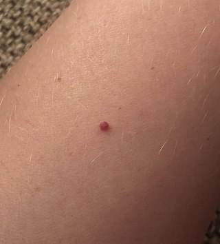
Cherry angioma, also called cherry hemangioma, is a small bright red dome-shaped bump on the skin. It ranges between 0.5 – 6 mm in diameter and usually several are present, typically on the chest and arms, and increasing in number with age. If scratched, they may bleed.

<span class="mw-page-title-main">Hemangioendothelioma</span> Medical condition

Hemangioendotheliomas are a family of vascular neoplasms of intermediate malignancy.

<span class="mw-page-title-main">Hemangiopericytoma</span> Medical condition

A hemangiopericytoma is a type of soft-tissue sarcoma that originates in the pericytes in the walls of capillaries. When inside the nervous system, although not strictly a meningioma tumor, it is a meningeal tumor with a special aggressive behavior. It was first characterized in 1942.

<span class="mw-page-title-main">Lymphangioma</span> Malformations of the lymphatic system characterized by lesions that are thin-walled cysts

Lymphangiomas are malformations of the lymphatic system characterized by lesions that are thin-walled cysts; these cysts can be macroscopic, as in a cystic hygroma, or microscopic. The lymphatic system is the network of vessels responsible for returning to the venous system excess fluid from tissues as well as the lymph nodes that filter this fluid for signs of pathogens. These malformations can occur at any age and may involve any part of the body, but 90% occur in children less than 2 years of age and involve the head and neck. These malformations are either congenital or acquired. Congenital lymphangiomas are often associated with chromosomal abnormalities such as Turner syndrome, although they can also exist in isolation. Lymphangiomas are commonly diagnosed before birth using fetal ultrasonography. Acquired lymphangiomas may result from trauma, inflammation, or lymphatic obstruction.

<span class="mw-page-title-main">Angiomatosis</span> Medical condition

Angiomatosis is a non-neoplastic condition characterised by nests of proliferating capillaries arranged in a lobular pattern, displacing adjacent muscle and fat. It consists of many angiomas.

<span class="mw-page-title-main">Focal nodular hyperplasia</span> Medical condition

Focal nodular hyperplasia (FNH) is a benign tumor of the liver, which is the second most prevalent tumor of the liver. It is usually asymptomatic, rarely grows or bleeds, and has no malignant potential. This tumour was once often resected because it was difficult to distinguish from hepatic adenoma, but with modern multiphase imaging is usually now diagnosed by strict imaging criteria and not resected.

<span class="mw-page-title-main">Vascular disease</span> Medical condition

Vascular disease is a class of diseases of the vessels of the circulatory system in the body, including blood vessels – the arteries and veins, and the lymphatic vessels. Vascular disease is a subgroup of cardiovascular disease. Disorders in this vast network of blood and lymph vessels can cause a range of health problems that can sometimes become severe, and fatal. Coronary heart disease for example, is the leading cause of death for men and women in the United States.

A vascular malformation is a blood vessel or lymph vessel abnormality. Vascular malformations are one of the classifications of vascular anomalies, the other grouping is vascular tumors. They may cause aesthetic problems as they have a growth cycle, and can continue to grow throughout life.

A vascular anomaly is any of a range of lesions from a simple birthmark to a large tumor that may be disfiguring. They are caused by a disorder of the vascular system. A vascular anomaly is a localized defect in blood or lymph vessels. These defects are characterized by an increased number of vessels, and vessels that are both enlarged and sinuous. Some vascular anomalies are congenital, others appear within weeks to years after birth, and others are acquired by trauma or during pregnancy. Inherited vascular anomalies are also described and often present with a number of lesions that increase with age. Vascular anomalies can also be a part of a syndrome.

Diffuse neonatal hemangiomatosis (DNH) is a potentially fatal disorder where multiple benign (non-cancerous) blood vessel tumors (hemangiomas) are present in the skin and other organs. The mortality rate of diffuse neonatal hemangiomatosis is 50-90%. This disease is normally found in female Caucasian infants. The most common site of internal organ damage, or lesions, is the liver, which can redirect blood away from the heart and cause arteriovenous shunting. This can cause high cardiac output, leading to further complications such as congestive heart failure. This condition affecting the liver is also known as infantile hepatic hemangioma (IHH). Other sites of internal organ damage can include the intestines, nervous system, lungs, and sometimes the skeletal system. Early detection and treatment with steroids results in most newborn babies with this disease remaining healthy, with serious problems developing for some individuals during the hemangioma's growth phase.

<span class="mw-page-title-main">Cavernous hemangioma</span> Human disease

Cavernous hemangioma, also called cavernous angioma, venous malformation, or cavernoma, is a type of venous malformation due to endothelial dysmorphogenesis from a lesion which is present at birth. A cavernoma in the brain is called a cerebral cavernous malformation or CCM. Despite its designation of a hemangioma, a cavernous hemangioma is not a tumor as it does not display endothelial hyperplasia. The abnormal tissue causes a slowing of blood flow through the cavities, or "caverns". The blood vessels do not form the necessary junctions with surrounding cells, and the structural support from the smooth muscle is hindered, causing leakage into the surrounding tissue. It is the leakage of blood, referred to as hemorrhage, that causes a variety of symptoms known to be associated with the condition.

<span class="mw-page-title-main">Cavernous liver hemangioma</span> Medical condition

A cavernous liver hemangioma or hepatic hemangioma is a benign tumor of the liver composed of hepatic endothelial cells. It is the most common benign liver tumour, and is usually asymptomatic and diagnosed incidentally on radiological imaging. Liver hemangiomas are thought to be congenital in origin. Several subtypes exist, including the giant hepatic haemangioma, which can cause significant complications.

<span class="mw-page-title-main">Hemangioma</span> Medical condition

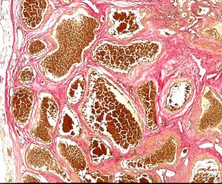
A hemangioma or haemangioma is a usually benign vascular tumor derived from blood vessel cell types. The most common form, seen in infants, is an infantile hemangioma, known colloquially as a "strawberry mark", most commonly presenting on the skin at birth or in the first weeks of life. A hemangioma can occur anywhere on the body, but most commonly appears on the face, scalp, chest or back. They tend to grow for up to a year before gradually shrinking as the child gets older. A hemangioma may need to be treated if it interferes with vision or breathing or is likely to cause long-term disfigurement. In rare cases internal hemangiomas can cause or contribute to other medical problems. Most of the time they tend to disappear in 10 years. The first line treatment option is beta blockers, which are highly effective in the majority of cases. Ones that form at birth are called congenital hemangiomas while ones that form later in life are called infantile hemangiomas.

<span class="mw-page-title-main">Vertebral hemangioma</span> Medical condition

Vertebral hemangiomas or haemangiomas (VHs) are a common vascular lesion found within the vertebral body of the thoracic and lumbar spine. These are predominantly benign lesions that are often found incidentally during radiology studies for other indications and can involve one or multiple vertebrae. Vertebral hemangiomas are a common etiology estimated to be found in 10-12% of humans at autopsy. They are benign in nature and frequently asymptomatic. Symptoms, if they do occur, are usually related to large hemangiomas, trauma, the hormonal and hemodynamic changes of pregnancy (causing intra-spinal bleeding), or osseous expansion and extra-osseous extension into surround soft tissues or epidural region of the spinal canal.

Diffuse capillary malformation with overgrowth (DCMO) is a subset of capillary malformations (CM) associated with hypertrophy, i.e. increased size of body structures. CM can be considered an umbrella term for various vascular anomalies caused by increased diameter or number of capillary blood vessels. It is commonly referred to as "port-wine stain", and is thought to affect approximately 0.5% of the population. Typically capillaries in the papillary dermis are involved, and this gives rise to pink or violaceous colored lesions. The majority of DCMO lesions are diffuse, reticulated pale-colored stains.

References

  1. "NCI Dictionary of Cancer Terms". National Cancer Institute. 2 February 2011. Retrieved 15 November 2019.
  2. "Dorlands Medical Dictionary:angioma". 2 February 2009. Archived from the original on 2 February 2009.
  3. Johnson, EF; Davis, DM; Tollefson, MM; Fritchie, K; Gibson, LE (April 2018). "Vascular Tumors in Infants: Case Report and Review of Clinical, Histopathologic, and Immunohistochemical Characteristics of Infantile Hemangioma, Pyogenic Granuloma, Noninvoluting Congenital Hemangioma, Tufted Angioma, and Kaposiform Hemangioendothelioma". The American Journal of Dermatopathology. 40 (4): 231–239. doi:10.1097/DAD.0000000000000983. PMID   29561329. S2CID   4033341.
  4. 1 2 3 4 5 6 7 8 Steiner, JE; Drolet, BA (September 2017). "Classification of Vascular Anomalies: An Update". Seminars in Interventional Radiology. 34 (3): 225–232. doi:10.1055/s-0037-1604295. PMC   5615389 . PMID   28955111.
  5. 1 2 3 Sadick, M; Müller-Wille, R; Wildgruber, M; Wohlgemuth, WA (September 2018). "Vascular Anomalies (Part I): Classification and Diagnostics of Vascular Anomalies". RöFo: Fortschritte auf dem Gebiete der Röntgenstrahlen und der Nuklearmedizin. 190 (9): 825–835. doi: 10.1055/a-0620-8925 . PMID   29874693.
  6. 1 2 3 4 5 Darrow, DH; Greene, AK; Mancini, AJ; Nopper, AJ; SECTION ON DERMATOLOGY, SECTION ON OTOLARYNGOLOGY–HEAD AND NECK SURGERY, and SECTION ON PLASTIC, SURGERY. (October 2015). "Diagnosis and Management of Infantile Hemangioma". Pediatrics. 136 (4): e1060-104. doi: 10.1542/peds.2015-2485 . PMID   26416931.{{cite journal}}: CS1 maint: multiple names: authors list (link)
  7. 1 2 3 Wildgruber, M; Sadick, M; Müller-Wille, R; Wohlgemuth, WA (13 March 2019). "Vascular tumors in infants and adolescents". Insights into Imaging. 10 (1): 30. doi:10.1186/s13244-019-0718-6. PMC   6419671 . PMID   30868300.
  8. "Congenital haemangioma". www.gosh.nhs.uk.
  9. van Vugt, LJ; van der Vleuten, CJM; Flucke, U; Blokx, WAM (June 2017). "The utility of GLUT1 as a diagnostic marker in cutaneous vascular anomalies: A review of literature and recommendations for daily practice". Pathology, Research and Practice. 213 (6): 591–597. doi:10.1016/j.prp.2017.04.023. PMID   28552538.
  10. 1 2 Kumar, V; Abbas, A; Aster, J (2018). Robbins basic pathology (Tenth ed.). pp. 392–396. ISBN   9780323353175.
  11. 1 2 "MeSH Browser". meshb.nlm.nih.gov. Retrieved 22 November 2019.
  12. 1 2 3 4 5 Victoria Martínez, AM; Cubells Sánchez, L; Esteve Martínez, A; Estela Cubells, JR; Febrer Bosch, I; Alegre de Miquel, V; Oliver Martínez, V (September 2015). "[Tufted angiomas in childhood: A series of 9 cases and a literature review]". Anales de Pediatria. 83 (3): 201–8. doi:10.1016/j.anpedi.2014.10.018. PMID   25468452.
  13. Vivas-Colmenares, GV; Ramirez-Villar, GL; Bernabeu-Wittel, J; Matute de Cardenas, JA; Fernandez-Pineda, I (January 2015). "The importance of early diagnosis and treatment of kaposiform hemangioendothelioma complicated by Kasabach-Merritt phenomenon". Dermatology Practical & Conceptual. 5 (1): 91–3. doi:10.5826/dpc.0501a18. PMC   4325701 . PMID   25692091.
  14. 1 2 3 4 Putra, J; Gupta, A (June 2017). "Kaposiform haemangioendothelioma: a review with emphasis on histological differential diagnosis". Pathology. 49 (4): 356–362. doi:10.1016/j.pathol.2017.03.001. PMID   28438388.
  15. Croteau, SE; Gupta, D (September 2016). "The clinical spectrum of kaposiform hemangioendothelioma and tufted angioma". Seminars in Cutaneous Medicine and Surgery. 35 (3): 147–52. doi:10.12788/j.sder.2016.048. PMID   27607323.
  16. Ghose, A; Guha, G; Kundu, R; Tew, J; Chaudhary, R (June 2017). "CNS Hemangiopericytoma: A Systematic Review of 523 Patients". American Journal of Clinical Oncology. 40 (3): 223–227. doi:10.1097/COC.0000000000000146. PMID   25350465. S2CID   12236073.