Lymphangiomatosis

Last updated
Lymphangiomatosis
Other namesLYMF
Lung biopsy showing infiltration of lymphatic tissue.png
Lung biopsy showing infiltration of lymphatic tissue.

Lymphangiomatosis is a condition where a lymphangioma is not present in a single localised mass, but in a widespread or multifocal manner. It is a rare type of tumor which results from an abnormal development of the lymphatic system. [1]

Contents

It is thought to be the result of congenital errors of lymphatic development occurring prior to the 20th week of gestation. [2] Lymphangiomatosis is a condition marked by the presence of cysts that result from an increase both in the size and number of thin-walled lymphatic channels that are abnormally interconnected and dilated. [2] [3] [4] 75% of cases involve multiple organs. [2] It typically presents by age 20 and, although it is technically benign, these deranged lymphatics tend to invade surrounding tissues and cause problems due to invasion and/or compression of adjacent structures. [2] The condition is most common in the bones and lungs [2] and shares some characteristics with Gorham’s disease. Up to 75% of patients with lymphangiomatosis have bone involvement, leading some to conclude that lymphangiomatosis and Gorham’s disease should be considered as a spectrum of disease rather than separate diseases. [2] [5] When it occurs in the lungs, lymphangiomatosis has serious consequences and is most aggressive in the youngest children. [2] [4] When the condition extends into the chest it commonly results in the accumulation of chyle in the linings of the heart and/or lungs. [2] [4]

Chyle is composed of lymph fluid and fats that are absorbed from the small intestine by specialized lymphatic vessels called lacteals during digestion. The accumulations are described based on location: chylothorax is chyle in the chest; chylopericardium is chyle trapped inside the sack surrounding the heart; chyloascites is chyle trapped in the linings of the abdomen and abdominal organs. The presence of chyle in these places accounts for many of the symptoms and complications associated with both lymphangiomatosis and Gorham’s disease. [2] [6] The incidence of lymphangiomatosis is unknown and it is often misdiagnosed. It is separate and distinct from lymphangiectasis, lymphangioleiomyomatosis (LAM), pulmonary capillary hemangiomatosis, Kaposi’s sarcoma, and kaposiform hemangioendothelioma. [4] Its unusual nature makes lymphangiomatosis (and Gorham’s disease) a diagnostic and therapeutic challenge. [4] [7] A multidisciplinary approach is generally necessary for optimal diagnosis and symptom management. The term literally means lymphatic system (lymph) vessel (angi) tumor or cyst (oma) condition (tosis).

Signs and symptoms

Lymphangiomatosis is a multi-system disorder. Symptoms depend on the organ system involved and, to varying degrees, the extent of the disease. Early in the course of the disease patients are usually asymptomatic, but over time the abnormally proliferating lymphatic channels that constitute lymphangiomatosis are capable of massive expansion and infiltration into surrounding tissues, bone, and organs. [2] Because of its slow course and often vague symptoms, the condition is frequently under-recognized or misdiagnosed. [8]

Early signs of disease in the chest include wheezing, cough, and feeling short of breath, which is often misdiagnosed as asthma. [2] The pain that accompanies bone involvement may be attributed to "growing pains" in younger children. With bone involvement the first indication for disease may be a pathological fracture. Symptoms may not raise concern, or even be noted, until the disease process has advanced to a point where it causes restrictive compression of vital structures. Further, the occurrence of chylous effusions seems to be unrelated to the pathologic "burden" of the disease, the extent of involvement in any particular tissue or organ, or the age of the patient. [9] This offers one explanation as to why, unfortunately, the appearance of chylous effusions in the chest or abdomen may be the first evidence of the disease.[ citation needed ]

Following are some of the commonly reported symptoms of lymphangiomatosis, divided into the regions/systems in which the disease occurs:

Heart and chest

Symptoms that arise from disease of the cardiothoracic region include a chronic cough, wheezing, dyspnea (shortness of breath)—especially serious when occurring at rest or when lying down—fever, chest pain, rapid heartbeat, dizziness, anxiety, and coughing up blood or chyle. [2] As the deranged lymphatic vessels invade the organs and tissues in the chest they put stress on the heart and lungs, interfering with their ability to function normally. Additionally, these lymphatic vessels may leak, allowing fluid to accumulate in the chest, which puts further pressure on the vital organs, thus increasing their inability to function properly. [2] Accumulations of fluid and chyle are named based on their contents and location: pulmonary edema (the presence of fluid and/or chyle in the lung), pleural effusions (fluid in the lung lining), pericardial effusions (fluid in the heart sack), chylothorax (chyle in the pleural cavity); and chylopericardium (chyle in the heart sack).[ citation needed ]

Abdominal

Lymphangiomatosis has been reported in every region of the abdomen, though the most reported sites involve the intestines and peritoneum; spleen, kidneys, and liver. Often there are no symptoms until late in the progression of the disease. When they do occur, symptoms include abdominal pain and/or distension; nausea, vomiting, diarrhea; decreased appetite and malnourishment. When the disease affects the kidneys the symptoms include flank pain, abdominal distension, blood in the urine, and, possibly, elevated blood pressure, which may result in it being confused with other cystic renal disease. [10] When lymphangiomatosis occurs in the liver and/or spleen it may be confused with polycystic liver disease. [11] Symptoms may include abdominal fullness and distension; anemia, disseminated intravascular coagulopathy (DIC), fluid accumulation in the abdomen(ascites), decreased appetite, weight loss, fatigue; late findings include liver failure. [2] [11] [12]

Bones

Symptoms of lymphangiomatosis in the skeletal system are the same as those of Gorham’s disease. Frequently asymptomatic, skeletal lymphangiomatosis may be discovered incidentally or when a pathological fracture occurs. Patients may experience pain of varying severity in areas around the effected bone. When the disease occurs in the bones of the spine, neurological symptoms such as numbness and tingling may occur due to spinal nerve compression. [13] Progression of disease in the spine may lead to paralysis. Lymphangiomatosis in conjunction with Chiari I malformation also has been reported. [14]

Causes

The cause of lymphangiomatosis is not yet known. As stated earlier, it is generally considered to be the result of congenital errors of lymphatic development occurring prior to the 20th week of gestation. [2] However, the root causes of these conditions remains unknown and further research is necessary.[ citation needed ]

Diagnosis

Because it is rare and has a wide spectrum of clinical, histological, and imaging features, diagnosing lymphangiomatosis can be challenging. [15] Plain x-rays reveal the presence of lytic lesions in bones, pathological fractures, interstitial infiltrates in the lungs, and chylous effusions that may be present even when there are no outward symptoms. [2] [5] [7]

The most common locations of lymphangiomatosis are the lungs and bones and one important diagnostic clue is the coexistence of lytic bone lesions and chylous effusion. [2] An isolated presentation usually carries a better prognosis than does multi-organ involvement; the combination of pleural and peritoneal involvement with chylous effusions and lytic bone lesions carries the least favorable prognosis. [16]

When lung involvement is suspected, high resolution computed tomography (HRCT) scans may reveal a diffuse liquid-like infiltration in the mediastinal and hilar soft tissue, resulting from diffuse proliferation of lymphatic channels and accumulation of lymphatic fluid; diffuse peribronchovascular and interlobular septal thickening; ground-glass opacities; and pleural effusion. [2] [17] Pulmonary function testing reveals either restrictive pattern or a mixed obstructive/restrictive pattern. [2] [4] While x-rays, HRCT scan, MRI, ultrasound, lymphangiography, bone scan, and bronchoscopy all can have a role in identifying lymphangiomatosis, biopsy remains the definitive diagnostic tool. [2] [5] [7] [17] [18] [19]

Microscopic examination of biopsy specimens reveals an increase in both the size and number of thin walled lymphatic channels along with lymphatic spaces that are interconnecting and dilated, lined by a single attenuated layer of endothelial cells involving the dermis, subcutis, and possibly underlying fascia and skeletal muscle. [3] Additionally, Tazelaar, et al., described a pattern of histological features of lung specimens from nine patients in whom no extrathoracic lesions were identified, which they termed "diffuse pulmonary lymphangiomatosis" (DPL). [4]

Recognition of the disease requires a high index of suspicion and an extensive workup. Because of its serious morbidity, lymphangiomatosis must always be considered in the differential diagnosis of lytic bone lesions accompanied by chylous effusions, in cases of primary chylopericardium, and as part of the differential diagnosis in pediatric patients presenting with signs of interstitial lung disease. [2] [18] [20] [21]

Treatment

There is no standard approach to the treatment of lymphangiomatosis and treatment often is aimed at reducing symptoms. [2] [17] Surgical intervention may be indicated when complications arise and a number of reports of response to surgical interventions, medications, and dietary approaches can be found in the medical literature. [2] [16] [17]

Unfortunately, there is no standardized treatment for lymphangiomatosis and no cure. Treatment modalities that have been reported in the medical literature, by system, include:

Heart and chest

Thoracocentesis, pericardiocentesis, pleurodesis, ligation of thoracic duct, pleuroperitoneal shunt, radiation therapy, pleurectomy, pericardial window, pericardiectomy, thalidomide, interferon alpha 2b, Total Parenteral Nutrition (TPN), medium chain triglyceride (MCT) and high protein diet, chemotherapy, sclerotherapy, transplant;[ citation needed ]

Abdominal

interferon alpha 2b, sclerotherapy, resection, percutaneous drainage, Denver shunt, Total Parenteral Nutrition (TPN), medium chain triglyceride (MCT) and high protein diet, transplant, splenectomy;[ citation needed ]

Bone

Interferon alpha 2b, bisphosphonates (i.e. pamidronate), surgical resection, radiation therapy, sclerotherapy, percutaneous bone cement, bone grafts, prosthesis, surgical stabilization.[ citation needed ]

Epidemiology

Lymphangiomatosis can occur at any age, but the incidence is highest in children and teenagers. Signs and symptoms are typically present before the age of 20 and the condition is often under-recognized in adults. [2]

It affects males and females of all races and exhibits no inheritance pattern. The medical literature contains case reports from every continent. Because it is so rare, and commonly misdiagnosed, it is not known exactly how many people are affected by this disease.[ citation needed ]

Related Research Articles

<span class="mw-page-title-main">Edema</span> Accumulation of excess fluid in body tissue

Edema, also spelled oedema, and also known as fluid retention, dropsy, hydropsy and swelling, is the build-up of fluid in the body's tissue. Most commonly, the legs or arms are affected. Symptoms may include skin which feels tight, the area may feel heavy, and joint stiffness. Other symptoms depend on the underlying cause.

<span class="mw-page-title-main">Lymphatic system</span> Organ system in vertebrates

The lymphatic system, or lymphoid system, is an organ system in vertebrates that is part of the immune system, and complementary to the circulatory system. It consists of a large network of lymphatic vessels, lymph nodes, lymphoid organs, lymphoid tissues and lymph. Lymph is a clear fluid carried by the lymphatic vessels back to the heart for re-circulation..

<span class="mw-page-title-main">Pleural cavity</span> Thin fluid-filled space between the two pulmonary pleurae (visceral and parietal) of each lung

The pleural cavity, pleural space, or interpleural space is the potential space between the pleurae of the pleural sac that surrounds each lung. A small amount of serous pleural fluid is maintained in the pleural cavity to enable lubrication between the membranes, and also to create a pressure gradient.

<span class="mw-page-title-main">Pleurisy</span> Disease of the lungs

Pleurisy, also known as pleuritis, is inflammation of the membranes that surround the lungs and line the chest cavity (pleurae). This can result in a sharp chest pain while breathing. Occasionally the pain may be a constant dull ache. Other symptoms may include shortness of breath, cough, fever, or weight loss, depending on the underlying cause. Pleurisy can be caused by a variety of conditions, including viral or bacterial infections, autoimmune disorders, and pulmonary embolism.

<span class="mw-page-title-main">Pleural effusion</span> Accumulation of excess fluid in the pleural cavity

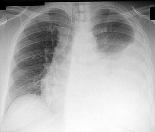
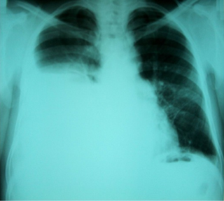
A pleural effusion is accumulation of excessive fluid in the pleural space, the potential space that surrounds each lung. Under normal conditions, pleural fluid is secreted by the parietal pleural capillaries at a rate of 0.6 millilitre per kilogram weight per hour, and is cleared by lymphatic absorption leaving behind only 5–15 millilitres of fluid, which helps to maintain a functional vacuum between the parietal and visceral pleurae. Excess fluid within the pleural space can impair inspiration by upsetting the functional vacuum and hydrostatically increasing the resistance against lung expansion, resulting in a fully or partially collapsed lung.

<span class="mw-page-title-main">Chyle</span> Milky bodily fluid consisting of lymph and emulsified fats

Chyle is a milky bodily fluid consisting of lymph and emulsified fats, or free fatty acids (FFAs). It is formed in the small intestine during digestion of fatty foods, and taken up by lymph vessels specifically known as lacteals. The lipids in the chyle are colloidally suspended in chylomicrons.

<span class="mw-page-title-main">Chest radiograph</span> Projection X-ray of the chest

A chest radiograph, called a chest X-ray (CXR), or chest film, is a projection radiograph of the chest used to diagnose conditions affecting the chest, its contents, and nearby structures. Chest radiographs are the most common film taken in medicine.

<span class="mw-page-title-main">Lymphangioleiomyomatosis</span> Medical condition

Lymphangioleiomyomatosis (LAM) is a rare, progressive and systemic disease that typically results in cystic lung destruction. It predominantly affects women, especially during childbearing years. The term sporadic LAM is used for patients with LAM not associated with tuberous sclerosis complex (TSC), while TSC-LAM refers to LAM that is associated with TSC.

<span class="mw-page-title-main">Miliary tuberculosis</span> Medical condition

Miliary tuberculosis is a form of tuberculosis that is characterized by a wide dissemination into the human body and by the tiny size of the lesions (1–5 mm). Its name comes from a distinctive pattern seen on a chest radiograph of many tiny spots distributed throughout the lung fields with the appearance similar to millet seeds—thus the term "miliary" tuberculosis. Miliary TB may infect any number of organs, including the lungs, liver, and spleen. Miliary tuberculosis is present in about 2% of all reported cases of tuberculosis and accounts for up to 20% of all extra-pulmonary tuberculosis cases.

<span class="mw-page-title-main">Chylothorax</span> Medical condition

A chylothorax is an abnormal accumulation of chyle, a type of lipid-rich lymph, in the space surrounding the lung. The lymphatics of the digestive system normally returns lipids absorbed from the small bowel via the thoracic duct, which ascends behind the esophagus to drain into the left brachiocephalic vein. If normal thoracic duct drainage is disrupted, either due to obstruction or rupture, chyle can leak and accumulate within the negative-pressured pleural space. In people on a normal diet, this fluid collection can sometimes be identified by its turbid, milky white appearance, since chyle contains emulsified triglycerides.

<span class="mw-page-title-main">Thoracentesis</span> Medical procedure

Thoracentesis, also known as thoracocentesis, pleural tap, needle thoracostomy, or needle decompression, is an invasive medical procedure to remove fluid or air from the pleural space for diagnostic or therapeutic purposes. A cannula, or hollow needle, is carefully introduced into the thorax, generally after administration of local anesthesia. The procedure was first performed by Morrill Wyman in 1850 and then described by Henry Ingersoll Bowditch in 1852.

<span class="mw-page-title-main">Respiratory disease</span> Disease of the respiratory system

Respiratory diseases, or lung diseases, are pathological conditions affecting the organs and tissues that make gas exchange difficult in air-breathing animals. They include conditions of the respiratory tract including the trachea, bronchi, bronchioles, alveoli, pleurae, pleural cavity, the nerves and muscles of respiration. Respiratory diseases range from mild and self-limiting, such as the common cold, influenza, and pharyngitis to life-threatening diseases such as bacterial pneumonia, pulmonary embolism, tuberculosis, acute asthma, lung cancer, and severe acute respiratory syndromes, such as COVID-19. Respiratory diseases can be classified in many different ways, including by the organ or tissue involved, by the type and pattern of associated signs and symptoms, or by the cause of the disease.

<span class="mw-page-title-main">Pericardial effusion</span> Medical condition

A pericardial effusion is an abnormal accumulation of fluid in the pericardial cavity. The pericardium is a two-part membrane surrounding the heart: the outer fibrous connective membrane and an inner two-layered serous membrane. The two layers of the serous membrane enclose the pericardial cavity between them. This pericardial space contains a small amount of pericardial fluid. The fluid is normally 15-50 mL in volume. The pericardium, specifically the pericardial fluid provides lubrication, maintains the anatomic position of the heart in the chest, and also serves as a barrier to protect the heart from infection and inflammation in adjacent tissues and organs.

<span class="mw-page-title-main">Yellow nail syndrome</span> Medical condition

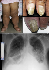
Yellow nail syndrome, also known as "primary lymphedema associated with yellow nails and pleural effusion", is a very rare medical syndrome that includes pleural effusions, lymphedema and yellow dystrophic nails. Approximately 40% will also have bronchiectasis. It is also associated with chronic sinusitis and persistent coughing. It usually affects adults.

<span class="mw-page-title-main">Mediastinitis</span> Inflammatory process affecting the mediastinum

Mediastinitis is inflammation of the tissues in the mid-chest, or mediastinum. It can be either acute or chronic. It is thought to be due to four different etiologies:

<span class="mw-page-title-main">Gorham's disease</span> Syndrome characterized by bone loss

Gorham's disease, also known as Gorham vanishing bone disease and phantom bone disease, is a very rare skeletal condition of unknown cause, characterized by the uncontrolled proliferation of distended, thin-walled vascular or lymphatic channels within bone, which leads to resorption and replacement of bone with angiomas and/or fibrosis.

Obstructive shock is one of the four types of shock, caused by a physical obstruction in the flow of blood. Obstruction can occur at the level of the great vessels or the heart itself. Causes include pulmonary embolism, cardiac tamponade, and tension pneumothorax. These are all life-threatening. Symptoms may include shortness of breath, weakness, or altered mental status. Low blood pressure and tachycardia are often seen in shock. Other symptoms depend on the underlying cause.

<span class="mw-page-title-main">Ground-glass opacity</span> Radiologic sign on radiographs and computed tomography scans

Ground-glass opacity (GGO) is a finding seen on chest x-ray (radiograph) or computed tomography (CT) imaging of the lungs. It is typically defined as an area of hazy opacification (x-ray) or increased attenuation (CT) due to air displacement by fluid, airway collapse, fibrosis, or a neoplastic process. When a substance other than air fills an area of the lung it increases that area's density. On both x-ray and CT, this appears more grey or hazy as opposed to the normally dark-appearing lungs. Although it can sometimes be seen in normal lungs, common pathologic causes include infections, interstitial lung disease, and pulmonary edema.

Tumor-like disorders of the lung pleura are a group of conditions that on initial radiological studies might be confused with malignant lesions. Radiologists must be aware of these conditions in order to avoid misdiagnosing patients. Examples of such lesions are: pleural plaques, thoracic splenosis, catamenial pneumothorax, pleural pseudotumor, diffuse pleural thickening, diffuse pulmonary lymphangiomatosis and Erdheim–Chester disease.

<span class="mw-page-title-main">Asbestos-related diseases</span> Medical condition

Asbestos-related diseases are disorders of the lung and pleura caused by the inhalation of asbestos fibres. Asbestos-related diseases include non-malignant disorders such as asbestosis, diffuse pleural thickening, pleural plaques, pleural effusion, rounded atelectasis and malignancies such as lung cancer and malignant mesothelioma.

References

  1. Marom, EM; Moran, CA; Munden, RF (April 2004). "Generalized lymphangiomatosis". American Journal of Roentgenology. 182 (4): 1068. doi:10.2214/ajr.182.4.1821068. PMID   15039189.
  2. 1 2 3 4 5 6 7 8 9 10 11 12 13 14 15 16 17 18 19 20 21 22 23 24 Faul J.L., Berry G.J., Colby T.V., Ruoss S.J., Walter M.B, Rosen G.D., Raffin T.A. (2000). "Thoracic Lymphangiomas, Lymphangiectasis, Lymphangiomatosis, and Lymphatic Dysplasia Syndrome". Am. J. Respir. Crit. Care Med. 161 (3): 1037–1046. doi:10.1164/ajrccm.161.3.9904056. PMID   10712360.{{cite journal}}: CS1 maint: multiple names: authors list (link)
  3. 1 2 Pernick, Nat. "Soft Tissue Tumors Part 3 Muscle, Vascular, Nerve, Other Lymphangiomatosis." PathologyOutlines.com. PathologyOutlines.com, Inc., 10/17/2009. Web. 6 Sep 2011. http://www.pathologyoutlines.com/topic/softtissue3lymphangiomatosis.html.
  4. 1 2 3 4 5 6 7 Tazelaar HD, Kerr D, Yousem SA, Saldana MJ, Langston C, Colby TV (Dec 1993). "Diffuse pulmonary lymphangiomatosis". Hum Pathol. 24 (12): 1313–22. doi: 10.1016/0046-8177(93)90265-i . PMID   8276379.{{cite journal}}: CS1 maint: multiple names: authors list (link)
  5. 1 2 3 Aviv RI, McHugh K, Hunt J. Angiomatosis of bone and soft tissue: a spectrum of disease from diffuse lymphangiomatosis to vanishing bone disease in young patients. Clin Radiol. 2001 Mar;56(3):184-90.
  6. Duffy B, Manon R, Patel R, Welsh JS, et al. A case of Gorham’s disease with chylothorax treated curatively with radiation therapy. Clin Med Res. 2005;3:83–
  7. 1 2 3 Yeager ND, Hammond S, Mahan J, Davis JT, Adler B. Unique diagnostic features and successful management of a patient with disseminated lymphangiomatosis and chylothorax. J Pediatr Hematol Oncol. 2008 Jan;30(1):66-9.
  8. Venkatramani R, Ma NS, Pitukcheewanont P, Malogolowkin MH, Mascarenhas L. Gorham’s disease and diffuse lymphangiomatosis in children and adolescents. Pediatr Blood Cancer. 2011 Apr;56(4):667-70
  9. Zisis C, Spiliotopoulos K, Patronis M, Filippakis G, Stratakos G, Tzelepis G, Bellenis I. Diffuse lymphangiomatosis: are there any clinical or therapeutic standards? J Thorac Cardiovasc Surg. 2007 Jun;133(6):1664-5.
  10. Hakeem A, Gojwari TA, Reyaz S, Rasool S, Shafi H, Mufti S (Jan 2010). "Computed tomography findings in bilateral perinephric lymphangiomatosis". Urol. Ann. 2 (1): 26–8. doi: 10.4103/0974-7796.62922 . PMC   2934585 . PMID   20842254.{{cite journal}}: CS1 maint: multiple names: authors list (link)
  11. 1 2 O’Sullivan DA, Torres VE, de Groen PC, Batts KP, King BF, Vockley J. "Hepatic lymphangiomatosis mimicking polycystic liver disease. Mayo Clin Proc. 1998 Dec;73(12):1188-92.
  12. Miller C, Mazzaferro V, Makowka L, ChapChap P, Demetris J, Tzakis A, Esquivel CO, Iwatsuki S, Starzl TE (Apr 1988). "Orthotopic liver transplantation for massive hepatic lymphangiomatosis". Surgery. 103 (4): 490–5. PMC   2963582 . PMID   3281302.{{cite journal}}: CS1 maint: multiple names: authors list (link)
  13. Watkins RG 4th, Reynolds RA, McComb JG, Tolo VT. Lymphangiomatosis of the spine: two cases requiring surgical intervention. Spine. 2003 Feb 1;28(3):E45-50.
  14. Jea A, McNeil A, Bhatia S, Birchansky S, Sotrel A, Ragheb J, Morrison G. A rare case of lymphangiomatosis of the craniocervical spine in conjunction with a Chiari I malformation. Pediatr Neurosurg. 2003 Oct;39(4):212-5.
  15. Shah V, Shah S, Barnacle A, Sebire NJ, Brock P, Harper JI, McHugh K. Mediastinal involvement in lymphangiomatosis: a previously unreported MRI sign. Pediatr Radiol. 2011 Aug;41(8):985-92.
  16. 1 2 CS Wong, TYC Chu. Clinical and radiological features of generalised lymphangiomatosis. Hong Kong Med J 2008;14:402-4
  17. 1 2 3 4 Ming-hua DU, Ruan-jian YE, Kun-kun SUN, Jian-feng LI, Dan-hua Shen, Jun Wang, Zhan-cheng GAO (2011). "Diffuse pulmonary lymphangiomatosis: a case report with literature review". Chinese Medical Journal. 124 (5): 797–800. PMID   21518582.{{cite journal}}: CS1 maint: multiple names: authors list (link)
  18. 1 2 Swensen SJ, Hartman TE, Mayo JR, Colby TV, Tazelaar HD, Müller NL (1995). "Diffuse pulmonary lymphangiomatosis: CT findings". J Comput Assist Tomogr. 19 (3): 348–52. doi:10.1097/00004728-199505000-00002. PMID   7790540. S2CID   13399987.{{cite journal}}: CS1 maint: multiple names: authors list (link)
  19. Wunderbaldinger P, Paya K, Partik B, Turetschek K, Hörmann M, Horcher E, Bankier AA (Mar 2000). "CT and MR imaging of generalized cystic lymphangiomatosis in pediatric patients". Am J Roentgenol. 174 (3): 827–32. doi:10.2214/ajr.174.3.1740827. PMID   10701634.{{cite journal}}: CS1 maint: multiple names: authors list (link)
  20. Martinez-Pajares JD, Rosa-Camacho V, Camacho-Alonso JM, Zabala-Arguelles I, Gil-Jaurena JM, Milano-Manso G. Spontaneous chylous pericardial effusion: report of two cases. An Pediatr (Barc). 2010 Jul;73(1):42-6.
  21. Lynch DA, Hay T, Newell JD (Sep 1999). "Jr, Divgi VD, Fan LL. Pediatric diffuse lung disease: diagnosis and classification using high-resolution CT". Am J Roentgenol. 173 (3): 713–8. doi:10.2214/ajr.173.3.10470910. PMID   10470910.{{cite journal}}: CS1 maint: multiple names: authors list (link)

Further reading