| Hemangioendothelioma | |
|---|---|
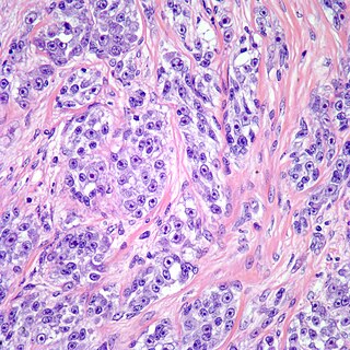
 | |
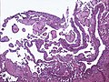
| Micrograph of a kaposiform hemangioendothelioma with "glomeruloid" nodules of endothelial cells. | |
| Specialty | Oncology |
Hemangioendotheliomas are a family of vascular neoplasms of intermediate malignancy.
| Hemangioendothelioma | |
|---|---|
| | |
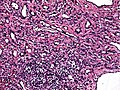
| Micrograph of a kaposiform hemangioendothelioma with "glomeruloid" nodules of endothelial cells. | |
| Specialty | Oncology |
Hemangioendotheliomas are a family of vascular neoplasms of intermediate malignancy.
They have been described as masses that fall between a hemangioma and angiosarcoma. They are vascular tumors that commonly present with an enlarging mass and most commonly involve the lungs, liver, and musculoskeletal system, although many other body sites have been reported, including the head and neck, intestines, lymph nodes, pleura, retroperitoneum, heel, stomach.[ citation needed ]
Possibly Bartonella spp bacteraemia[ citation needed ]
Hemangioendotheliomas may be classified as:

Treatment is varied and depends on the site and extent of tumor involvement, sites of metastasis, and specific individual factors. Surgical resection, radiotherapy, and chemotherapy have all been used to treat these masses, although studies on survival have yet to be conducted to delineate various treatment regimens. Kaposiform hemangioendothelioma might respond to chemotherapy or antiangiogenic therapies. Recently propanolol and steroids have been shown to be very effective in kaposiform hemangioendothelioma. [17]

Thyroid neoplasm is a neoplasm or tumor of the thyroid. It can be a benign tumor such as thyroid adenoma, or it can be a malignant neoplasm, such as papillary, follicular, medullary or anaplastic thyroid cancer. Most patients are 25 to 65 years of age when first diagnosed; women are more affected than men. The estimated number of new cases of thyroid cancer in the United States in 2010 is 44,670 compared to only 1,690 deaths. Of all thyroid nodules discovered, only about 5 percent are cancerous, and under 3 percent of those result in fatalities.

A sarcoma is a malignant tumor, a type of cancer that arises from cells of mesenchymal origin. Connective tissue is a broad term that includes bone, cartilage, fat, vascular, or other structural tissues, and sarcomas can arise in any of these types of tissues. As a result, there are many subtypes of sarcoma, which are classified based on the specific tissue and type of cell from which the tumor originates. Sarcomas are primary connective tissue tumors, meaning that they arise in connective tissues. This is in contrast to secondary connective tissue tumors, which occur when a cancer from elsewhere in the body spreads to the connective tissue. Sarcomas are one of five different types of cancer, classified by the cell type from which they originate. The word sarcoma is derived from the Greek σάρκωμα sarkōma 'fleshy excrescence or substance', itself from σάρξsarx meaning 'flesh'.

An infantile hemangioma (IH), sometimes called a strawberry mark due to appearance, is a type of benign vascular tumor or anomaly that affects babies. Other names include capillary hemangioma, strawberry hemangioma, strawberry birthmark and strawberry nevus. and formerly known as a cavernous hemangioma. They appear as a red or blue raised lesion on the skin. Typically, they begin during the first four weeks of life, growing until about five months of life, and then shrinking in size and disappearing over the next few years. Often skin changes remain after they shrink. Complications may include pain, bleeding, ulcer formation, disfigurement, or heart failure. It is the most common tumor of orbit and periorbital areas in childhood. It may occur in the skin, subcutaneous tissues and mucous membranes of oral cavities and lips as well as in extracutaneous locations including the liver and gastrointestinal tract.

A glomus tumor is a rare neoplasm arising from the glomus body and mainly found under the nail, on the fingertip or in the foot. They account for less than 2% of all soft tissue tumors. The majority of glomus tumors are benign, but they can also show malignant features. Glomus tumors were first described by Hoyer in 1877 while the first complete clinical description was given by Masson in 1924.

Fibrosarcoma is a malignant mesenchymal tumour derived from fibrous connective tissue and characterized by the presence of immature proliferating fibroblasts or undifferentiated anaplastic spindle cells in a storiform pattern. Fibrosarcomas mainly arise in people between the ages of 25 and 79. It originates in fibrous tissues of the bone and invades long or flat bones such as the femur, tibia, and mandible. It also involves the periosteum and overlying muscle.
The International Classification of Diseases for Oncology (ICD-O) is a domain-specific extension of the International Statistical Classification of Diseases and Related Health Problems for tumor diseases. This classification is widely used by cancer registries.

Focal nodular hyperplasia is a benign tumor of the liver, which is the second most prevalent tumor of the liver after hepatic hemangioma. It is usually asymptomatic, rarely grows or bleeds, and has no malignant potential. This tumour was once often resected because it was difficult to distinguish from hepatic adenoma, but with modern multiphase imaging it is usually now diagnosed by strict imaging criteria and not resected.
A vascular tumor is a tumor of vascular origin; a soft tissue growth that can be either benign or malignant, formed from blood vessels or lymph vessels. Examples of vascular tumors include hemangiomas, lymphangiomas, hemangioendotheliomas, Kaposi's sarcomas, angiosarcomas, and hemangioblastomas. An angioma refers to any type of benign vascular tumor.
Epithelioid may refer to:

Primary tumors of the heart are extremely rare tumors that arise from the normal tissues that make up the heart. The incidence of primary cardiac tumors has been found to be approximately 0.02%. This is in contrast to secondary tumors of the heart, which are typically either metastatic from another part of the body, or infiltrate the heart via direct extension from the surrounding tissues. Metastatic tumors to the heart are about 20 times more common than primary cardiac tumors.
Mast cell sarcoma is an extremely aggressive form of sarcoma made up of neoplastic mast cells. A sarcoma is a tumor made of cells from connective tissue. Mast cell sarcoma is an extremely rare tumor. Only three cases have been are reported so far. Prognosis is extremely poor. People with a mast cell sarcoma have no skin lesions, and pathology examination of the tumor shows it to be very malignant with an aggressive growth pattern. Mast cell sarcoma should not be confused with extracutaneous mastocytoma, a rare benign mast cell tumor without destructive growth. In the cases observed, mast cell sarcoma terminated quickly as mast cell leukemia; one of the most aggressive human cancers.

Pseudoangiomatous stromal hyperplasia (PASH) is an overgrowth of myofibroblastic cells in the breast. It has an appearance similar to fibroadenomatoid changes.

Epithelioid hemangioendothelioma (EHE) is a rare tumor, first characterized by Sharon Weiss and Franz Enzinger in 1982 that both clinically and histologically is intermediate between angiosarcoma and hemangioma. However, a distinct, disease-defining genetic alteration recently described for EHE indicates that it is an entirely separate entity from both angiosarcoma and hemangioma.
Clear cell sarcoma is a rare form of cancer called a sarcoma. It is known to occur mainly in the soft tissues and dermis. Rare forms were thought to occur in the gastrointestinal tract before they were discovered to be different and redesignated as gastrointestinal neuroectodermal tumors.
A vascular anomaly is any of a range of lesions from a simple birthmark to a large tumor that may be disfiguring. They are caused by a disorder of the vascular system. A vascular anomaly is a localized defect in blood or lymph vessels. These defects are characterized by an increased number of vessels, and vessels that are both enlarged and sinuous. Some vascular anomalies are congenital, others appear within weeks to years after birth, and others are acquired by trauma or during pregnancy. Inherited vascular anomalies are also described and often present with a number of lesions that increase with age. Vascular anomalies can also be a part of a syndrome.
Sharon Ann Whelan Weiss is an American pathologist who is best known for her contribution to the subspecialty of soft tissue pathology. She is the main author of Soft Tissue Tumors, one of the most widely used textbooks in the field of sarcoma and soft tissue pathology. She is also well known for her seminal descriptions of multiple soft tissue tumors, such as epithelioid hemangioendothelioma and pleomorphic hyalinizing angiectatic tumor of soft parts among others. She has also mentored and trained other well-known soft tissue pathologists.
Diffuse neonatal hemangiomatosis (DNH) is a potentially fatal disorder where multiple benign (non-cancerous) blood vessel tumors (hemangiomas) are present in the skin and other organs. The mortality rate of diffuse neonatal hemangiomatosis is 50-90%. This disease is normally found in female Caucasian infants. The most common site of internal organ damage, or lesions, is the liver, which can redirect blood away from the heart and cause arteriovenous shunting. This can cause high cardiac output, leading to further complications such as congestive heart failure. This condition affecting the liver is also known as infantile hepatic hemangioma (IHH). Other sites of internal organ damage can include the intestines, nervous system, lungs, and sometimes the skeletal system. Early detection and treatment with steroids results in most newborn babies with this disease remaining healthy, with serious problems developing for some individuals during the hemangioma's growth phase.

Proliferative fasciitis and proliferative myositis (PF/PM) are rare benign soft tissue lesions that increase in size over several weeks and often regress over the ensuing 1–3 months. The lesions in PF/PM are typically obvious tumors or swellings. Historically, many studies had grouped the two descriptive forms of PF/PM as similar disorders with the exception that proliferative fasciitis occurs in subcutaneous tissues while proliferative myositis occurs in muscle tissues. In 2020, the World Health Organization agreed with this view and defined these lesions as virtually identical disorders termed proliferative fasciitis/proliferative myositis or proliferative fasciitis and proliferative myositis. The Organization also classified them as one of the various forms of the fibroblastic and myofibroblastic tumors.
Fibroblastic and myofibroblastic tumors (FMTs) develop from the mesenchymal stem cells which differentiate into fibroblasts and/or the myocytes/myoblasts that differentiate into muscle cells. FMTs are a heterogeneous group of soft tissue neoplasms. The World Health Organization (2020) defined tumors as being FMTs based on their morphology and, more importantly, newly discovered abnormalities in the expression levels of key gene products made by these tumors' neoplastic cells. Histopathologically, FMTs consist of neoplastic connective tissue cells which have differented into cells that have microscopic appearances resembling fibroblasts and/or myofibroblasts. The fibroblastic cells are characterized as spindle-shaped cells with inconspicuous nucleoli that express vimentin, an intracellular protein typically found in mesenchymal cells, and CD34, a cell surface membrane glycoprotein. Myofibroblastic cells are plumper with more abundant cytoplasm and more prominent nucleoli; they express smooth muscle marker proteins such as smooth muscle actins, desmin, and caldesmon. The World Health Organization further classified FMTs into four tumor forms based on their varying levels of aggressiveness: benign, intermediate, intermediate, and malignant.