Related Research Articles

Oral leukoplakia is a potentially malignant disorder affecting the oral mucosa. It is defined as "essentially an oral mucosal white lesion that cannot be considered as any other definable lesion." Oral leukoplakia is a white patch or plaque that develops in the oral cavity and is strongly associated with smoking. Leukoplakia is a firmly attached white patch on a mucous membrane which is associated with increased risk of cancer. The edges of the lesion are typically abrupt and the lesion changes with time. Advanced forms may develop red patches. There are generally no other symptoms. It usually occurs within the mouth, although sometimes mucosa in other parts of the gastrointestinal tract, urinary tract, or genitals may be affected.

Lichen planus (LP) is a chronic inflammatory and autoimmune disease that affects the skin, nails, hair, and mucous membranes. It is not an actual lichen, but is named for its appearance. It is characterized by polygonal, flat-topped, violaceous papules and plaques with overlying, reticulated, fine white scale, commonly affecting dorsal hands, flexural wrists and forearms, trunk, anterior lower legs and oral mucosa. The hue may be gray-brown in people with darker skin. Although there is a broad clinical range of LP manifestations, the skin and oral cavity remain as the major sites of involvement. The cause is unknown, but it is thought to be the result of an autoimmune process with an unknown initial trigger. There is no cure, but many different medications and procedures have been used in efforts to control the symptoms.

A precancerous condition is a condition, tumor or lesion involving abnormal cells which are associated with an increased risk of developing into cancer. Clinically, precancerous conditions encompass a variety of abnormal tissues with an increased risk of developing into cancer. Some of the most common precancerous conditions include certain colon polyps, which can progress into colon cancer, monoclonal gammopathy of undetermined significance, which can progress into multiple myeloma or myelodysplastic syndrome. and cervical dysplasia, which can progress into cervical cancer. Bronchial premalignant lesions can progress to squamous cell carcinoma of the lung.
Ocular melanosis (OM) is a blue-gray and/or brown lesion of the conjunctiva that can be separated into benign conjunctival epithelial melanosis (BCEM) and primary acquired melanosis (PAM), of which the latter is considered a risk factor for uveal melanoma. The disease is caused by an increase of melanocytes in the iris, choroid, and surrounding structures. Overproduction of pigment by these cells can block the trabecular meshwork through which fluid drains from the eye. The increased fluid in the eye leads to increased pressure, which can lead to glaucoma. In humans, this is sometimes known as pigment dispersion syndrome.
The oral mucosa is the mucous membrane lining the inside of the mouth. It comprises stratified squamous epithelium, termed "oral epithelium", and an underlying connective tissue termed lamina propria. The oral cavity has sometimes been described as a mirror that reflects the health of the individual. Changes indicative of disease are seen as alterations in the oral mucosa lining the mouth, which can reveal systemic conditions, such as diabetes or vitamin deficiency, or the local effects of chronic tobacco or alcohol use. The oral mucosa tends to heal faster and with less scar formation compared to the skin. The underlying mechanism remains unknown, but research suggests that extracellular vesicles might be involved.

Geographic tongue, also known by several other terms, is a condition of the mucous membrane of the tongue, usually on the dorsal surface. It is a common condition, affecting approximately 2–3% of the general population. It is characterized by areas of smooth, red depapillation which migrate over time. The name comes from the map-like appearance of the tongue, with the patches resembling the islands of an archipelago. The cause is unknown, but the condition is entirely benign, and there is no curative treatment. Uncommonly, geographic tongue may cause a burning sensation on the tongue, for which various treatments have been described with little formal evidence of efficacy.

Fordyce spots are harmless and painless visible sebaceous glands typically appearing as white/yellow small bumps or spots on the inside of lips or cheeks, gums, or genitalia. They are common, and are present in around 80% of adults. Treatment is generally not required and attempts to remove them typically result in pain and scarring.

Vernal keratoconjunctivitis is a recurrent, bilateral, and self-limiting type of conjunctivitis having a periodic seasonal incidence.

Episcleritis is a benign, self-limiting inflammatory disease affecting part of the eye called the episclera. The episclera is a thin layer of tissue that lies between the conjunctiva and the connective tissue layer that forms the white of the eye (sclera). Episcleritis is a common condition, and is characterized by the abrupt onset of painless eye redness.

An Eye neoplasm is a tumor of the eye. A rare type of tumor, eye neoplasms can affect all parts of the eye, and can either be benign or malignant (cancerous), in which case it is known as eye cancer. Eye cancers can be primary or metastatic cancer. The two most common cancers that spread to the eye from another organ are breast cancer and lung cancer. Other less common sites of origin include the prostate, kidney, thyroid, skin, colon and blood or bone marrow.

Endometrial intraepithelial neoplasia (EIN) is a premalignant lesion of the uterine lining that predisposes to endometrioid endometrial adenocarcinoma. It is composed of a collection of abnormal endometrial cells, arising from the glands that line the uterus, which have a tendency over time to progress to the most common form of uterine cancer—endometrial adenocarcinoma, endometrioid type.
Leukoedema is a blue, grey or white appearance of mucosae, particularly the buccal mucosa ; it may also occur on the mucosa of the larynx or vagina. It is a harmless and very common condition. Because it is so common, it has been argued that it may in fact represent a variation of the normal appearance rather than a disease, but empirical evidence suggests that leukoedema is an acquired condition caused by local irritation. It is found more commonly in black skinned people and tobacco users. The term is derived from the Greek words λευκός leukós, "white" and οἴδημα oídēma, "swelling".
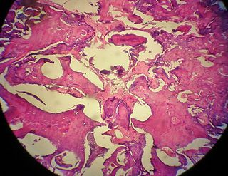
An odontoma, also known as an odontome, is a benign tumour linked to tooth development. Specifically, it is a dental hamartoma, meaning that it is composed of normal dental tissue that has grown in an irregular way. It includes both odontogenic hard and soft tissues. As with normal tooth development, odontomas stop growing once mature which makes them benign.

White sponge nevus (WSN) is an autosomal dominant condition of the oral mucosa. It is caused by a mutations in certain genes coding for keratin, which causes a defect in the normal process of keratinization of the mucosa. This results in lesions which are thick, white and velvety on the inside of the cheeks within the mouth. Usually, these lesions are present from birth or develop during childhood. The condition is entirely harmless, and no treatment is required.

A pinguecula is a common type of conjunctival stromal degeneration in the eye. It appears as an elevated yellow-white plaque in the bulbar conjunctiva near the limbus. Calcification may also seen occasionally.

Gingival enlargement is an increase in the size of the gingiva (gums). It is a common feature of gingival disease. Gingival enlargement can be caused by a number of factors, including inflammatory conditions and the side effects of certain medications. The treatment is based on the cause. A closely related term is epulis, denoting a localized tumor on the gingiva.

Genodermatosis is a hereditary skin disease with three inherited modes including single gene inheritance, multiple gene inheritance and chromosome inheritance. There are many different types of genodermatosis, the prevalence of genodermatosis ranges from 1 per 6000 people to 1 per 500,000 people. Genodermatosis has influence on the texture, color and structure of skin cuticle and connective tissue, specific lesion site and clinical manifestations on the body vary depending on the type. In the spite of the variety and complexity of genodermatosis, there are still some common methods that can help people diagnose. After diagnosis, different types of genodermatosis require different levels of therapy including interventions, nursing interventions and treatments. Among that, research of therapy for some new, complex and rare types are still in the developing stage. The impact of genodermatosis not only can be seen in body but also can be seen in all aspects of patients' life, including but not limited to psychological, family life, economic conditions and social activities. Accordingly, the patients need treatment, support and help in these areas.

A squamous cell papilloma is a generally benign papilloma that arises from the stratified squamous epithelium of the skin, lip, oral cavity, tongue, pharynx, larynx, esophagus, cervix, vagina or anal canal. Squamous cell papillomas are typically associated with human papillomavirus (HPV) while sometimes the cause is unknown.

Warty dyskeratoma, also known as an Isolated dyskeratosis follicularis, is a benign epidermal proliferation with distinctive histologic findings that may mimic invasive squamous cell carcinoma and commonly manifests as an umbilicated lesion with a keratotic plug, WD have some histopathologic similarities to viral warts but it's not caused by HPV and the majority of these lesions display overall histopathologic features consistent with a follicular adnexal neoplasm. Usually limited to the head, neck, scalp or face and vulva. Lesions are generally solitary and sporadic and may be associated with a follicular unit. Oral involvement, particularly the hard palate, and genital involvement have been reported. it can also be thought of as one of the manifestations of focal acantholytic dyskeratosis, an epidermal reaction pattern that can be seen in several disorders, including Darier's disease and Grover's disease. But the main Difference between Darier disease and Warty dyskeratoma, is that Darier disease inherited dermatosis consisting of multiple keratotic papules on the face, trunk, and extremities, while WD occurs as an isolated, noninherited, single keratotic nodule mainly confined to the head and neck as mentioned earlier.
Familial disseminated comedones without dyskeratosis (FDCWD) is a rare autosomal dominant skin disorder characterized by the presence of numerous comedones on the face, trunk, and extremities. The comedones are typically asymptomatic and do not lead to scarring. The disorder is thought to be caused by a mutation in the gene encoding the protein involucrin.
References
- ↑ Bui, T; Young, JW; Frausto, RF; Markello, TC; Glasgow, BJ; Aldave, AJ (20 February 2014). "Hereditary Benign Intraepithelial Dyskeratosis: Report of a Case and Re-examination of the Evidence for Locus Heterogeneity". Ophthalmic Genetics. 37 (1): 76–80. doi:10.3109/13816810.2014.889169. PMC 4139474 . PMID 24555743.
- 1 2 3 Woo SB (2012). Oral Pathology: A Comprehensive Atlas and Text. Elsevier Health Sciences. p. 9. ISBN 978-1-4377-2226-0.
| | This genetic disorder article is a stub. You can help Wikipedia by expanding it. |