This article needs more reliable medical references for verification or relies too heavily on primary sources .(June 2012) |  |
| Calcinosis | |
|---|---|
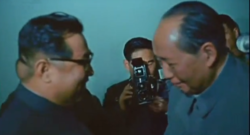 | |
| North Korean President Kim Il Sung's calcium deposit is noticeable on the back of his head in this rare newsreel still image during a diplomatic meeting between him and Chinese leader Mao Zedong in Beijing, 1970.[ medical citation needed ] | |
| Specialty | Endocrinology |
Calcinosis is the formation of calcium deposits in any soft tissue. [1] It is a rare condition that has many different causes. These range from infection and injury to systemic diseases like kidney failure.