Calcinosis cutis

Last updated
Calcinosis cutis
Other namesCutaneous calcification
Calcinosis cutis -- low mag.jpg
Micrograph of calcinosis cutis. The calcification is purple (bottom of image). H&E stain.
Specialty Dermatology   OOjs UI icon edit-ltr-progressive.svg

Calcinosis cutis is an uncommon condition marked by calcium buildup in the skin and subcutaneous tissues. Calcinosis cutis can range in intensity from little nodules in one area of the body to huge, crippling lesions affecting a vast portion of the body. [1] Five kinds of the condition are typically distinguished: calciphylaxis, idiopathic calcification, iatrogenic calcification, dystrophic calcification, and metastatic calcification. [2]

Contents

Tumors, inflammation, varicose veins, infections, connective tissue disease, hyperphosphatemia, and hypercalcemia can all lead to calcinosis. Systemic sclerosis is linked to calcineuris cutis. [3] Calcinosis is seen in Limited Cutaneous Systemic Sclerosis, also known as CREST syndrome (the "C" in CREST). [4]

Signs and symptoms

Calcinosis cutis new image.jpg

Lesions might be more severe and widespread, or they can develop gradually and show no symptoms. The nodules may cause pain and hinder function in addition to having a variety of sizes and shapes. The underlying condition determines the localization of the lesions in dystrophic calcification. The elbows, fingers, knees, and forearms are the most often affected regions in people with systemic sclerosis. Elbows, knees, and regions of prior inflammatory lesions in dermatomyositis are affected by calcification. Lupus erythematosus affects the limbs, buttocks, area beneath lupus lesions, and periarticular areas. Periarticular lesions are found in metastatic calcification. In tumoral calcinosis, the lesions are found around joints, but in idiopathic calcification, the lesions are found on children's faces as subepidermal calcified nodules. In iatrogenic calcification, the calcification is found at venipuncture sites. [3]

Causes

Calcinosis may result from a variety of causes such as: [3]

Calcinosis cutis is associated with systemic sclerosis. [3]

Diagnosis

Classification

Calcinosis cutis may be divided into the following types: [5] :527–530

Dystrophic calcinosis cutis

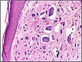
Dystrophic calcinosis cutis is the most prevalent kind of calcification on the skin. [2] The ectopic calcified mass usually consists of amorphous calcium phosphate and hydroxyapatite. [6] Dystrophic calcification is linked to a number of illnesses, such as infections, hereditary diseases, cutaneous neoplasms, and connective tissue diseases. [7] The clinical manifestation can be as minor as an accidental radiography imaging finding or as severe as subcutaneous nodules or plaques. [8]

Metastatic calcinosis cutis

Metastatic calcinosis cutis is the consequence of calcium salts precipitating in normal tissue due to an underlying abnormality in the metabolism of phosphate and/or calcium. [2] Metastatic calcification can result from any systemic condition raising serum calcium and/or phosphate levels. Chronic renal failure is the most frequent underlying cause. [9]

Iatrogenic calcinosis cutis

Iatrogenic calcinosis cutis is characterized by firm nodules in the subcutis or dermis, which are caused by calcium salts precipitating quickly in the skin. [9] This occurrence typically manifests as a warm, sensitive swelling at the site of venipuncture, [2] and it most frequently happens following the extravasation of intravenous calcium chloridecalcium gluconate, or phosphate-containing solutions. [10] Iatrogenic calcification, which manifests as soft yellow-white epidermal plaques, [2] has also been linked to calcium salt exposure via electroencephalography or electromyographic electrode compounds. [11]

Traumatic calcinosis cutis

Traumatic calcinosis cutis is a cutaneous condition characterized by calcification of the skin resulting from the deposition of calcium and phosphorus often resulting from occupational exposure, as in cases reported in oil-field workers and coal miners. [12] :528

Idiopathic calcinosis cutis

Skin calcification that is not linked to a systemic illness or an underlying tissue injury is referred to as idiopathic calcification. [2] Most often, the calcification is restricted to a single general location, yet there has been one case of calcinosis cutis that is exceptionally broad. [13]

Idiopathic scrotal calcinosis

Idiopathic scrotal calcinosis is a cutaneous condition characterized by calcification of the skin resulting from the deposition of calcium and phosphorus occurring on the scrotum. [14] :528 However, the levels of calcium and phosphate in the blood are normal. [15] Idiopathic scrotal calcinosis typically affects young males, with an onset between adolescence and early adulthood. [15] The scrotal calcinosis appears, without any symptoms, as yellowish nodules that range in size from 1 mm to several centimeters. [16]

Subepidermal calcified nodule

Subepidermal calcified nodule is characterized by calcification of the skin resulting from the deposition of calcium and phosphorus, occurring most frequently as one or a few skin lesions on the scalp or face of children. [17] :528

Tumoral calcinosis

Tumoral calcinosis is distinguished by the accumulation of calcific masses surrounding the main joints. It mainly affects teens who are otherwise in good health. Joint function may be hampered by the subcutaneous or intramuscular calcified deposits. [2]

Osteoma cutis

Osteoma cutis is a cutaneous condition characterized by the presence of bone within the skin in the absence of a preexisting or associated lesion. [18] :529 Osteoma cutis often manifests as solid, varying-sized, skin-colored subcutaneous nodules. [19]

Treatment

Diltiazem, a calcium channel blocker, has been a mainstay of medical treatment for calcinosis cutis. [1] It is thought to work by modifying intracellular calcium levels, which reduces the capacity for the production and crystallization of calcium nidus. [20]

Colchicine is an antimicrotubule drug with anti-inflammatory properties that has been used for gouty arthritis treatment for a long time. [1] Calcinosis cutis inflammation brought on by a foreign body-like response aggravates the illness's symptoms. [21] Colchicine, therefore, has been used to treat calcinosis cutis, albeit with varying degrees of success. [1]

See also

References

  1. 1 2 3 4 Gutierrez Jr, Albert; Wetter, David A. (2012). "Calcinosis cutis in autoimmune connective tissue diseases: Calcinosis cutis and connective tissue disease". Dermatologic Therapy. 25 (2): 195–206. doi: 10.1111/j.1529-8019.2012.01492.x . PMID   22741938.
  2. 1 2 3 4 5 6 7 Reiter, Nadine; El-Shabrawi, Laila; Leinweber, Bernd; Berghold, Andrea; Aberer, Elisabeth (2011). "Calcinosis cutis". Journal of the American Academy of Dermatology. 65 (1): 1–12. doi:10.1016/j.jaad.2010.08.038. PMID   21679810.
  3. 1 2 3 4 Le, Cuong; Bedocs, Paul M. (2023-07-10). "Calcinosis Cutis". StatPearls Publishing. PMID   28846311 . Retrieved 2024-02-28.
  4. "CREST syndrome: MedlinePlus Medical Encyclopedia Image". medlineplus.gov. Retrieved 2023-02-12.
  5. James, William D.; Berger, Timothy G.; et al. (2006). Andrews' Diseases of the Skin: clinical Dermatology. Saunders Elsevier. ISBN   978-0-7216-2921-6.
  6. Wang, Wen-Jen (1988-11-01). "Calcinosis Cutis in Juvenile Dermatomyositis: Remarkable Response to Aluminum Hydroxide Therapy". Archives of Dermatology. 124 (11): 1721. doi:10.1001/archderm.1988.01670110077022. ISSN   0003-987X. PMID   3178257.
  7. Walsh, John S; Fairley, Janet A (1995). "Calcifying disorders of the skin". Journal of the American Academy of Dermatology. 33 (5): 693–706. doi:10.1016/0190-9622(95)91803-5. PMID   7593766.
  8. Boulman, Nina; Slobodin, Gleb; Rozenbaum, Michael; Rosner, Itzhak (2005). "Calcinosis in Rheumatic Diseases". Seminars in Arthritis and Rheumatism. 34 (6). Elsevier BV: 805–812. doi:10.1016/j.semarthrit.2005.01.016. ISSN   0049-0172. PMID   15942915.
  9. 1 2 "UpToDate". UpToDate. Retrieved 2024-02-29.
  10. Amati, C.; Pesce, V.; Armenio, A.; Solarino, G.; Moretti, B. (2015-04-08). "Tumoral calcinosis of the hand". Journal of Surgical Case Reports. 2015 (4). Oxford University Press (OUP): rjv036. doi:10.1093/jscr/rjv036. hdl: 11586/171759 . ISSN   2042-8812. PMC   4390718 . PMID   25858267.
  11. Touart, Diane M.; Sau, Purnima (1998). "Cutaneous deposition diseases. Part II". Journal of the American Academy of Dermatology. 39 (4). Elsevier BV: 527–546. doi:10.1016/s0190-9622(98)70001-5. ISSN   0190-9622. PMID   9777759.
  12. James, William D.; Berger, Timothy G.; et al. (2006). Andrews' Diseases of the Skin: clinical Dermatology. Saunders Elsevier. ISBN   978-0-7216-2921-6.
  13. Guermazi, Ali; Grigoryan, Mikayel; Cordoliani, Florence; Kérob, Delphine (2006-12-21). "Unusually diffuse idiopathic calcinosis cutis". Clinical Rheumatology. 26 (2): 268–270. doi:10.1007/s10067-005-0135-8. ISSN   0770-3198. PMID   16416033.
  14. James, William D.; Berger, Timothy G.; et al. (2006). Andrews' Diseases of the Skin: clinical Dermatology. Saunders Elsevier. ISBN   978-0-7216-2921-6.
  15. 1 2 Grenader, Tal; Shavit, Linda (Aug 18, 2011). "Scrotal Calcinosis". New England Journal of Medicine. 365 (7): 647. doi:10.1056/NEJMicm1013803. PMID   21848465.
  16. Khallouk A, Yazami OE, Mellas S, Tazi MF, El Fassi J, Farih MH (2011). "Idiopathic scrotal calcinosis: a non-elucidated pathogenesis and its surgical treatment". Reviews in Urology. 13 (2): 95–7. PMC   3176555 . PMID   21935341.
  17. James, William D.; Berger, Timothy G.; et al. (2006). Andrews' Diseases of the Skin: clinical Dermatology. Saunders Elsevier. ISBN   978-0-7216-2921-6.
  18. James, William D.; Berger, Timothy G.; et al. (2006). Andrews' Diseases of the Skin: clinical Dermatology. Saunders Elsevier. ISBN   978-0-7216-2921-6.
  19. Niebel, Dennis; Poortinga, Sietske; Wenzel, rg (2020-09-22). "Osteoma Cutis and Calcinosis Cutis: "Similar but Different"". The Journal of Clinical and Aesthetic Dermatology. 13 (11). Matrix Medical Communications: 28–31. PMC   7716738 . PMID   33282099.
  20. Torralba, Tito P.; Li-Yu, Julie; Navarra, Sandra T.G.V. (1999). "Successful Use of Diltiazem in Calcinosis Caused by Connective Tissue Disease". JCR: Journal of Clinical Rheumatology. 5 (2). Ovid Technologies (Wolters Kluwer Health): 74–78. doi:10.1097/00124743-199904000-00007. ISSN   1076-1608. PMID   19078360.
  21. Fuchs, D; Fruchter, L; Fishel, B; Holtzman, M; Yaron, M (December 1986). "Colchicine suppression of local inflammation due to calcinosis in dermatomyositis and progressive systemic sclerosis". Clinical Rheumatology. 5 (4): 527–530. PMID   3816102.

Further reading