Related Research Articles
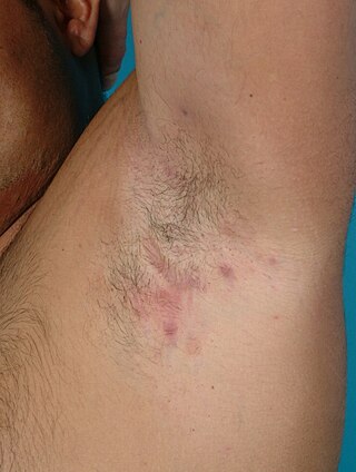
Hidradenitis suppurativa (HS), sometimes known as acne inversa or Verneuil's disease, is a long-term dermatological condition characterized by the occurrence of inflamed and swollen lumps. These are typically painful and break open, releasing fluid or pus. The areas most commonly affected are the underarms, under the breasts, and the groin. Scar tissue remains after healing. HS may significantly limit many everyday activities, for instance, walking, hugging, moving, and sitting down. Sitting disability may occur in patients with lesions in sacral, gluteal, perineal, femoral, groin or genital regions; and prolonged periods of sitting down itself can also worsen the condition of the skin of these patients.

Biliary atresia, also known as extrahepatic ductopenia and progressive obliterative cholangiopathy, is a childhood disease of the liver in which one or more bile ducts are abnormally narrow, blocked, or absent. It can be congenital or acquired. It has an incidence of one in 10,000–15,000 live births in the United States, and a prevalence of one in 16,700 in the British Isles. Biliary atresia is most common in East Asia, with a frequency of one in 5,000.

Pityriasis rosea is a type of skin rash. Classically, it begins with a single red and slightly scaly area known as a "herald patch". This is then followed, days to weeks later, by an eruption of many smaller scaly spots; pinkish with a red edge in people with light skin and greyish in darker skin. About 20% of cases show atypical deviations from this pattern. It usually lasts less than three months and goes away without treatment. Sometimes malaise or a fever may occur before the start of the rash or itchiness, but often there are few other symptoms.

The word Lichen comes from the Greek word 'lichen' (λειχην), meaning also lichen, lichen like, rash; the word sclerosus comes from the Greek word 'skleros' (σκλερóς), meaning dry, hard; the Latin suffix -osus makes an adjective in Latin from it. Lichen sclerosus (LS) is a chronic, inflammatory skin disease of unknown cause which can affect any body part of any person but has a strong preference for the genitals and is also known as balanitis xerotica obliterans (BXO) when it affects the penis. Lichen sclerosus is not contagious. There is a well-documented increase of skin cancer risk in LS, potentially improvable with treatment. LS in adult age women is normally incurable, but improvable with treatment, and often gets progressively worse if not treated properly. Most males with mild or intermediate disease restricted to foreskin or glans can be cured by either medical or surgical treatment.

Actinic keratosis (AK), sometimes called solar keratosis or senile keratosis, is a pre-cancerous area of thick, scaly, or crusty skin. Actinic keratosis is a disorder of epidermal keratinocytes that is induced by ultraviolet (UV) light exposure. These growths are more common in fair-skinned people and those who are frequently in the sun. They are believed to form when skin gets damaged by UV radiation from the sun or indoor tanning beds, usually over the course of decades. Given their pre-cancerous nature, if left untreated, they may turn into a type of skin cancer called squamous cell carcinoma. Untreated lesions have up to a 20% risk of progression to squamous cell carcinoma, so treatment by a dermatologist is recommended.

Blaschko's lines, also called the lines of Blaschko, are lines of normal cell development in the skin. These lines are only visible in those with a mosaic skin condition or in chimeras where different cell lines contain different genes. These lines may express different amounts of melanin, or become visible due to a differing susceptibility to disease. In such individuals, they can become apparent as whorls, patches, streaks or lines in a linear or segmental distribution over the skin. They follow a V shape over the back, S-shaped whirls over the chest and sides, and wavy shapes on the head. Not all mosaic skin conditions follow Blaschko's lines.

Degos disease, also known as Köhlmeier-Degos disease or malignant atrophic papulosis, is an extremely rare condition caused by blockage of arteries and veins. Individuals with this condition will develop papules. Those diagnosed with this disease may also develop complications due to impairment of internal organs. The exact underlying mechanism is still unknown, and an effective treatment is still being developed. There are fewer than 50 living patients presently known worldwide, and fewer than 200 reported in medical literature. However, many individuals may go undiagnosed due to rarity of the disease. Most individuals develop symptoms between the ages of 20–50; however, cases outside of this age range have been reported as well.
Pityriasis alba is a skin condition, a type of dermatitis, commonly seen in children and young adults as dry, fine-scaled, pale patches on the face. It is self-limiting and usually only requires use of moisturizer creams.
Amelanotic melanoma is a type of skin cancer in which the cells do not make any melanin. They can be pink, red, purple or of normal skin color, and are therefore difficult to diagnose correctly. They can occur anywhere on the body, just as a typical melanoma can.
Chilblain lupus erythematosus was initially described by Hutchinson in 1888 as an uncommon manifestation of chronic cutaneous lupus erythematosus. Chilblain lupus erythematosus is characterized by a rash that primarily affects acral surfaces that are frequently exposed to cold temperatures, such as the toes, fingers, ears, and nose. The rash is defined by oedematous skin, nodules, and tender plaques with a purple discoloration.
Anetoderma is a benign but uncommon disorder that causes localized areas of flaccid or herniated sac-like skin due to a focal reduction of dermal elastic tissue. Anetoderma is subclassified as primary anetoderma, secondary anetoderma, iatrogenic anetoderma of prematurity, congenital anetoderma, familial anetoderma, and drug-induced anetoderma.

Psoriatic onychodystrophy or psoriatic nails is a nail disease. It is common in those with psoriasis, with reported incidences varying from 10% to 78%. Elderly patients and those with psoriatic arthritis are more likely to have psoriatic nails.
Generalized pustular psoriasis (GPP) is an extremely rare type of psoriasis that can present in a variety of forms. Unlike the most general and common forms of psoriasis, GPP usually covers the entire body and with pus-filled blisters rather than plaques. GPP can present at any age, but is rarer in young children. It can appear with or without previous psoriasis conditions or history, and can reoccur in periodic episodes.
Annular elastolytic giant-cell granuloma is a cutaneous condition characterized histologically by a dermal infiltrate of macrophages.
Indeterminate cell histiocytosis(LCH) is an uncommon proliferative illness where the predominant cells have characteristics from both non-Langerhans cell histiocytosis (NLCH) and Langerhans cell histiocytosis (LCH) in terms of morphology and immunophenotypic characteristics. Wood et al. originally described ICH in 1985 as a neoplastic disease arising from dermal indeterminate cells that lack Birbeck granules but are characteristically positive for S-100 and CD1a.
Centrofacial lentiginosis is a cutaneous condition characterized by lentigines on the nose and adjacent cheeks.
Halogenodermas are skin eruptions that result after exposure to halogen-containing drugs or substances. This may last several weeks after drug use is discontinued. This is because of the slow elimination rate of iodides and bromides. Fluoroderma is a particular type of halogenoderma which is caused by fluoride. Fluoride is present in oral hygiene products such as toothpastes and mouth washes, hence this type of acne is seen mostly around the mouth and jawline. Acute fluoroderma has been observed in patients exposed to anaesthetics containing fluoride such as sevoflurane.

Arthur James Rook FRCP was a leading British dermatologist and the principal author of Rook's Textbook of Dermatology (1968), known as "Rook's", which reached its ninth edition in 2016.

Familial cutaneous collagenoma is an autosomal dominant genetic disorder characterized by the presence of multiple symmetric nodules on the trunk and upper arms in multiple members of the same family. The nodules are flesh-colored, asymptomatic, and they start appearing during adolescent years. It has been described in 10 families worldwide.
Aplasia cutis-myopia syndrome is a rare genetic disorder characterized by a combination of aplasia cutis congenita, high myopia, and dysfunction of the cone-rods. Other findings include congenital nystagmus, atrophy of the iris and pigment epithelium, easily scarred skin and keratoconus. Only 4 cases have been described in medical literature. Transmission is either autosomal dominant or autosomal recessive.
References
- ↑ Kim, En Hyung; Kang, Hee Young (2008). "A Case of Atrophia Maculosa Varioliformis Cutis". Annals of Dermatology. XMLink. 20 (4): 244–246. doi:10.5021/ad.2008.20.4.244. ISSN 1013-9087. PMC 4903990 . PMID 27303203.
- 1 2 Sethy, Mitanjali; Sachan, Suvigya; Srinivas, ChakravarthiR; Sahu, Satyajit (2021). "Atrophia maculosa varioliformis cutis: A rare case report". Indian Dermatology Online Journal. Medknow. 12 (2): 346–348. doi: 10.4103/idoj.idoj_270_20 . ISSN 2229-5178. PMC 8088183 . PMID 33959542.
- 1 2 Gordon, P.M.; Doherty, V.R. (1996). "Familial atrophia maculosa varioliformis cutis". British Journal of Dermatology. Oxford University Press (OUP). 134 (5): 982–983. doi:10.1111/j.1365-2133.1996.tb06345.x. ISSN 0007-0963. PMID 8736355. S2CID 1850815.
- ↑ Criado, PR; Pegas, JR; Tebecherani, A; Souza, AC; Sueto, M; Pires, MC (May 5, 2005). "Atrophia maculosa varioliformis cutis: a case with extrafacial involvement and familial facial lesions". Journal of the European Academy of Dermatology and Venereology. Wiley. 19 (6): 764–766. doi:10.1111/j.1468-3083.2005.01251.x. ISSN 0926-9959. PMID 16268891. S2CID 7485371.
- ↑ MARKS, V.J.; MILLER, O.F. (1986). "Atrophia maculosa varioliformis cutis". British Journal of Dermatology. Oxford University Press (OUP). 115 (1): 105–109. doi:10.1111/j.1365-2133.1986.tb06227.x. ISSN 0007-0963. PMID 3730277. S2CID 45573880.
- ↑ Venencie, Pierre Y.; Foldès, Christine; Cuny, Michèle; Samuel, Didier; Bismuth, Henri (1989). "Atrophia maculosa varioliformis cutis with extrahepatic biliary atresia". Journal of the American Academy of Dermatology. Elsevier BV. 21 (2): 309. doi:10.1016/s0190-9622(89)70185-7. ISSN 0190-9622. PMID 2768583.
- ↑ Callot, V.; Wechsler, J.; Hovnanian, A.; Revuz, J. (1995). "Pachydermodactyly and Atrophia maculosa varioliformis cutis". Dermatology. S. Karger AG. 190 (1): 56–58. doi:10.1159/000246636. ISSN 1018-8665. PMID 7894099.
- ↑ Nakayama, H; Mihara, M (May 1, 1995). "Atrophia maculosa varioliformis cutis". Acta Dermato-Venereologica. Medical Journals Sweden AB. 75 (3): 252. doi:10.2340/0001555575252. ISSN 1651-2057. PMID 7653197.