Related Research Articles

The intercostal nerves are part of the somatic nervous system, and arise from the anterior rami of the thoracic spinal nerves from T1 to T11. The intercostal nerves are distributed chiefly to the thoracic pleura and abdominal peritoneum and differ from the anterior rami of the other spinal nerves in that each pursues an independent course without plexus formation.

The iliohypogastric nerve is a nerve that originates from the lumbar plexus that supplies sensation to skin over the lateral gluteal region and motor to the internal and transverse abdominal muscles.

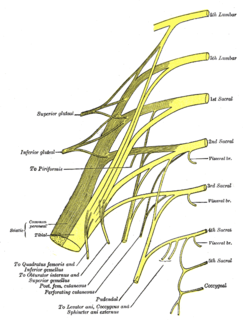
The perforating cutaneous nerve is a cutaneous nerve that supplies skin over the gluteus maximus muscle.

The inferior gluteal artery, the smaller of the two terminal branches of the anterior trunk of the internal iliac artery, is distributed chiefly to the buttock and back of the thigh.

The zygomaticofacial nerve or zygomaticofacial branch of zygomatic nerve passes along the infero-lateral angle of the orbit, emerges upon the face through the zygomaticofacial foramen in the zygomatic bone, and, perforating the orbicularis oculi to reach the skin of the malar area.

The supraclavicular nerves arise from the third and fourth cervical nerves; they emerge beneath the posterior border of the sternocleidomastoideus, and descend in the posterior triangle of the neck beneath the platysma and deep cervical fascia.

The transverse cervical nerve arises from the second and third spinal nerves, turns around the posterior border of the sternocleidomastoideus about its middle, and, passing obliquely forward beneath the external jugular vein to the anterior border of the muscle, it perforates the deep cervical fascia, and divides beneath the Platysma into ascending and descending branches, which are distributed to the antero-lateral parts of the neck. It provides cutaneous innervation to this area.

The posterior cutaneous nerve of forearm is a nerve found in humans and other animals. It is also known as the dorsal antebrachial cutaneous nerve, the external cutaneous branch of the musculospiral nerve, and the posterior antebrachial cutaneous nerve. It is a cutaneous nerve of the forearm.

The dorsal branch of ulnar nerve arises about 5 cm. proximal to the wrist; it passes backward beneath the Flexor carpi ulnaris, perforates the deep fascia, and, running along the ulnar side of the back of the wrist and hand, divides into two dorsal digital branches; one supplies the ulnar side of the little finger; the other, the adjacent sides of the little and ring fingers.

The anterior divisions of the seventh, eighth, ninth, tenth, and eleventh thoracic intercostal nerves are continued anteriorly from the intercostal spaces into the abdominal wall; hence they are named thoraco-abdominal nerves.

The anterior cutaneous branches of the femoral nerve consist of the following nerves: intermediate cutaneous nerve and medial cutaneous nerve.

The medial calcaneal branches of the tibial nerve perforate the laciniate ligament, and supply the skin of the heel and medial side of the sole of the foot.

Cutaneous innervation refers to the area of the skin which is supplied by a specific nerve.

Elastosis perforans serpiginosa is a unique perforating disorder characterized by transepidermal elimination of elastic fibers and distinctive clinical lesions, which are serpiginous in distribution and can be associated with specific diseases.
Acquired perforating dermatosis is clinically and histopathologically similar to perforating folliculitis, also associated with chronic kidney failure, with or without hemodialysis or peritoneal dialysis, and/or diabetes mellitus, but not identical to Kyrle disease.
Reactive perforating collagenosis is a rare, familial, nonpuritic skin disorder characterized by papules that grow in a diameter of 4 to 6mm and develop a central area of umbilication to which keratinous material is lodged. The cause of reactive perforating collagenosis is unknown.
Perforating calcific elastosis is an acquired, localized cutaneous disorder, most frequently found in obese, multiparous, middle-aged women, characterized by lax, well-circumscribed, reticulated or cobble-stoned plaques occurring in the periumbilical region with keratotic surface papules.
The perforating branches of the internal thoracic artery pierce through the internal intercostal muscles of the superior six intercostal spaces. These small arteries run with the anterior cutaneous branches of the intercostal nerves.
Perforator flap surgery is a technique used in reconstructive surgery where skin and/or subcutaneous fat are removed from a distant or adjacent part of the body to reconstruct the excised part. The vessels that supply blood to the flap are isolated perforator(s) derived from a deep vascular system through the underlying muscle or intermuscular septa. Some perforators can have a mixed septal and intramuscular course before reaching the skin. The name of the particular flap is retrieved from its perforator and not from the underlying muscle. If there is a potential to harvest multiple perforator flaps from one vessel, the name of each flap is based on its anatomical region or muscle. For example, a perforator that only traverses through the septum to supply the underlying skin is called a septal perforator. Whereas a flap that is vascularised by a perforator traversing only through muscle to supply the underlying skin is called a muscle perforator. According to the distinct origin of their vascular supply, perforators can be classified into direct and indirect perforators. Direct perforators only pierce the deep fascia, they don't traverse any other structural tissue. Indirect perforators first run through other structures before piercing the deep fascia.
References
- ↑ Freedberg, et al. (2003). Fitzpatrick's Dermatology in General Medicine. (6th ed.). McGraw-Hill. ISBN 0-07-138076-0.
| This condition of the skin appendages article is a stub. You can help Wikipedia by expanding it. |